Introduction
Hemangioma is the most common benign tumor of
infancy. The incidence in newborns is 2–3% and this increases to
∼10% by the age of 1 year (1).
Typically, hemangioma lesions appear in the early postnatal period
(1–2 weeks), rapidly proliferate for up to 12 months and gradually
involute over 5–10 years (2,3).
Infantile hemangioma has been suggested to arise from the clonal
expansion of endothelial cells, based on the analysis of
X-chromosome inactivation patterns (2,4). It
may also be a result of somatic mutations in one or more genes that
affect endothelial cell growth (5). However, the pathogenesis of
hemangiomas remains largely unknown. Although the majority of
hemangiomas are benign and resolve spontaneously, ∼10% may cause
cosmetic and life-threatening complications and thus require
treatment (6).
Several pharmacological therapies are currently
available for patients with problematic hemangiomas, including
steroids, interferon-α, bleomycin, vincristine and β-blockers
(7,8). Induction of apoptosis represents an
important mechanism for drug-induced hemangioma regression
(9). Generally, there are two main
apoptotic pathways: The extrinsic or death receptor pathway and the
intrinsic or mitochondrial pathway (10,11).
The two pathways converge on the activation of caspases, which
comprise a family of cysteine proteases and play a central role in
the execution of apoptosis. In the mitochondrial-initiated pathway,
caspase activation is triggered by the formation of a multimeric
Apaf-1/cytochrome c complex, which is implicated in the
activation of procaspase-9. Activated caspase-9 then cleaves and
activates downstream caspases, including caspase-3, -6 and -7,
ultimately culminating in apoptosis.
Propranolol is a non-selective β-blocker commonly
used in the treatment of cardiovascular diseases, including heart
failure and hypertension. The therapeutic benefit of propranolol in
hemangiomas was accidentally identified in 2008 by Léauté-Labrèze
et al (12) who noted the
regression of an infantile hemangioma following the administration
of propranolol for steroid-induced hypertrophic cardiomyopathy.
Since then, several other studies have also reported the
effectiveness of propranolol in the treatment of problematic
infantile hemangiomas (13,14).
In the current study, we aimed to investigate the molecular
mechanism(s) underlying the therapeutic effects of propranolol
against hemangiomas, using primary infantile hemangioma endothelial
cells (IHECs).
Materials and methods
Cell culture
Human IHECs isolated from a proliferating infantile
hemangioma were obtained from the Department of Pediatric Surgery,
Second Hospital of Xi’an Jiaotong University (Xi’an, China). Cells
were cultured in RPMI-1640 medium supplemented with 10% fetal
bovine serum, 10 μg/ml streptomycin and 100 U/ml penicillin
(Invitrogen Life Technologies, Carlsbad, CA, USA) at 37°C under 5%
CO2. Confluent cells were routinely subcultured using
trypsin-ethylenediaminetetraacetic acid (EDTA) solution (0.05%;
Invitrogen Life Technologies) and cells at passages 5–6 were used
in this study. The study was approved by the medical ethics
committee of Xi’an Jiaotong University and informed consent was
obtained from the patient’s family.
Drug treatment
Cells were seeded in quadruplicate at a density of
1×10⁵ cells per well into 24-well plates. After
incubation overnight at 37°C, cells were left untreated as the
controls or treated with propranolol (Tianjin Lisheng
Pharmaceutical Co., Ltd., Tianjin, China) at 40, 50 or 60
μg/ml for 24 h. Morphological and biochemical changes of the
cells were then examined using the following methods.
Morphological changes detected by light
microscopy
Cell morphology was examined every 1 h until 40 h
after indicated treatments under a contrast-phase microscope
(Leica, Bensheim, Germany).
Transmission electron microscopy
(TEM)
For TEM examination, cells were harvested 24 h after
drug treatment. The cells were prefixed in 2.5% glutaraldehyde,
postfixed in 1% osmium tetroxide and dehydrated in an ascending
series of ethanol to 100%. The cell samples were then embedded and
cut into ultrathin sections (50–70 nm). Sections were stained with
0.5% uranyl acetate and saturated lead citrate and examined using
an electron microscope (H-600, Hitachi, Tokyo, Japan).
Apoptosis analysis
After drug treatment for 24 h, cells were collected,
washed and subjected to apoptosis analysis using an Annexin
V-fluorescein isothiocyanate (FITC) kit (Trevigen, Gaithersburg,
MD, USA), according to the manufacturer’s instructions. Cells were
analyzed using a FACScan flow cytometer with CellQuest software (BD
Biosciences, Franklin lakes, NJ, USA).
Western blot analysis
The primary antibodies used were as follows:
Anti-caspase-8 (1:1,000 dilution), anti-cleaved caspase-3 (1:1,000
dilution), anti-lamin B1 (1:1,000 dilution), anti-cytochrome
c (1:500 dilution), anti-apoptosis-inducing factor (AIF;
1:500 dilution), anti-poly (ADP-ribose) polymerase 1 (PARP1;
1:1,000 dilution) and anti-glyceralde-hyde-3-phosphate
dehydrogenase (GAPDH; 1:1,000 dilution). These antibodies were
purchased from ProteinTech (Chicago, IL, USA). Samples of protein
extracts were resolved by sodium dodecyl sulfate-polyacrylamide gel
electrophoresis and transferred to nitrocellulose membranes (Pierce
Biotechnology, Inc., Rockford, IL, USA). After blocking with 5%
fat-free milk solution, the membranes were incubated overnight at
4°C with primary antibodies, followed by incubation with
horseradish peroxidase-linked secondary antibodies (Pierce
Biotechnology, Inc.) for 1 h at 37°C. Bound antibodies were
visualized by an enhanced chemiluminescence detection kit (Toyobo
Co., Ltd., Osaka, Japan). Signal intensities were quantitated using
Quantity One Software (Bio-Rad, Hercules, CA, USA).
Quantitative polymerase chain reaction
(qPCR)
Total RNA was extracted with TRIzol according to the
manufacturer’s instructions (Invitrogen Life Technologies). Reverse
transcription was performed using the First Strand cDNA Synthesis
kit (MBI Fermentas, Vilnius, Lithuania). qPCR amplification was
conducted using the IQ5.0 Real-Time PCR System (Bio-Rad) and
SYBR-Green PCR Master mix (Toyobo Co., Ltd.). The PCR primers used
are listed in Table I. The PCR
conditions were as follows: initial denaturation at 95°C for 1 min,
followed by 40 cycles of denaturation (95°C for 15 sec), annealing
(60°C for 15 sec) and extension (72°C for 45 sec) and then a last
extension at 72°C for 10 min. All assays were performed in
triplicate and the threshold cycle (Ct) was calculated. The
relative mRNA expression level normalized by that of β-actin (used
as an internal control) was then determined using the
2−ΔΔCt method (15).
 | Table I.Primers used for quantitative PCR
assays. |
Table I.
Primers used for quantitative PCR
assays.
| Gene | Primer sequence |
|---|
| Cytochrome
c | Sense:
GGTGATGTTGAGAAAGGCAAGAAG |
| Antisense:
GGCGGCTGTGTAAGAGTATCC |
| Caspase-3 | Sense:
CTGGACTGTGGCATTGAGAC |
| Antisense:
ACAAAGCGACTGGATGAACC |
| Caspase-8 | Sense:
ATTAGGGACAGGAATGGAACACAC |
| Antisense:
GGAGAGGATACAGCAGATGAAGC |
| AIF | Sense:
CCAGGCAACTTGTTCCAG |
| Antisense:
TTCATAGTCTTGTAGGCATAGG |
| Lamin B1 | Sense:
AATCGTTGTCAGAGCCTTAC |
| Antisense:
CCTTATACAGCCTCACTTGG |
| PARP1 | Sense:
ACCACTTCTCCTGCTTCTG |
| Antisense:
TCTTCTCTGCCTTGCTACC |
| β-actin | Sense:
ATCGTGCGTGACATTAAGGAGAAG |
| Antisense:
AGGAAGGAAGGCTGGAAGAGTG |
Statistical analysis
Significant differences between groups were
calculated using the Student’s t-test. One-way analysis of variance
followed by the Tukey’s post hoc test was used to examine
differences among multiple groups. P<0.05 was considered to
indicate a statistically significant difference.
Results
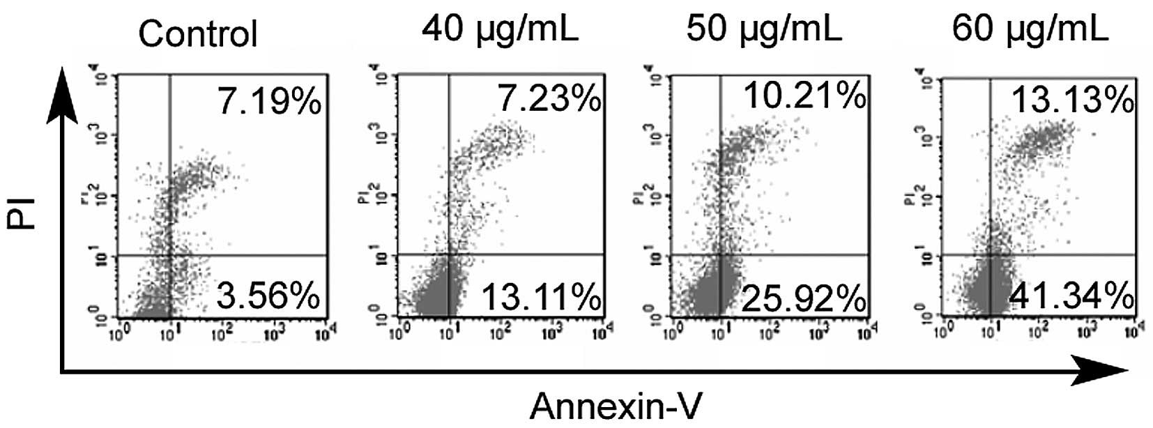
Effects of propranolol on the apoptosis
of IHECs
As shown in Fig. 1,
treatment with propranolol for 24 h resulted in a significant
increase in the apoptosis of IHECs, which occurred in a
concentration-dependent manner. Propranolol at 60 μg/ ml
caused ∼3-fold more apoptotic cells than propranolol at 40
μg/ml (54 vs. 20%).
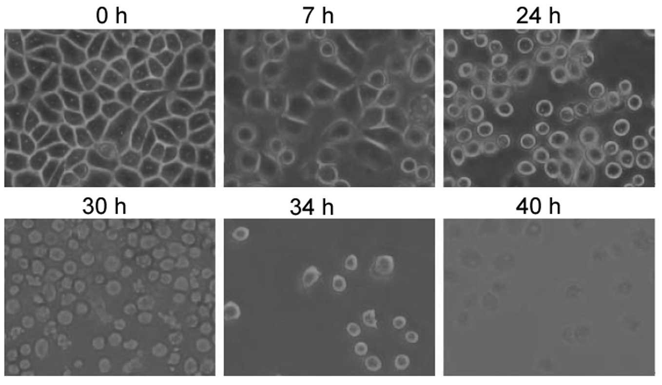
Effects of propranolol on the morphology
of IHECs
Morphological examination using phase-contrast
microscopy revealed that the IHECs became round and a few were
detached from the culture plates at 24 h after exposure to
propranolol at a concentration of 50 mg/ml (Fig. 2). At 30 h after drug treatment, the
IHECs presented a shrunken cytoplasm, with an intact plasma
membrane, indicative of the typical morphology of apoptosis.
Apoptosis was observed in the majority of IHECs exposed to
propranolol for 40 h. Consistent with the results of light
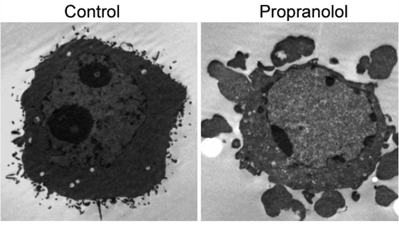
microscopy, TEM examination confirmed the induction of apoptosis in
the IHECs treated with 50 μg/ml propranolol for 24 h
(Fig. 3). Control cells presented
a normal morphology (Fig. 3A). By
contrast, the propranolol-treated cells exhibited typical
characteristics of apoptosis, including an intact cell membrane,
chromatin condensation, fragmentation of nuclei and the formation
of apoptotic bodies (Fig. 3B).
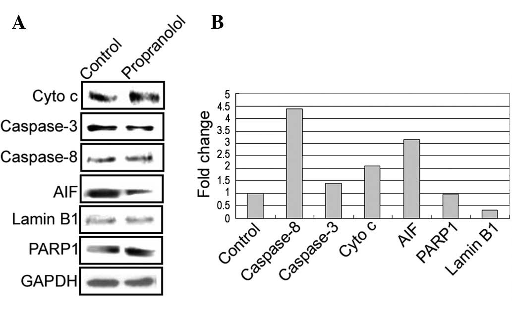
Protein and mRNA levels of
apoptosis-related genes
Western blot analysis revealed that the
propranolol-treated cells had a marked increase in protein levels
of caspase-8, cytochrome c, apoptosis-inducing factor,
caspase-3 and poly (ADP-ribose) polymerase 1, as well as a
concomitant reduction in lamin B1, (Fig. 4A). The qPCR assay further confirmed
similar changes at the mRNA level upon exposure to propranolol
(Fig. 4B).
Discussion
Propranolol is gaining increasing attention in the
treatment of infantile hemangiomas. Bertrand et al (13) reviewed a series of 35 consecutive
patients with infantile hemangioma and reported that oral
propranolol was effective in controlling the proliferative phase of
problematic infantile hemangioma, without causing serious adverse
effects. A meta-analysis study conducted by Izadpanah et al
(16) revealed that propranolol
achieves a greater response rate in the treatment of infantile
hemangiomas in comparison with corticosteroids (99 vs. <90%).
Propranolol has been reported to have the ability to block cell
proliferation and migration of IHECs (17). Our results further elucidated the
biological role of propranolol in IHECs and demonstrated that
exposure to propranolol caused significant apoptosis in IHECs.
Moreover, this pro-apoptotic effect was shown to be
concentration-dependent. Apoptosis is regarded as an active
suicidal response, which is characterized by nuclear condensation
and fragmentation, cellular shrinkage without loss of plasma
membrane integrity and the formation of apoptotic bodies. Forty
hours after treatment with a moderate dose (50 μg/ml) of
propranolol, almost all the cells were observed to go apoptosis.
These data provide an explanation for the excellent performance of
propranolol in the treatment of infantile hemangiomas.
Propranolol-induced apoptosis of hemangioma endothelial cells has
been documented in previous studies (18,19).
It is well accepted that apoptosis is induced by two
distinct pathways, extrinsic and intrinsic (20). The extrinsic pathway involves the
ligation of death receptors, the activation of caspase-8 and the
cleavage and activation of effector caspases, particularly
caspase-3. The intrinsic signaling pathways involve a diverse array
of non-receptor-mediated stimuli, which initiate different
intracellular signal cascades that converge at the level of the
mitochondria, leading to mitochondrial membrane permeabilization
and the release of pro-apoptotic factors, including cytochrome
c and AIF, into the cytoplasm. Propranolol has been shown to
exert growth-suppressive effects on pancreatic cancer cells through
the induction of caspase-9-and caspase-3-mediated apoptosis
(17). Similarly, Ji et al
(19) demonstrated that treatment
with propranolol induces apoptosis in hemangioma-derived
endothelial cells, which involves the activation of caspase-9 and
caspase-3 of the intrinsic pathway. In agreement with these
observations, our data revealed that treatment with propranolol
initiated the intrinsic apoptotic pathway in IHECs, as shown by the
increased release of cytochrome c and AIF into the cytoplasm
and the enhanced cleavage of caspase-3 and PARP1. Moreover,
propranolol treatment also increased the level of caspase-8,
suggesting an involvement of the extrinsic apoptotic pathway. Lamin
B1 is a member of the nuclear lamin family of proteins and is
thought to be involved in nuclear stability and chromatin
structure. Lamin B1 degradation is recognized as an early feature
of apoptosis (21). Our data
indicated a marked reduction of the levels of lamin B1 following
propranolol treatment, suggesting that the downregulation of this
gene is involved in propranolol-induced apoptosis.
However, there are a number of limitations of the
present study that should be noted. Firstly, the detailed signaling
pathways involved in the propranolol-induced apoptosis of IHECs
continue to require definition. Additionally, it remains unclear
whether the findings of this study may be translated into an in
vivo setting.
The induction of apoptosis has been regarded as a
preferred and superior strategy for clearing tumor cells. The
retention of plasma membrane integrity during apoptosis prevents
the onset of an inflammatory response that favors tumor progression
(22). Our data collectively
demonstrate that propranolol is capable of inducing apoptosis in
IHECs through activation of the intrinsic and extrinsic apoptotic
pathways, which represents an important mechanism for its
therapeutic effects against infantile hemangiomas.
Acknowledgements
This study was supported by the
National Natural Science Foundation of China (No. 30700954). The
authors appreciate technical assistance from Shanghai Zhongke New
Life Science & Technology (Shanghai, China).
References
|
1.
|
Jacobs AH and Walton RG: The incidence of
birthmarks in the neonate. Pediatrics. 58:218–222. 1976.PubMed/NCBI
|
|
2.
|
Boye E, Yu Y, Paranya G, Mulliken JB,
Olsen BR and Bischoff J: Clonality and altered behavior of
endothelial cells from hemangiomas. J Clin Invest. 107:745–752.
2001. View
Article : Google Scholar : PubMed/NCBI
|
|
3.
|
Mulliken JB: Cutaneous vascular anomalies.
Semin Vasc Surg. 6:204–218. 1993.PubMed/NCBI
|
|
4.
|
Bischoff J: Monoclonal expansion of
endothelial cells in hemangioma: an intrinsic defect with extrinsic
consequences? Trends Cardiovasc Med. 12:220–224. 2002. View Article : Google Scholar : PubMed/NCBI
|
|
5.
|
Walter JW, North PE, Waner M, Mizeracki A,
Blei F, Walker JW, Reinisch JF and Marchuk DA: Somatic mutation of
vascular endothelial growth factor receptors in juvenile
hemangioma. Genes Chromosomes Cancer. 33:295–303. 2002. View Article : Google Scholar : PubMed/NCBI
|
|
6.
|
Mendiratta V and Jabeen M: Infantile
hemangioma: an update. Indian J Dermatol Venereol Leprol.
76:469–475. 2010. View Article : Google Scholar : PubMed/NCBI
|
|
7.
|
Mabeta P and Pepper MS: Hemangiomas -
current therapeutic strategies. Int J Dev Biol. 55:431–437. 2011.
View Article : Google Scholar : PubMed/NCBI
|
|
8.
|
Itinteang T, Withers AH, Leadbitter P, Day
DJ and Tan ST: Pharmacologic therapies for infantile hemangioma: is
there a rational basis? Plast Reconstr Surg. 128:499–507. 2011.
View Article : Google Scholar : PubMed/NCBI
|
|
9.
|
Peng Q, Liu W, Zhou F, Wang Y and Ji Y: An
experimental study on the therapy of infantile hemangioma with
recombinant interferon γ. Pediatr Surg. 46:496–501. 2011.PubMed/NCBI
|
|
10.
|
Elmore S: Apoptosis: a review of
programmed cell death. Toxicol Pathol. 35:495–516. 2007. View Article : Google Scholar : PubMed/NCBI
|
|
11.
|
Budihardjo I, Oliver H, Lutter M, et al:
Biochemical pathways of caspase activation during apoptosis. Annu
Rev Cell Dev Biol. 15:269–290. 1999. View Article : Google Scholar : PubMed/NCBI
|
|
12.
|
Léauté-Labrèze C, Dumas de la Roque E,
Hubiche T, Boralevi F, Thambo JB and Taïeb A: Propranolol for
severe hemangiomas of infancy. N Engl J Med. 358:2649–2651.
2008.PubMed/NCBI
|
|
13.
|
Bertrand J, Sammour R, McCuaig C, Dubois
J, Hatami A, Ondrejchak S, Boutin C, Bortoluzzi P, Laberge LC and
Powell J: Propranolol in the treatment of problematic infantile
hemangioma: review of 35 consecutive patients from a vascular
anomalies clinic. J Cutan Med Surg. 16:317–323. 2012.
|
|
14.
|
Zegpi-Trueba MS, Abarzúa-Araya A,
Silva-Valenzuela S, Navarrete-Dechent C, Uribe-González P and
Nicklas-Díaz C: Oral propranolol for treating infantile
hemangiomas: a case series of 57 patients. Actas Dermosifiliogr.
103:708–717. 2012. View Article : Google Scholar : PubMed/NCBI
|
|
15.
|
Livak KJ and Schmittgen TD: Analysis of
relative gene expression data using real-time quantitative PCR and
the 2(−Delta Delta C(T)) method. Methods. 25:402–408. 2001.
|
|
16.
|
Izadpanah A, Izadpanah A, Kanevsky J,
Belzile E and Schwarz K: Propranolol versus corticosteroids in the
treatment of infantile hemangioma: a systematic review and
meta-analysis. Plast Reconstr Surg. 131:601–613. 2013. View Article : Google Scholar : PubMed/NCBI
|
|
17.
|
Zhang D, Ma Q, Shen S and Hu H: Inhibition
of pancreatic cancer cell proliferation by propranolol occurs
through apoptosis induction: the study of beta-adrenoceptor
antagonist’s anticancer effect in pancreatic cancer cell. Pancreas.
38:94–100. 2009.PubMed/NCBI
|
|
18.
|
Wong A, Hardy KL, Kitajewski AM, Shawber
CJ, Kitajewski JK and Wu JK: Propranolol accelerates adipogenesis
in hemangioma stem cells and causes apoptosis of hemangioma
endothelial cells. Plast Reconstr Surg. 130:1012–1021. 2012.
View Article : Google Scholar : PubMed/NCBI
|
|
19.
|
Ji Y, Li K, Xiao X, Zheng S, Xu T and Chen
S: Effects of propranolol on the proliferation and apoptosis of
hemangioma-derived endothelial cells. J Pediatr Surg. 47:2216–2223.
2012. View Article : Google Scholar : PubMed/NCBI
|
|
20.
|
Kroemer G, Galluzzi L and Brenner C:
Mitochondrial membrane permeabilization in cell death. Physiol Rev.
87:99–163. 2007. View Article : Google Scholar : PubMed/NCBI
|
|
21.
|
Neamati N, Fernandez A, Wright S, Kiefer J
and McConkey DJ: Degradation of lamin B1 precedes oligonucleosomal
DNA fragmentation in apoptotic thymocytes and isolated thymocyte
nuclei. J Immunol. 154:3788–3795. 1995.PubMed/NCBI
|
|
22.
|
Sethi G, Shanmugam MK, Ramachandran L,
Kumar AP and Tergaonkar V: Multifaceted link between cancer and
inflammation. Biosci Rep. 32:1–15. 2012. View Article : Google Scholar : PubMed/NCBI
|