Introduction
Bock greenbrier rhizome (1), also known as Smilax china L.,
induces heat clearing and detoxification and dispels wind dampness
according to traditional Chinese medicine. It is used to treat pain
associated with rheumatic arthritis and injuries from falls,
fractures, contusions and strains in traditional Chinese medicine.
There have been numerous studies showing that Smilax china
L. has certain antitumor effects against various types of cancer,
including lung, stomach, liver and colon cancer. The researchers
screened 50 kinds of Chinese herbals by MTT method and they found
out Smilax China L has antitumor effects (2). Compositions of Smilax China L
inclusions are complex, including: steroidal saponins, flavonoids,
polyphenols, stilbene type and tannins. Which have antitumor
effects (3,4). Study found out apigenin have induced
effect on C50 and 308 skin cells in mice and human leukemia HL 60
cell cycle arrest at G2 / M phase (5). However, the underlying mechanism of
its antitumor activity remains to be elucidated (6). DNA polymerase delta δ of B family
(DNA polymerase δ, pol δ) is the only one enzyme With the relevant
to cell cycle, and plays an important role in DNA replication,
repair, restructuring, and cell cycle regulation (7). POLD1 gene is the key of DNA
polymerase delta catalytic subunit gene, and plays an important
role on cell growth and differentiation. OU (8) detected POLD1 gene expression by
RT-PCR, and found out that the expression in liver cancer tissue
was significantly higher than tissue adjacent to carcinoma. The
research showed that primary liver cancer associated with POLD1
gene. Basis on these studies, we have done deep research.
In the present study, serum pharmacological methods,
3-(4,5-dimethylthiazol-2-yl)-2,5-diphenyltetrazolium bromide (MTT)
assay, flow cytometry and fluorescence quantitative polymerase
chain reaction (PCR) were used to determine the effects of bock
greenbrier rhizome on human hepatocellular carcinoma (HCC) cell
growth and the expression of DNA polymerase δ catalytic subunit
gene 1 (POLD1). The mechanism underlying the antitumor activity of
Smilax china L. in HCC cells was also investigated.
Materials and methods
Drugs
Smilax china L. was purchased from the
Guangxi Institute of National Medicine (Nanning, China). The
Smilax china L. (200 g) was soaked in water for 30 min
followed by gentle boiling for 30 min. The mixture was then cooled
for 30 min and the residue was filtered to obtain the extract. This
procedure was repeated twice, and the resulting products were
mixed. The drug extracts were reduced to a volume of 100 ml, which
contained 2 g/ml crude drug, and stored. Cyclophosphamide (CTX) was
provided by Shanghai Fahrenheit Pharmaceutical Co., Ltd. (Shanghai,
China).
Animals and cell lines
Adult Sprague Dawley (SD) rats were purchased from
the Experimental Animal Center of Guangxi Medical University
(Nanning, China). The SMMC-7721 HCC cell line was obtained from the
Cancer Institute of Guangxi Medical University (Nanning, China).
The study was approved by the Ethicical Review Committee of The
First Affiliated Hospital of Guangxi Medical University (Nanning,
China).
Animal model and treatment
Healthy SD rats (weight, 220–250 g) were allocated
to the following three groups: blank control (group A; n=10), CTX
(group B; n=10) and Smilax china L. (group C; n=10). Group A
was given a gavage of saline, and group C received a gavage of
Smilax china L. at a dose of 1.5 ml/100 g of body weight.
The rats of group B were treated with an intramuscular injection of
CTX at a dose of 25 mg/kg. According to the serum pharmacological
flux method (9), the animals were
dosed twice a day for 3 days. The animals were given water while
fasting for 12 h. Blood was drawn following the last intramuscular
injection or intra-gastric administration. Following treatment,
blood samples were obtained from the abdominal aorta when the rats
were under anesthesia with pentobarbital sodium (0.35 ml/100 g).
All the animals were treated humanely according to the
institutional guidelines of the Ethics Committee of The First
Affiliated Hospital Of Guangxi Medical University. The blood
samples were centrifuged at 1,006.2 x g for 15 min at 4°C to
completely remove the cellular components. After incubating in a
water bath at 56°C for 30 min (to achieve inactivation), the sample
was sterile filtered through a 0.22-μm microporous membrane
filter. The supernatant was collected and stored at −20°C.
Cell culture
All the cells were cultured in Roswell Park Memorial
Institute (RPMI)-1640 (Thermo Fisher Scientific, West Palm Beach,
FL, USA) supplemented with 10% fetal bovine serum (FBS; Beijing
Solarbio Science & Technology Co., Ltd., Beijing, China) in a
humidified incubator with 5% CO2 at 37°C. The culture
solution was replaced every 1–2 days, and the cells were passaged
once. Exponentially growing cells were selected for the
experiments.
MTT assay
Exponentially growing cells were harvested by
trypsin digestion and made into single-cell suspension after cell
counting. After adjusting the cell concentration to
1×10⁴/ml, the cells were added to a 96-well plate (100
μl per well). The cells were supplemented with 10% FBS and
stored in a humidified incubator with 5% CO2 at 37°C.
After the cells had been cultured for 24 h, the culture medium was
removed. The wells were divided into a blank control low
concentration group (10 μl blank rat serum + 90 μl
RPMI-1640 per well), a blank control high concentration group (30
μl blank rat serum + 70 μl RPMI-1640 per well), a CTX
low concentration group (10 μl blank rat serum + 90
μl RPMI-1640 per well), a CTX high concentration group (30
μl blank rat serum + 70 μl RPMI-1640 per well), a
Smilax china L. low concentration group (10 μl blank
rat serum + 90 μl RPMI-1640 per well) and a Smilax
china L. high concentration group (30 μl blank rat serum
+ 70 μl RPMI-1640 per well). After continuous culture for 24
h, 10 μl MTT (Sigma, St. Louis, MO, USA) was added to each
well. This mixture was allowed to incubate for 4 h. Subsequently,
100 μl dimethylsulfoxide (DMSO) was added to each well. The
plate was oscillated for 10 min until the MTT-formazan was
completely dissolved. The absorbance was measured at 490 nm to
determine the cell viability. Each experiment was performed three
times.
Apoptosis detected by flow cytometry
The exponentially grown SMMC-7721 cells were
harvested (using a 2.5 g/ml trypsin digestion), made into a single
cell suspension and counted. After the cell concentration was
adjusted to 4×10⁴/ml, the cells were added to a 6-well
plate (2 ml per well) supplemented with 10% FBS and grown in a
humidified incubator with 5% CO2 at 37°C. After 24 h,
the culture medium was removed. For the apoptosis analysis, the
SMMC-7721 cells were plated into 6-well plates and treated with
various quantities (10–30%) of blank serum, and serum containing
either CTX and or Smilax china L. for 24 h. The cell culture
fluid was then collected. The cells were washed once with
phosphate-buffered saline (PBS) at 4°C, subjected to trypsin
digestion (2.5 g/l) and then placed in cell nutrient solution. The
cells were then blended and centrifuged at 447.2 × g for 5 min.
Then, the supernatant was removed. The cells were incubated in the
dark for 10 min at room temperature before adding 500 μl
binding buffer, 5 μl Annexin V-fluorescein isothiocyanate
(FITC) and 5 μl propidium iodide (PI; Beijing DingGuo
ChangSheng Biotechnology Co., Ltd., Beijing, China). The apoptotic
rates were determined by flow cytometry (BD Biosciences, Franklin
Lakes, NJ, USA). The percentage of early apoptotic cells was
calculated through the determination of Annexin V-positive and
PI-negative cells, and the percentage of late apoptotic cells was
calculated through the determination of Annexin V-positive and
PI-positive cells.
Cell cycle detection by flow
cytometry
The cells (plated at a density of
4×10⁴/ml per well in 6-well plates) were collected by
centrifugation. The cells were washed twice with PBS at 4°C.
Subsequently, the cells were fixed in 1 ml cold 70% ethanol and
fixed at −20°C overnight. Centrifugation was repeated. The fixed
cells were washed once with PBS at 4°C, treated with 0.2 ml PBS and
RNase A (50 μg/ml), and incubated for 30–45 min at 37°C
after a 1-min ice bath. The cells were stained with PI (65
μg/ml; Beijing Tripod Countries Biological Technology Co.,
Ltd.) in the dark for 30 min at 4°C. The cell cycle was analyzed by
flow cytometry (BD Biosciences).
Quantitative PCR
The total RNA was extracted using TRIzol solution
(Invitrogen, Carlsbad, CA, USA), according to the manufacturer’s
instructions. RNA was then converted to cDNA by reverse
transcription. The reactions were placed in a 96-well plate using a
preheated real-time instrument (ABI 7500 HT; Applied Biosystems,
Foster City, CA, USA). The POLD1 specific primers were:
5′-GACTACACGGGA GCCACTGTCA-3′ (sense) and 5′-GTAACACAGGTTGTG
GGCCATC-3′ (antisense), with an amplicon length of 117 bp. β-actin
(Gene ID: 60) was used as an internal control for RNA integrity
with the following specific primers: 5′-AACTCCATCATGAAGTGTGA-3′
(sense) and 5′-ACTCC TGCTTGCTGATCCAC-3′ (antisense) (length, 247
bp). Each 10 μl reaction volume contained 1 μl cDNA,
which was synthesized as described above. The reaction also
contained 10 μl Power SYBR®-Green Master mix and
1 μl each pair of the oligonucleotide primers described
above. Each reaction was performed thrice. The cycling parameters
were as follows: 95°C for 10 min, followed by 40 cycles of PCR
amplification performed at 95°C for 15 sec, annealing at 62°C for
31 sec, and extension at 95°C for 15 sec. The Ct value was measured
during the exponential amplification phase. The data were analyzed
using the comparative threshold cycle
(2−ΔΔCt) method.
Statistical analysis
Data are expressed as the mean ± SEM. The
differences among the groups were determined by ANOVA analysis, and
comparisons between two groups were analyzed by SNK analysis.
Differences between two groups were determined using Student’s
t-test. P<0.05 was considered to indicate a statistically
significant difference.
Results
Cell growth inhibition
The growth of SMMC-7721 human hepatoma cells was
inhibited by bock greenbrier rhizome, as detected by MTT assay
(Table I). Serum containing CTX or
Smilax china L., particularly in high concentrations, was
demonstrated to inhibit the proliferation of the SMMC-7721 human
HCC cell line. The highest rate of growth inhibition was observed
in the cells treated with CTX-containing serum. However, the growth
inhibition of the cells treated with Smilax china
L.-containing serum was not significantly different compared with
that of the cells treated with CTX-containing serum.
 | Table I.Effect of Smilax china
L.-containing serum on the proliferation of human HCC SMMC-7721
cells (mean ± SEM, n=6). |
Table I.
Effect of Smilax china
L.-containing serum on the proliferation of human HCC SMMC-7721
cells (mean ± SEM, n=6).
| Group | MTT (OD value) | Inhibition ratio
(%) |
|---|
| Blank | | |
| Low
concentration | 0.832±0.014 | - |
| High
concentration | 0.722±0.011 | - |
| CTX | | |
| Low
concentration | 0.774±0.017 | 6.97a |
| High
concentration | 0.563±0.019 | 22.02b |
| Smilax china
L. | | |
| Low
concentration | 0.807±0.022 | 3a |
| High
concentration | 0.588±0.017 | 18.56b |
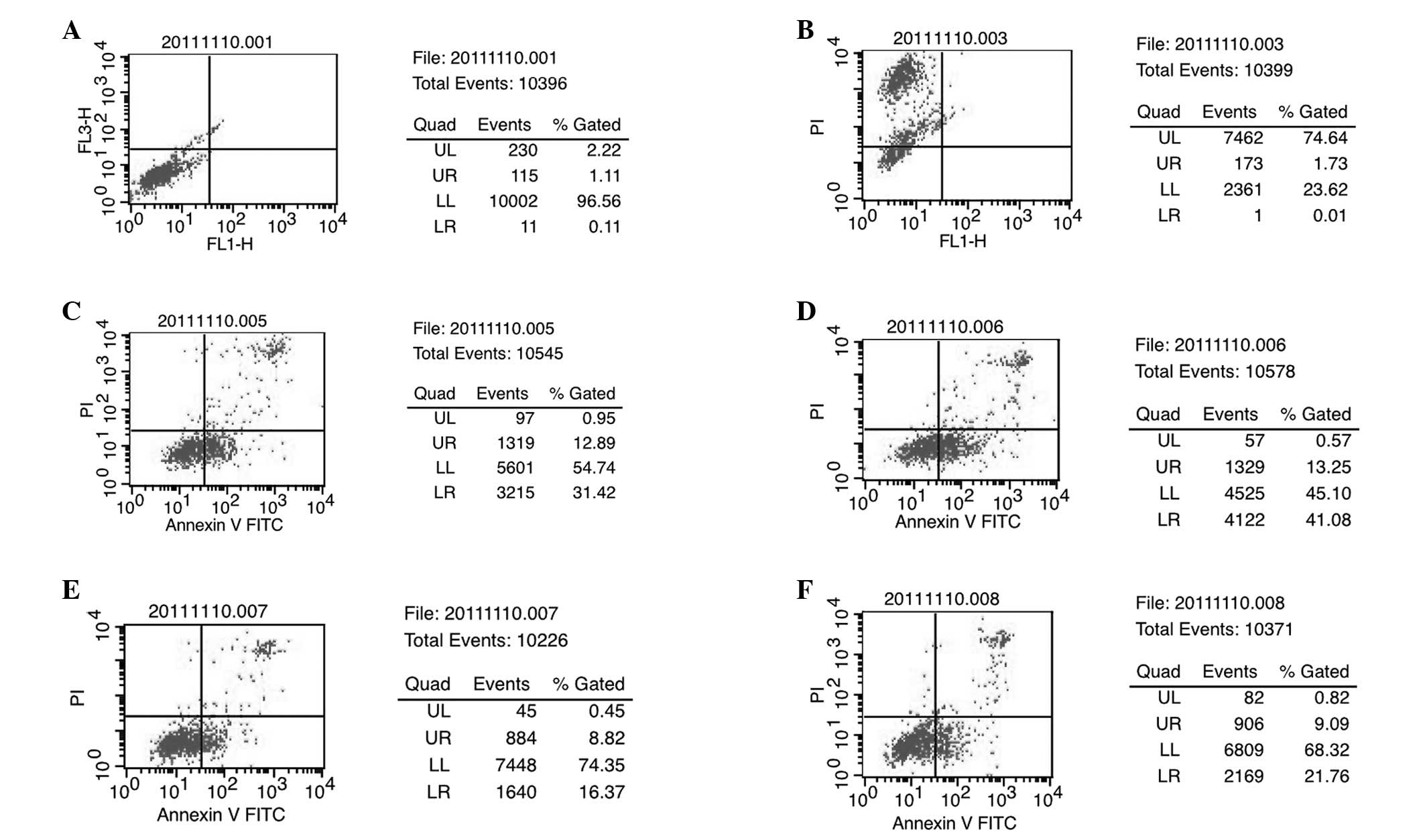
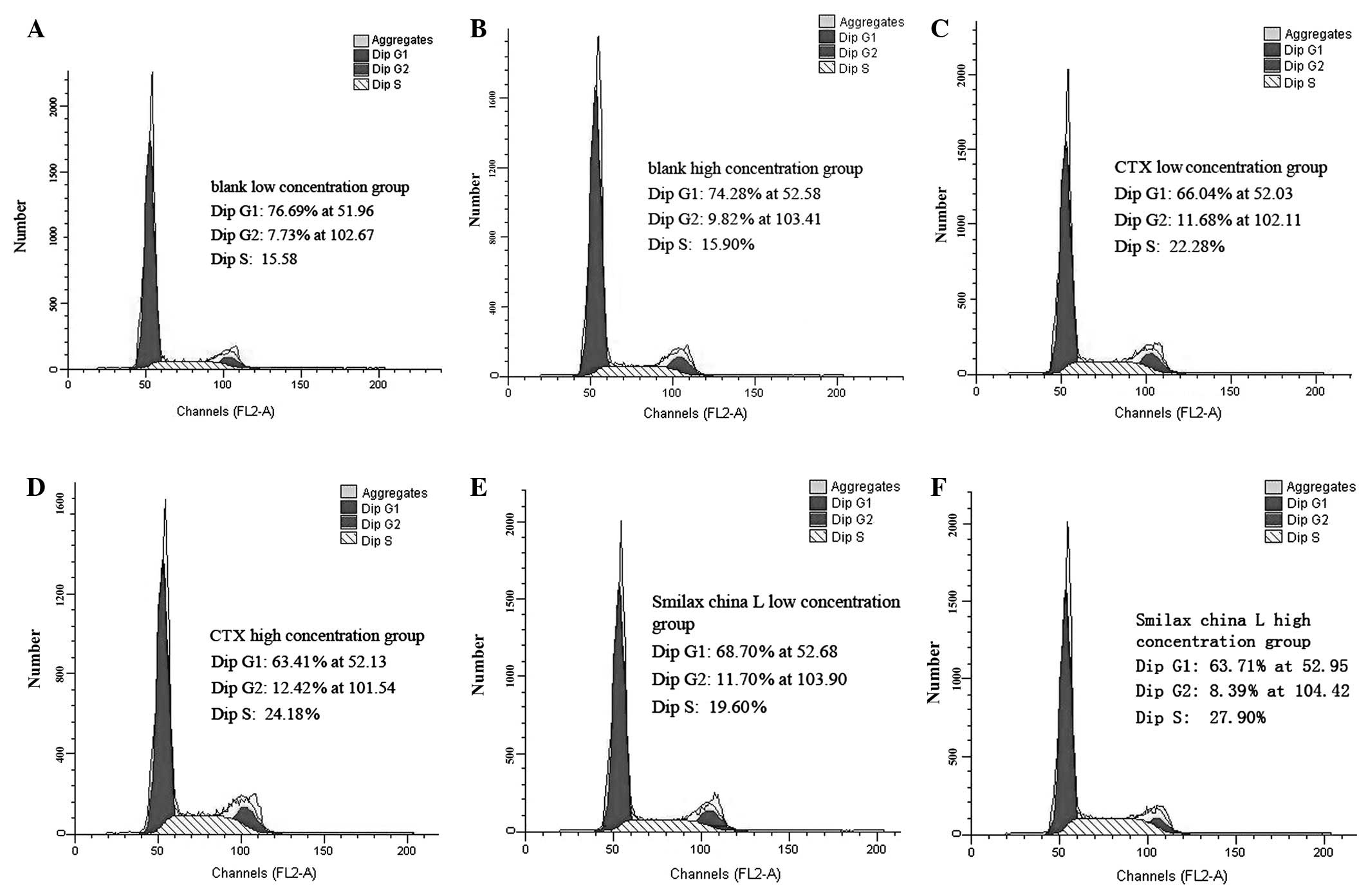
Cell cycle and apoptosis detection
Treatment with serum containing Smilax china
L. or CTX inhibited the proliferation of SMMC-7721 cells
(P<0.05). A particularly marked effect was observed with a high
concentration of CTX-containing serum. The percentages of cells in
the S phase increased following treatment with CTX- and Smilax
china L.-containing serum (P<0.05). However, no significant
differences were observed between the CTX- and Smilax china
L. groups (Table II, Figs. 1 and 2).
| Table II.Effect of serum from each group on the
apoptosis and cell cycle distribution of SMMC-7721 human HCC cells
(mean ± SEM, n=3). |
Table II.
Effect of serum from each group on the
apoptosis and cell cycle distribution of SMMC-7721 human HCC cells
(mean ± SEM, n=3).
| Group | Apoptosis ratio
(%) | Cell cycle (%)
|
|---|
| G0/G1 | S | G2/M |
|---|
| Blank | | | | |
| Low
concentration | 1.22±0.31 | 76.69±1.63 | 15.58±1.82 | 7.73±0.19 |
| High
concentration | 1.74±0.27 | 74.28±1.42 | 15.9±1.2 | 9.82±0.22 |
| CTX | | | | |
| Low
concentration | 44.31±2.14a | 66.04±1.53 | 22.28±1.3a | 11.68±0.23 |
| High
concentration | 54.33±3.43b | 63.41±1.21 | 24.18±1.3b | 12.41±0.09 |
| Smilax china
L. | | | | |
| Low
concentration |
25.19±2.2a,c | 68.7±1.8 | 19.6±2.1a | 11.7±0.3 |
| High
concentration |
30.85±3.1b,d | 63.71±3.1 | 27.9±2.7b | 8.39±0.4 |
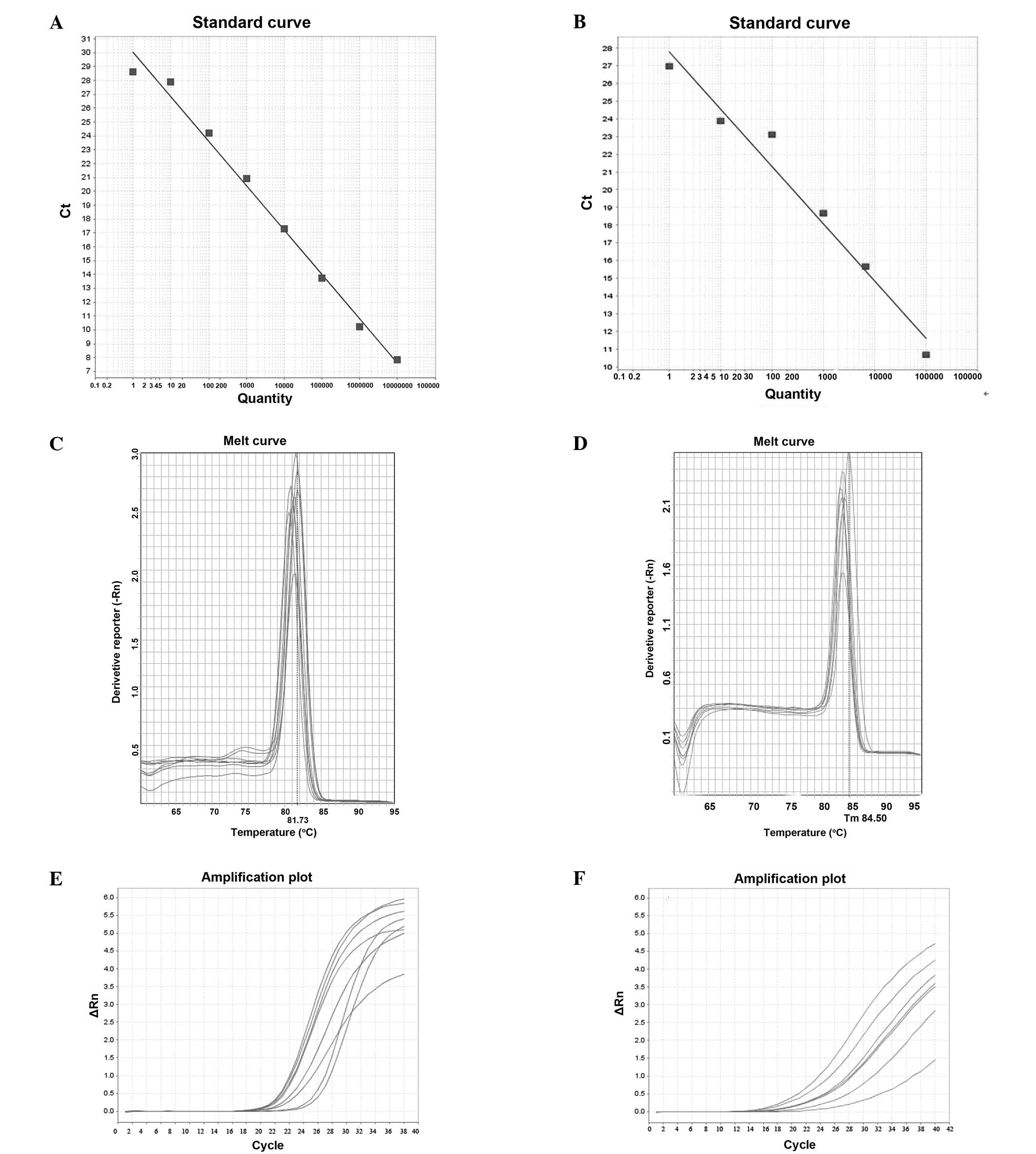
POLD1 mRNA expression
The mRNA expression of POLD1 was detected in HCC
cells cultured with serum containing Smilax china L. The
expression levels of POLD1 mRNA in the high and low concentration
groups (0.45±0.1 and 0.28±0.06) were significantly different from
the POLD1 mRNA expression levels in the blank group (P<0.05;
Table III and Fig. 3).
| Table III.Expression of POLD1 mRNA in each
group of SMMC-7721 human HCC cells (mean ± SEM). |
Table III.
Expression of POLD1 mRNA in each
group of SMMC-7721 human HCC cells (mean ± SEM).
| Group | Ct value
|
2−ΔΔCt |
|---|
| POLD1 | β-actin |
|---|
| Blank | 26.53±0.21 | 25.08±0.17 | 1 |
| Smilax china
L. high concentration | 26.70±0.11 | 24.47±0.19 | 0.45±0.1a |
| Smilax china
L. low concentration | 28.42±0.32 | 25.87±0.29 | 0.28±0.06a |
Discussion
HCC is one of the most frequent malignant tumors
worldwide. Particularly, HCC is the fifth most common malignant
tumor (10). Multiple gene
interactions lead to primary HCC. Several relevant genes, including
cancer (e.g., c-myc, c-fos, BC-12, cets2, Ras), tumor-suppressor
(e.g., p16, p53, p21), cell cycle regulatory and cell apoptosis
genes, as well as genes that maintain stability of the cell genome
(11). The malignant proliferation
of cancer cells is associated with the DNA bulk copy. DNA
polymerase δ (pol δ) is the only DNA replication enzyme that is
related to the cell cycle and plays a prominent role in DNA
replication (12). The POLD1 gene
is a key gene for the DNA polymerase δ catalytic subunit and plays
an important role in cell growth and differentiation. Recent
studies have shown that genetic mutations in the POLD1 gene are
related to lymphoma, liver (8),
colon and gastric cancer (13,14).
Serum pharmacology (15) uses medicated serum as an object of
research. Serum pharmacology not only eliminates the interference
of the physical and chemical properties of traditional Chinese
medicine preparations, but it also reflects the last process of
digesting and absorbing Chinese traditional medicine in the
gastrointestinal tract. Moreover, serum pharmacology further
reflects the biological transformation and production of a
biological effect, and represents the real effective function of
drug components that affect the body. Therefore, it is appropriate
for Chinese medicine, which has complex chemical components.
In a previous study conducted by our group (16), HCC cells were cultured in
vitro with serum containing 10 different concentrations of a
Chinese medicine. MTT assay was then used to determine whether the
medicated serum affected cancer cell proliferation. According to
the results obtained, the growth of HCC cells was inhibited
following Smilax china L. and CTX treatment. Based on these
previous findings, a more detailed study was then conducted by our
group.
Smilax china L. is an herb with a complex
chemical composition and is classified as a Chinese herbal
medicine. It has a complex set of ingredients including steroid
saponins, flavonoids, polyphenols, stilbenes and tannins. Smilax
china L. is known to expel wind and dampness as well as to
scatter detoxification stasis (17,18).
Previous studies have shown that Smilax china L. has an
inhibitory effect on cancer cell growth (14,19).
However, its mechanism remains unknown and further study is
needed.
In the present study, we investigated the
antineoplastic mechanism by which Smilax china L. affects
SMMC-7721 cells using serum pharmacological methods. Tumor
pathogenesis has a close association with suppression of apoptosis.
In malignant tumor tissues, there are abnormal increases in the
proliferation of malignant cells and marked reductions in cell
apoptosis. Apoptosis defects constitute one of the pathogenic
mechanisms of tumors. Therefore, the induction of apoptosis has the
potential to inhibit and block tumor occurrence and development,
thereby increasing the ratio of cell apoptosis to
proliferation.
Apoptosis is a complicated cell death network and
involves many components. Numerous studies have shown that one of
the antineoplastic mechanisms of traditional Chinese medicine is
the induction of apoptosis (20,21).
In the present study, the serum of rats treated with high and low
concentrations of Smilax china L. induced the inhibition of
HCC cell proliferation and apoptosis in vitro. Thus,
Smilax china L.-containing serum induced apoptosis, which
inhibited SMMC-7721 cell proliferation in vitro.
The results obtained following cell cycle analysis
showed that the number of cells in the S+G2/M phase increased
following treatment with the serum from the Smilax china
L.-treated rats (the number of cells in the S phase was
significant) compared with the control cells. However, the number
of cells in the G0/G1 phase decreased. Therefore, Smilax
china L. is suggested to effectively mobilize cells from the
G0/G1 to the S phase. Statistical analysis also indicated that the
numbers of cells in the S+G2/M phase observed in the Smilax
china L.-treated groups were not significantly different from
those in the CTX-treated groups. Therefore, Smilax china L.
is suggested to be a cell cycle non-specific drug, similar to CTX.
Smilax china L. interferes with DNA and RNA function,
particularly DNA function (22,23).
Smilax china L. crosslinks with DNA and inhibits DNA
synthesis. The effect on the number of cells in the S phase was the
most evident. A study has confirmed that POLD gene expression is
strictly regulated by the cell cycle (13). In the G1/S phase, or even in the
early S phase, pol δ synthesis and POLD1 promoter activity
increases, suggesting that POLD1 gene regulation occurs mainly in
the G1/S phase.
Fluorescent quantitative PCR results showed that a
clear downregulation of POLD1 mRNA expression was observed in the
Smilax china L. high concentration group. The regulation of
POLD1 mRNA expression by the tumor suppressors p53 and Sp1 through
competing for the POLD1 promoter binding site is suggested to be a
potential mechanism underlying the activity of Smilax china
L. (24). It has been reported
that p53 binds directly to the POLD1 promoter in the cell (25). Another potential mechanism is p21
(a p53 transcription product) binding. p21 restrains E2F1 release
through binding to the POLD1 promoter at the E2F1 binding site,
thereby inhibiting POLD1 activity (19). Moreover, p21 binds to proliferation
cell antigen (PCNA), thus affecting the binding of PCNA and pol δ
and hindering DNA replication (25). Furthermore, cyclin E/CDK2
indirectly regulates POLD1 promoter activity through the regulation
of the downstream E2F1 family, which influences the expression of
POLD1 mRNA (26). Song et
al (27) demonstrated that the
upregulation of POLD1 mRNA expression by CDE/CHR gene mutations is
directly or indirectly regulated by p21 and E2F1. The regulation of
POLD and the associations among p53, p21, CDE/CDK, the cyclin/CDK
complex and POLD1 requires further study.
In conclusion, Smilax china L.-containing
serum inhibits HCC cell proliferation and induces apoptosis.
Smilax china L. also causes S phase cell cycle arrest in HCC
cells. This mechanism is suggested to be associated with the
inhibition of POLD1 gene expression.
Acknowledgements
The present study was funded by
Guangxi Health Plan Project Funds (project no. Z2012115).
References
|
1.
|
Liang QC and Zhong M: China Zhuang
nationality medicine. Guangxi Nationalities Publishing House;
Nanning: pp. 2262005
|
|
2.
|
Wei JY, Li Y, Wei T, et al: Drug screening
study of 50 kinds of Chinese herbal and zhuang nationality medicine
in guangxi. J Guangxi Traditional Chinese Med Univ. 4:3–7. 2003.(In
Chinese).
|
|
3.
|
Xu Y, Wang HY, Jiang JY, et al: Studies on
steroidal saponins from Smilax china and their cytotoxic
activities. Chinese J Experimental Traditional Med Formulae.
11:92–96. 2011.(In Chinese).
|
|
4.
|
Jiang LY and Liu YM: Study on the
relationship between structures and anti-tumor activity of
flavonoids compounds. Computers and Applied Chemistry. 4:21–24.
2005.
|
|
5.
|
Kohn K W, Aladjem MI, Weinstein JN and
Pommier Y: Chromatin challenges during DNA replication: a systems
representation. Mol Biol Cell. 19:1–7. 2008.PubMed/NCBI
|
|
6.
|
Zhang K and Ma SL: Progress on anticancer
molecule mechanism of naturally occurring drugs. China Journal of
Traditional Chinese Medicine and Pharmacy. 10:2344–2347. 2011.(In
Chinese).
|
|
7.
|
Lepley DM, Li B, Birt DF and Pelling JC:
The chemopreventive flavonoid apigenin induces G2/M arrest in
keratinocytes. Carcinogenesis. 17:2367–2375. 1996. View Article : Google Scholar : PubMed/NCBI
|
|
8.
|
Ou XH, Liao LF, Liu HG and Xu H:
Significance of POLD1 expression in primary hepatocellular
carcinoma. World Chin J Digestology. 19:151–155. 2011.(In
Chinese).
|
|
9.
|
Yan YQ: The Ideas and Methods of Modern
Research of Traditional Chinese Medicine. Chemical Industry Press;
Beijing: pp. 340–341. 2006
|
|
10.
|
Li F, Li SL and Yin YG: Construction of
NK4 recombinant lentiviral vector and its expression in HCCLM3
cells. J Chin Med Univ. 40:1081–1084. 2011.(In Chinese).
|
|
11.
|
Zhao YQ and Zhe ZF: Advances in gene
Treatment of Primary hepatic carcinoma. Disease Monitor and
Control. 8:482–484. 2011.(In Chinese).
|
|
12.
|
Lin XY, Zhang SR and Xu H: Advances of
Eukaryotic DNA polymerase δ. Progress in Natural Science. 1:3–9.
2005.(In Chinese).
|
|
13.
|
Ruan XL, Li YL and Wu Q: P21 suppresses
cell proliferation and downregulates POLD1 expression in human
gastric cancer cell line MGC-803. World Chin J Digestology.
19:1990–1995. 2011.(In Chinese).
|
|
14.
|
Liao ZJ, Zhang XM, Guo YH and Sun HF:
Study on the effect of proliferation and apoptosis of
sarsasapogenin to human gastric cancer line BGC-823. Modern
Oncology. 18:1085–1087. 2010.(In Chinese).
|
|
15.
|
Huang CH, Lu Y, Gao XJ, et al: Advances of
serum pharmacology of Chinese medicine. Chin J Exp Trad Med
Formulae. 17:266–271. 2011.(In Chinese).
|
|
16.
|
10 kinds of Zhuang medicine-containing
serum against human hepatocellular carcinoma cells’ effect and the
study of Jingangteng-containing serum affect the tumor suppressor
gene WWOX’s expression. Dissertation of Guangxi Med Univ.
14:2012.(In Chinese).
|
|
17.
|
Chinese Pharmacopoeia Commission:
Pharmacopoeia of People’s Republic of China. Beijing Chemical
Industry Press; pp. 2162005
|
|
18.
|
Zeng XX and Chen XL: Advances of
traditional application and modern research of Smilax china
L. Chinese Traditional and Herbal Drugs. 6:952–954. 2008.
|
|
19.
|
Wang XJ: Phenolic constituents from
Smilax china L and their antitumor activities. Dissertation
of Nanjing University of Science & Technology. 23:2009.(In
Chinese).
|
|
20.
|
Li Y, Yao LB, Wang LF, et al: Inducing
effect of resveratrol on anoikis of gastric cancer cells. Chinese
Traditional and Herbal Drugs. 10:64–66. 2004.
|
|
21.
|
Luo Q, Song GB, Qin J, et al: Progress of
research on hepato-cellular carcinoma cells apoptosis induced by
Chinese traditional medicine. Journal of Chongqing Univ. 7:115–117.
2005.
|
|
22.
|
Li GC, Dong KL and Hu CH: Effect of
compound Rhizoma Smilacinus granules on the expression of Bcl-2 and
Bax gene in A549 cell lines of non-small cell lung cancers. Zhong
Nan Da Xue Xue Bao Yi Xue Ban. 32:312–315. 2007.(In Chinese).
|
|
23.
|
Xu W: Anti-proliferative effect on tumor
cells of kaempferol-7-O-β-D-glucoside isolated from Smilax
china L. Rhizome. Dissertation of East China University of
Science and Technology. 42:2008.(In Chinese).
|
|
24.
|
Sanefuji K, Taketomi A, Iguchi T, et al:
Significance of DNA polymerase delta catalytic subunit p125 induced
by mutant p53 in the invasive potential of human hepatocellular
carcinoma. Oncology. 79:229–237. 2010. View Article : Google Scholar : PubMed/NCBI
|
|
25.
|
Xiaoyan L, Shuren Z and Heng X: Eukaryotic
DNA polymerase δ research present situation. J Prog Nat Sci.
17:716–723. 2007.
|
|
26.
|
Hansen K, Farkas T, Lukas J, et al:
Phosphorylation-dependent and -independent functions of p130
cooperate to evoke a sustained G1 block. EMBO J. 20:422–432. 2001.
View Article : Google Scholar : PubMed/NCBI
|
|
27.
|
Song N, Zhu X, Shi L, et al:
Identification and functional analysis of a CDE/CHR element in the
POLD1 promoter. Sci China C Life Sci. 52:551–559. 2009. View Article : Google Scholar : PubMed/NCBI
|