Aging-associated changes in L-type calcium channels in the left atria of dogs

  • Authors:
    • Tian-Yi Gan
    • Weiwei Qiao
    • Guo-Jun Xu
    • Xian-Hui Zhou
    • Bao-Peng Tang
    • Jian‑Guo Song
    • Yao-Dong Li
    • Jian Zhang
    • Fa-Peng Li
    • Ting Mao
    • Tao Jiang
  • View Affiliations

  • Published online on: August 20, 2013     https://doi.org/10.3892/etm.2013.1266
  • Pages: 919-924
Metrics: Total Views: 0 (Spandidos Publications: | PMC Statistics: )
Total PDF Downloads: 0 (Spandidos Publications: | PMC Statistics: )


Abstract

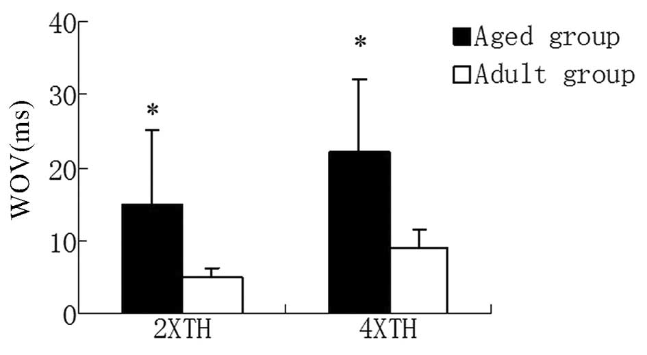
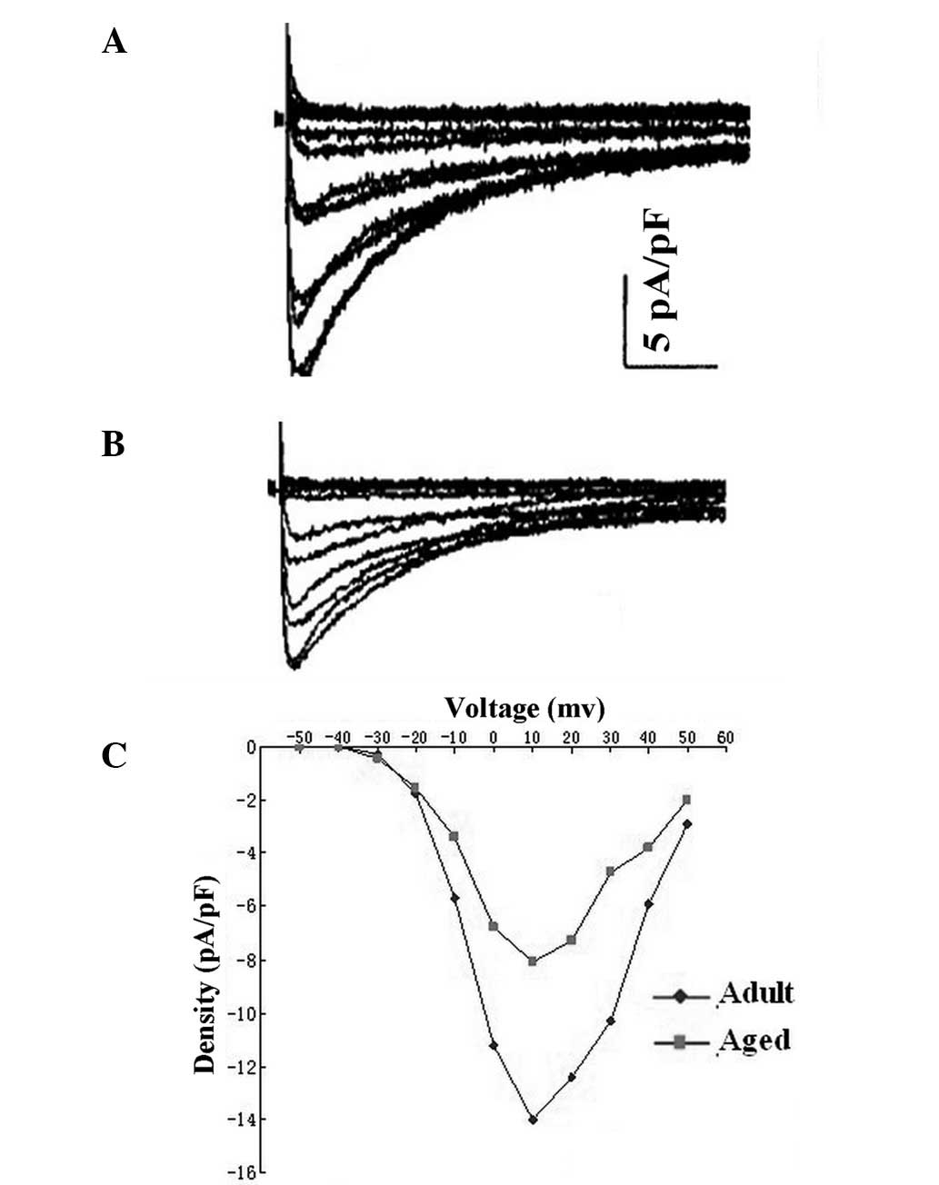
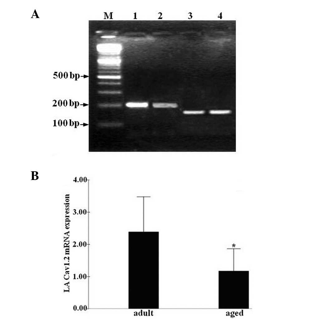
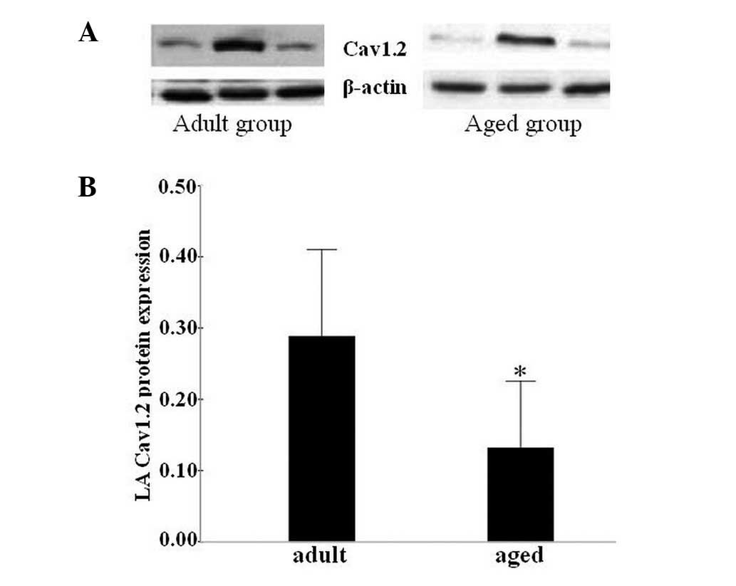
Action potential (AP) contours vary considerably between the fibers of normal adult and aged left atria. The underlying ionic and molecular mechanisms that mediate these differences remain unknown. The aim of the present study was to investigate whether the L-type calcium current (ICa.L) and the L-type Ca2+ channel of the left atria may be altered with age to contribute to atrial fibrillation (AF). Two groups of mongrel dogs (normal adults, 2-2.5 years old and older dogs, >8 years old) were used in this study. The inducibility of AF was quantitated using the cumulative window of vulnerability (WOV). A whole-cell patch-clamp was used to record APs and ICa.L in left atrial (LA) cells obtained from the two groups of dogs. Protein and mRNA expression levels of the a1C (Cav1.2) subunit of the L-type calcium channel were assessed using western blotting and quantitative PCR (qPCR), respectively. Although the resting potential, AP amplitude and did not differ with age, the plateau potential was more negative and the APD90 was longer in the aged cells compared with that in normal adult cells. Aged LA cells exhibited lower peak ICa.L current densities than normal adult LA cells (P<0.05). In addition, the Cav1.2 mRNA and protein expression levels in LA cells were decreased in the aged group compared with those in the normal adult group. The lower AP plateau potential and the decreased ICa.L of LA cells in aged dogs may contribute to the slow and discontinuous conduction of the left atria. Furthermore, the reduction of the expression levels of Cav1.2 with age may be the molecular mechanism that mediates the decline in ICa.L with increasing age.
View Figures
View References

Related Articles

Journal Cover

October 2013
Volume 6 Issue 4

Print ISSN: 1792-0981
Online ISSN:1792-1015

Sign up for eToc alerts

Recommend to Library

Copy and paste a formatted citation
x
Spandidos Publications style
Gan T, Qiao W, Xu G, Zhou X, Tang B, Song JG, Li Y, Zhang J, Li F, Mao T, Mao T, et al: Aging-associated changes in L-type calcium channels in the left atria of dogs. Exp Ther Med 6: 919-924, 2013
APA
Gan, T., Qiao, W., Xu, G., Zhou, X., Tang, B., Song, J. ... Jiang, T. (2013). Aging-associated changes in L-type calcium channels in the left atria of dogs. Experimental and Therapeutic Medicine, 6, 919-924. https://doi.org/10.3892/etm.2013.1266
MLA
Gan, T., Qiao, W., Xu, G., Zhou, X., Tang, B., Song, J., Li, Y., Zhang, J., Li, F., Mao, T., Jiang, T."Aging-associated changes in L-type calcium channels in the left atria of dogs". Experimental and Therapeutic Medicine 6.4 (2013): 919-924.
Chicago
Gan, T., Qiao, W., Xu, G., Zhou, X., Tang, B., Song, J., Li, Y., Zhang, J., Li, F., Mao, T., Jiang, T."Aging-associated changes in L-type calcium channels in the left atria of dogs". Experimental and Therapeutic Medicine 6, no. 4 (2013): 919-924. https://doi.org/10.3892/etm.2013.1266