Introduction
Polysaccharides are macromolecules that are able to
transfer large amounts of biological information between cells
(1). The biological activities of
polysaccharides may be developed and enhanced by means of chemical
modifications, which broaden their application and clinical use.
Sulfated modification may produce high activity, good functional
polysaccharides and polysaccharide derivatives, and the
introduction of sulfate groups may change the physicochemical
properties, three-dimensional conformation and activities of the
polysaccharides (2,3). Previous studies revealed that certain
polysaccharides without antitumor activity showed antitumor
activity following sulfated modification, while certain sulfated
polysaccharides with antitumor activity demonstrated a reduction or
loss of antitumor activity following the removal of sulfate groups
(4,5). The main methods used for the sulfated
modification of polysaccharides include the sulfuric acid method,
chlorosulfonic acid-pyridine method, chlorosulfonic
acid-carboxamide method, sulfur trioxide-pyridine method and sulfur
trioxide-dimethylformamide method. The chlorosulfonic acid-pyridine
method is widely used due to the high yields and degree of
substitution of the products (6,7).
Laminarin, also known as brown algae starch, is an
active component in kelp that shows numerous activities, including
antitumor, immunomodulatory, antibacterial, antiviral, blood lipid
regulating, anti-oxidation, anticlotting, antiradiation and
hypoglycemic activities (8,9). Ji
et al (10) reported that
laminarin was able to inhibit LoVo human colon cancer cell
proliferation and induce LoVo cell apoptosis through a
mitochondrial pathway. In the present study, we selected to modify
laminarin by the chlorosulfonic acid-pyridine method, and then
investigated the differences in the structures and antitumor
activities of laminarin and laminarin sulfate (LAMS). This may
provide a scientific basis for further studies concerning
polysaccharide modifications and the comprehensive developments and
utilizations of polysaccharides.
Materials and methods
Main reagents
Laminarin, dextrans, MTT and DMSO were purchased
from Sigma-Aldrich (St. Louis, MO, USA). The chlorosulfonic acid,
pyridine, trichloroacetic acid (TCA), potassium sulfate
(K2SO4), gelatin, barium chloride and
dimethylformamide (DMF) were purchased from Tianjin Kemiou Chemical
Reagent Co., Ltd. (Tianjin, China). DMEM/F12 culture medium was
purchased from Thermo Scientific (Waltham, MA, USA). Fetal bovine
serum was purchased from Hangzhou Sijiqing Biological Engineering
Materials Co., Ltd. (Hangzhou, China). Trypsin was purchased from
Invitrogen (Carlsbad, CA, USA).
Main apparatus
The UV1000 UV-VIS spectrophotometer was purchased
from Techcomp Ltd. (Shanghai, China), The 2695 HPLC system was
purchased from Waters (Milford, MA, USA), the FTS-3100 FT-IR
spectrometer was purchased from Varian Medical Systems, Inc. (Salt
Lake City, UT, USA), the AVANCE III 400 MHz spectrometer was
purchased from Bruker (Karlsruhe, Germany), the QUANTA 250 FEG
scanning electron microscope was purchased from FEI (Hillsboro, OR,
USA), the Model 680 microplate reader was purchased from Bio-Rad
(Hercules, CA, USA) and the CB-150 CO2 incubator was
purchased from New Brunswick Scientific (Edison, NJ, USA).
Sulfated modification of laminarin
Pyridine (3 ml) was added to a bottle and cooled in
an ice-bath. Chlorosulfonic acid (1 ml) was slowly dropped into the
pyridine solution. After 1 h, a primrose-yellow esterification
product appeared and the bottle was removed from the ice-bath.
Laminarin (100 mg) was dissolved in 10 ml dimethylformamide (DMF)
and agitated with a magnetic stirrer for 20 min. The mixture was
added to the esterification product bottle. The bottle was placed
into a hot water bath (75°C) for 1.5 h. Then, the bottle was cooled
and 25 ml ice-water was added. The solution was adjusted to pH 7.0
with 2.5 mol/l NaOH and 75 ml ethanol was added. The solution was
centrifuged, and the sediment was dissolved in water and dialyzed
with distilled water for 3 days. The solution was concentrated and
freeze-dried to provide LAMS.
Preparation of sulfate standard
curve
K2SO4 (108.75 mg) was weighed
and dissolved in 1 mol/l hydrochloric acid to a volume of 100 ml.
K2SO4 solution (0.0, 0.08, 0.16, 0.24, 0.32
and 0.40 ml) was extracted with a pipette and made up to a final
volume of 0.40 ml with 1 mol/l hydrochloric acid in a test tube.
TCA (7.6 ml, 3% w/v) and 2.0 ml barium chloride (1% w/v, 10 g/l)
and gelatin (0.5% w/v, 5 g/l) solution were added to the test tubes
separately. After 15 min the absorbance of each solution was
determined at λ=360 nm with a UV1000 UV-VIS spectrophotometer and
the absorbance value A1 was obtained. Gelatin solution (2.0 ml;
0.5% w/v, 5 g/l), instead of barium chloride-gelatin solution, was
added to the test tubes and the absorbance value A2 was determined
at λ=360 nm. A standard curve was drawn with the sulfate
concentration (μg/ml) and A1-A2 as horizontal and vertical
coordinates, respectively.
Determination of sulfate content in
LAMS
LAMS (5 mg) was accurately weighed and dissolved in
5 ml 1 mol/l hydrochloric acid. The resulting solution was
incubated in a water bath at 100°C for 6 h. The cooled solution was
put into a 5-ml volumetric flask and diluted with 1 mol/l
hydrochloric acid to the 5-ml mark. A 0.4-ml sample of the solution
was removed and placed into a test tube. TCA (7.6 ml, 3% w/v) and
2.0 ml barium chloride (1% w/v, 10 g/l) and gelatin (0.5% w/v, 5
g/l) solution were added to the test tubes separately. After 15 min
the absorbance of each solution was determined at λ=360 nm with a
UV1000 UV-VIS spectrophotometer and the absorbance value A1 was
obtained. Gelatin solution (2.0 ml; 0.5% w/v, 5 g/l), instead of
barium chloride-gelatin solution, was added to the test tubes and
the absorbance was measured at λ=360 nm to determine A2. The
concentration of sulfate in the samples was calculated according to
a linear regression equation.
Measurement of molecular weight
The molecular weight was measured by high
high-performance liquid chromatography using the 2695 system with a
refractive index detector. The column and the detector were
maintained at 40°C. The mobile phase was 0.2 mol/l ammonium acetate
buffering at a flow rate of 0.8 ml/min. Standard dextrans (2 mg)
were dissolved in 1 ml 0.2 mol/l ammonium acetate. The dextrans
were measured by HPLC and their retention time was recorded. A
standard curve was drawn with the logarithm of standard dextrans
molecular weight (lgMW) and retention time (tR) as horizontal and
vertical coordinates, respectively. Laminarin and LAMS (2 mg) were
dissolved in 1 ml 0.2 mol/l ammonium acetate, measured by HPLC and
their retention time was recorded. The molecular weights of
laminarin and LAMS were calculated according to a linear regression
equation.
IR spectra
The sample (5 mg) and KBr (400 mg) were homogenized
for 5–10 min, pressed into a tablet and then scanned at wavelengths
of 4,000-400 cm−1 with a FTS-3100 FT-IR
spectrometer.
NMR spectra
The sample (10 mg) was dissolved in 1.0 ml
D2O, followed by centrifugation and lyophilization. The
process was repeated twice and the final sample was dissolved in
0.5 ml D2O. The 1H and 13C NMR
spectra were recorded on an AVANCE III 400 spectrometer at
25°C.
Observation under scanning electron
microscope
The sample powder was fixed on a conductive
adhesive. The three-dimensional images were produced by a QUANTA
250 FEG-scanning electron microscope at 20 kV.
LoVo cell culture
The LoVo human colon cancer cell line was provided
by The Center of Research and Development on Life Sciences and
Environmental Sciences of Harbin University of Commerce (Harbin,
China). The LoVo cells were cultured in DMEM/F12 medium containing
10% fetal bovine serum at 37°C in a 5% CO2 humidified
incubator. Research carried out on human cells followed
international and national regulations. The Biomedical Research
Ethics Committee in the Engineering Research Center of Natural
Anticancer Drugs, Ministry of Education of China (Harbin, China)
had approved the experiments undertaken.
Antitumor activity
The exponentially growing cells were washed,
digested with trypsin and resuspended in DMEM/F12 medium to a
concentration of 1×10⁵ cells per ml. In a 96-well plate,
100 μl cell suspension per well was cultured for 24 h. The
cells were then incubated with varying concentrations of laminarin
or LAMS (400, 800 or 1,600 mg/l) for a further 72 h with 12 wells
per concentration. The cells were incubated with 100 μl
DMEM/F12 culture medium only as a control group. MTT was dissolved
in PBS to provide a solution with a final concentration of 0.5
mg/ml. After 72 h, the cell suspension was discarded, then 200
μl of the 0.5 mg/ml MTT solution was added into each well
and incubated at 37°C for 4 h. All media were then removed and 150
μl DMSO was added to each well to dissolve the purple
formazan crystals. The plate was agitated for 3 min and the
spectrophotometric absorbance at 570 nm was read using a Model 680
microplate reader. The inhibition rate was used to evaluate the
cytotoxicity.
Statistical analysis
Statistical comparisons within groups were carried
out by one way ANOVA. P<0.05 was considered to indicate a
statistically significant result.
Results
Sulfate content of LAMS
The linear regression equation of the sulfate
standard curve was A=2.2856C-0.0205 (R2=0.999) and the
content of SO42− showed a positive linear
correlation in the range of 12–120 mg. According to the formula:
f=W/C, the conversion factor f was 0.969. The content of
SO42− in LAMS was 45.92%, and according to
the formula: DS=(1.62×S%)/(32–1.02×S%), where W = weighing quality
of sulfate, C = calculated quality of sulfate, S% = the content of
sulfur (15.3% in LAMS). The substitution degree (DS) was 1.51.
Molecular weight
The linear regression equation of the molecular
weight standard curve was calculated as:
lgMW=−0.787tR+12.542 (R2= 0.993), and the
molecular weights of laminarin and LAMS were calculated to be 5,000
and 16,000, respectively.
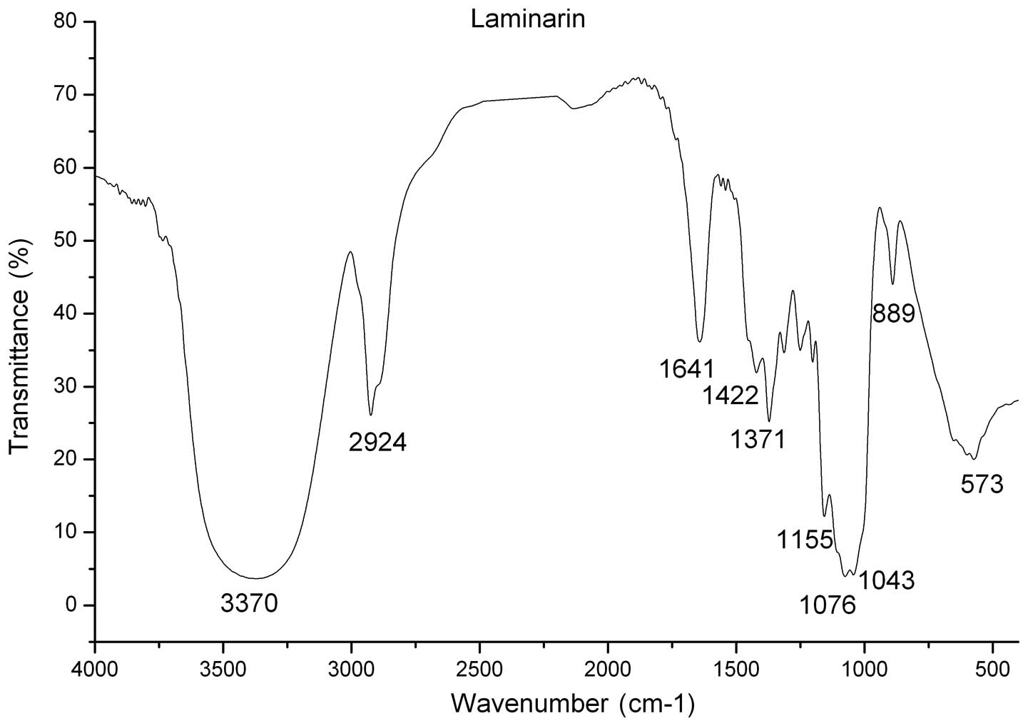
IR analysis
The IR spectrum of laminarin showed the
characteristic absorption peak of -OH stretching vibration at 3,370
cm−1 and the peak of C-H stretching vibration in
-CH3 or -CH2 groups appeared at 2,924
cm−1, which are the characteristic absorption peaks of
sugar. The characteristic pyranose absorption peaks were also
visible. The characteristic absorption peak of C-H scissor
vibration appeared at 889 cm−1, which indicated that the
glycosidic bond in laminarin was β type. Therefore, the laminarin
had a pyranose skeleton with a β-glucosidic bond. These results are
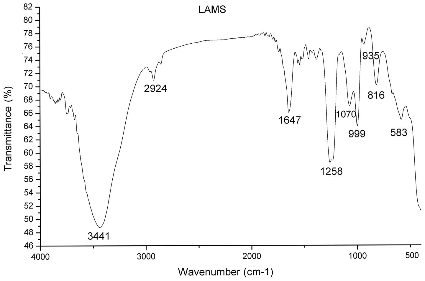
shown in Fig. 1 and Table I. By comparison, the IR spectrum of
LAMS showed the characteristic absorption peaks of S=O and C-O-S
stretching vibrations at 1,258 cm−1 and 816
cm−1, respectively, which indicated that the sulfates
were introduced into the sugar molecules. The IR spectrum also
showed that LAMS has a pyranose skeleton with a β-glucosidic bond.
The IR data for LAMS are shown in Fig.
2 and Table I.
 | Table I.IR analysis of laminarin and LAMS. |
Table I.
IR analysis of laminarin and LAMS.
| Group | Vibration mode | Peak
(cm−1)
|
|---|
| Laminarin | LAMS |
|---|
| O-H | O-H stretching
vibration | 3370 | 3441 |
| -CH2- | C-H stretching
vibration | 2924 | 2978 |
| C=O | Symmetric and
asymmetric stretching vibration | 1641 | 1649 |
| C-O | C-O stretching
vibration | 1043, 1076 | 1070 |
|
-O-SO3 | S=O stretching
vibration | - | 1258 |
|
-O-SO3 | C-O-S stretching
vibration | - | 816 |
NMR analysis
The 1H-NMR spectrum of laminarin showed
two absorption peaks in the range of δ4.5–5.5 ppm; one was from
D2O and the other was from the anomeric hydrogen of
glucose. Since the chemical shift of the anomeric hydrogen was
δ4.758 ppm, which was <5.0 ppm, this indicated the glycosidic
bond in laminarin was β type. The 1H-NMR spectrum of
LAMS showed that the chemical shifts of hydrogen generally moved
downfield, which indicated that some of the hydroxyl groups in
laminarin had been sulfated. The spectrum also showed that LAMS had
β-glycosidic bonds. The 13C-NMR spectrum of laminarin
showed that the chemical shifts of C1 and C3 had moved downfield
compared with those of glucose, indicating that the glucosidic bond
in laminarin was β-(1→3) type. The 13C-NMR spectrum of
LAMS showed that the chemical shifts of C2 and C6 had moved
downfield and the peak intensity had weakened, whereas the chemical
shifts of C1 and C3 had moved upfield, and the chemical shifts of
C4 and C5 showed no change, thus indicating that the substitution
positions of the sulfate groups were the hydroxyl groups of C2 and
C6. Laminarin and LAMS have a pyranose skeleton with β-(1→3)
glucosidic bonds, as shown in Table
II.
| Table II.Change of chemical shifts of laminarin
after sulfation. |
Table II.
Change of chemical shifts of laminarin
after sulfation.
| Sugar carbon | Chemical shift (ppm)
|
|---|
| Laminarin | LAMS |
|---|
| C1 | 102.5 | 101.2 |
| C2 | 75.6 | 77.9–78.7 |
| C3 | 84.2 | 83.0 |
| C4 | 68.1 | 67.7 |
| C5 | 73.3 | 72.7–73.4 |
| C6 | 60.7 | 67.0 |
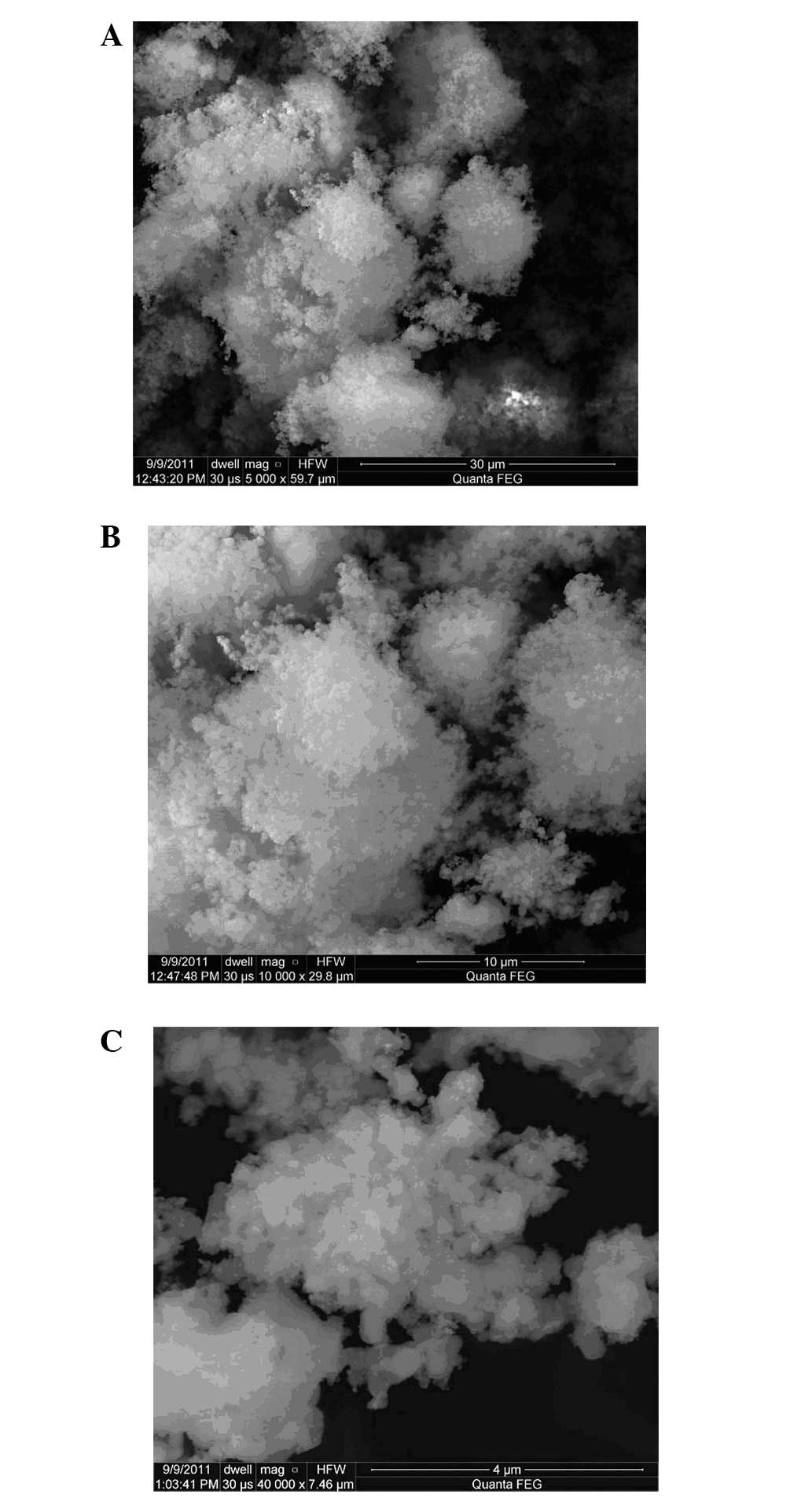
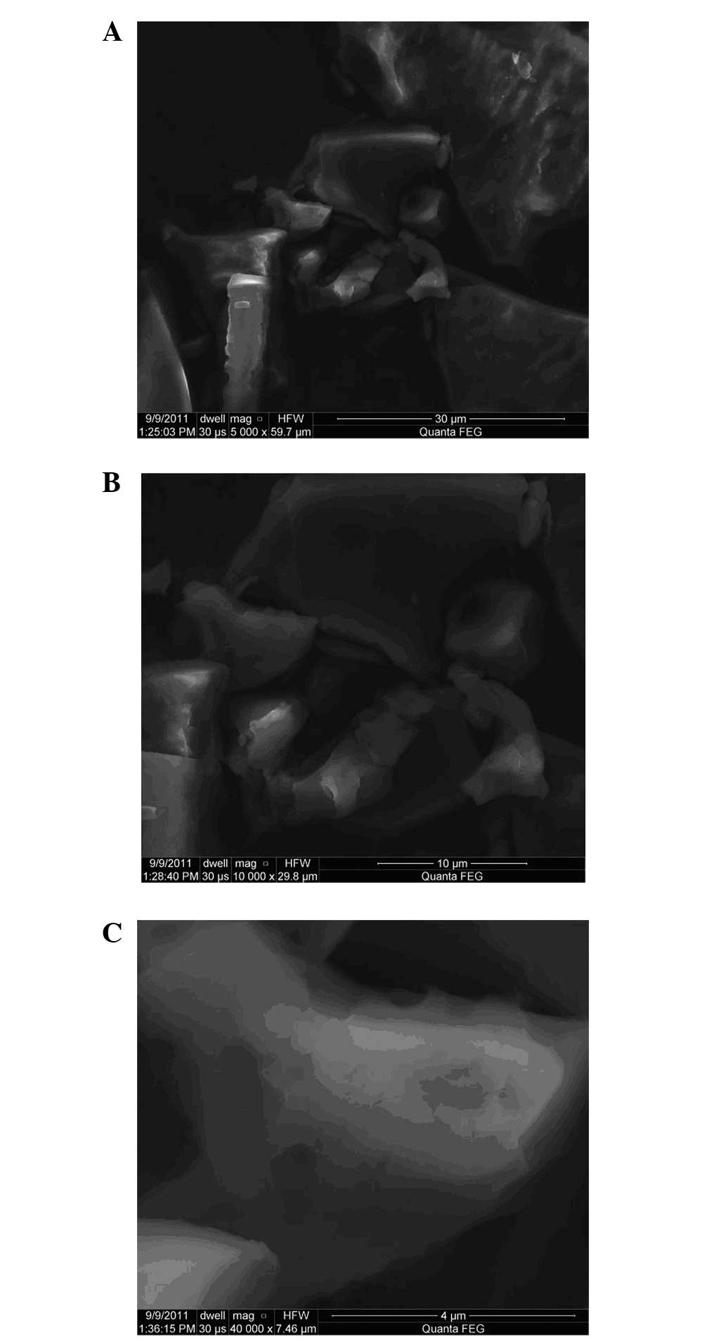
Conformation observation
Under a scanning electron microscope, marked
differences were observed in surface structure between laminarin
and LAMS. Laminarin was spongy and cloud-like, and contained
numerous dispersed sugar particles. LAMS was flakey and block-like,
and contained few dispersed sugar particles, as shown in Figs. 3 and 4, respectively.
Antitumor activity
After treatment with various concentrations of
laminarin and LAMS for 72 h, MTT assay results showed that the two
compounds inhibited LoVo proliferation. The inhibitory effects were
significantly different from that in the control group (P<0.05)
and concentration-dependent. After sulfated modification, the
antitumor activity of laminarin was enhanced, and the inhibition
rate of LAMS was significantly greater than that of the same
concentration of laminarin (P<0.01; Table III).
| Table III.Inhibitory effects of laminarin and
LAMS on LoVo cells by MTT assay. |
Table III.
Inhibitory effects of laminarin and
LAMS on LoVo cells by MTT assay.
| Samples | Concentration
(μg/ml) | OD (mean ± SD) | IR (%) |
|---|
| Control | 0 | 0.923±0.065 | - |
| Laminarin | 400 | 0.727±0.053a | 21.24 |
| 800 | 0.674±0.061b | 26.98 |
| 1600 | 0.564±0.072b | 38.89 |
| LAMS | 400 |
0.247±0.055bc | 73.24 |
| 800 |
0.184±0.028bc | 80.06 |
| 1600 |
0.124±0.042bc | 86.57 |
Discussion
Numerous pharmacological experiments have shown that
certain polysaccharides have direct cytotoxic activity against
tumor cells and directly kill cancer cells in vitro; the
majority of polysaccharides have an antitumor effect in
vivo, which enhances the activity of T and B lymphocytes,
macrophages, natural killer (NK) cells and other immune cells,
activates the complement system and promotes the production of
cytokines, which then regulates the immune system (10,11).
Sulfated polysaccharides are polysaccharide
derivatives in which the hydroxyl groups in the monosaccharide
moieties of the polysaccharide chains are substituted by sulfate
groups. These polysaccharides have complex chemical structures and
biological activities. Through the induction of apoptosis,
inhibition of cell proliferation, inhibition of tumor angiogenesis,
regulation of immune function and improvement of the effects of
chemotherapy drugs on tumor cells, the sulfated polysaccharides may
be effective in cancer therapy. The biological activities of
sulfated polysaccharides are closely associated with their
structures and physicochemical properties. The steric and
electrostatic repulsion effects of sulfate groups may change the
spatial structure of the polysaccharide and the flexion of the
sugar chain, thus increasing water-solubility and leading to
changes in biological activity (12–14).
In the current study, LAMS with a substitution
degree of 1.5 was synthesized by the chlorosulfonic acid-pyridine
method. The IR spectrum of LAMS showed that the characteristic
absorption peak of -OH weakened (3,441 cm−1), and the
characteristic symmetric stretching vibration peak of C-O-S (816
cm−1) and the characteristic asymmetric stretching
vibration peak of S=O (1,258 cm−1) appeared. The
1H-NMR spectrum of LAMS showed that the chemical shift
of the hydrogen generally moved downfield, which indicated that
some of the hydroxyl moieties in laminarin were sulfated. The
13C-NMR spectrum of LAMS showed that the chemical shifts
of C2 and C6 moved downfield (by 3 and 6.5 ppm, respectively) and
the peak intensity weakened, while the chemical shifts of C1 and C3
moved upfield, and the chemical shifts of C4 and C5 showed no
change compared with those of laminarin, thus indicating that the
substitution positions of the sulfate group were the hydroxyl
groups of C2 and C6. Laminarin and LAMS both have a pyranose
skeleton with a β-(1→3) glucosidic bond. Under a scanning electron
microscope, there were clear differences in surface structure
between laminarin and LAMS. Laminarin was spongy and cloud-like,
whereas LAMS was block-like and flaky. Changes of shape indicate a
chemical change in the majority of cases. In LAMS, the substitution
of the hydroxyl groups of the sugar units by sulfate groups may
lead to a change in the conformation of the sugar chain, and
repulsion between sulfuric acid groups may result in a conformation
showing extended or rigid structure.
Colon cancer is a common malignant tumor in the
digestive tract and one of the four most common malignant tumors
throughout the world. In a previous study (10), laminarin was shown to be a potent
agent for cancer prevention and inhibited LoVo human colon cancer
cell proliferation. The antitumor experiment showed that after
sulfated modification, the anti-tumor activity of laminarin was
significantly enhanced, and the inhibition rate of LAMS was
significantly greater than that of laminarin at the same
concentration. This may be due to the sulfated modification
changing the molecular structure and spatial conformation of the
polysaccharide, leading to changes in biological activity. When the
hydroxyl group of a laminarin sugar unit is substituted by a
sulfate group, the conformation of the sugar chains distorts or
changes, and the formation of a non-covalent bond becomes easier.
In addition, repulsions between the anionic groups elongate the
sugar chain, and some of the sulfuric acid and hydroxyl groups on
the sugar chain may form hydrogen bonds. This may result in the
chain forming a helical structure and adopting an active
conformation, thus causing the increase in its activity.
Sulfated modifications may help to produce high
activity, good functional polysaccharides and polysaccharide
derivatives. How the introduction of sulfate moieties affects the
properties and conformation of the polysaccharide, and thus affects
the biological activity, has yet to be investigated. Thus, it is
necessary to carry out in-depth studies in order to investigate
polysaccharide structure-activity relationships, which may provide
a theoretical basis for polysaccharide research and
development.
Acknowledgements
This study was supported by grants
from the China Postdoctoral Fund (No. 2012M520761) and the Youth
Science Foundation of Heilongjiang Province (No. QC2011C100).
References
|
1.
|
Morris G, Kök S, Harding S and Adams G:
Polysaccharide drug delivery systems based on pectin and chitosan.
Biotechnol Genet Eng Rev. 27:257–284. 2010. View Article : Google Scholar : PubMed/NCBI
|
|
2.
|
Yuan X, Lv J, Xiao J and Ma Q: Research
progress of laminarin. Journal of Anhui Agricultural Sciences.
38:15447–15448. 154522010.(In Chinese).
|
|
3.
|
Sun ZW, He YL, Liang ZH, et al: Sulfation
of (1→3)-β-D-glucan from the fruiting bodies of Russula
virescens and antitumor activities of the modifiers. Carbohyd
Polym. 77:628–633. 2009.
|
|
4.
|
Vishchuk OS, Ermakova SP and Zvyagintseva
TN: Sulfated polysaccharides from brown seaweeds Saccharina
japonica and Undaria pinnatifida: isolation, structural
characteristics, and antitumor activity. Carbohydr Res.
346:2769–2776. 2011.PubMed/NCBI
|
|
5.
|
Assreuy AM, Gomes DM, da Silva MS, et al:
Biological effects of a sulfated-polysaccharide isolated from the
marine red algae Champia feldmannii. Biol Pharm Bull.
31:691–695. 2008. View Article : Google Scholar : PubMed/NCBI
|
|
6.
|
Wang J, Hu Y, Wang D, et al: Sulfated
modification can enhance the immune-enhancing activity of Lycium
barbarum polysaccharides. Cell Immunol. 263:219–223. 2010.
View Article : Google Scholar : PubMed/NCBI
|
|
7.
|
Chen T, Wang J, Li Y, et al: Sulfated
modification and cytotoxicity of Portulaca oleracea L.
polysaccharides. Glycoconj J. 27:635–642. 2010. View Article : Google Scholar
|
|
8.
|
Gao YX, Yang S, Zhou XJ, et al:
Preparation and characterization of sulfated tamarind
polysaccharide. Sci Technol Food Ind. 32:142–143. 2011.(In
Chinese).
|
|
9.
|
Meng DY, Ji CF and Ji YB: Synthesis of
different degree of substitution of LAMS and preliminary study on
physical and chemical properties. Progress in Modern Biomedicine.
12:2298–2301. 2012.(In Chinese).
|
|
10.
|
Ji YB, Ji CF and Zhang H: Laminarin
induces apoptosis of human colon cancer LOVO cells through a
mitochondrial pathway. Molecules. 17:9947–9960. 2012. View Article : Google Scholar : PubMed/NCBI
|
|
11.
|
Xie HG, Chen MZ and Zhang YQ: Research
progress on structure-antitumor activity relationship of
polysaccharide and its mechanism. Food Sci. 32:329–333. 2011.(In
Chinese).
|
|
12.
|
Ouyang TZ, Li XD and Rong JH: Recent
studies on bioactivities of fungal polysaccharides. Nat Prod Res
Dev. 18:524–528. 2006.(In Chinese).
|
|
13.
|
Bao H, Choi WS and You S: Effect of
sulfated modification on the molecular characteristics and
biological activities of polysaccharides from Hypsizigus
marmoreus. Biosci Biotechnol Biochem. 74:1408–1414. 2010.
View Article : Google Scholar : PubMed/NCBI
|
|
14.
|
Zhang Y, Lu X, Zhang Y, et al: Sulfated
modification and immunomodulatory activity of water-soluble
polysaccharides derived from fresh Chinese persimmon fruit. Int J
Biol Macromol. 46:67–71. 2010. View Article : Google Scholar
|
|
15.
|
Zhao YY, Xu CY, Liang SX and Sun SX:
Preparation and anti-oxidation activity of selened Lycium
Barbarum polysaccharides sulfate and its inhibitory effect on
HeLa cell growth in vitro. Chin Pharm J. 47:423–426. 2012.(In
Chinese).
|