Introduction
Bone remodeling is a coupling process of bone
resorption and bone formation (1).
Resorption by osteoclasts and formation by osteoblasts, which leads
to the occurrence of a coupling mechanism, is a complex and
life-long process (2). This
remodeling process has been described as a ‘bone remodeling cycle’
consisting of activation, resorption, reversal and formation phases
(3). It is crucial for the normal
function of bone, including bone growth, bone repair and the
replacement of obsolete bone. Therefore, the molecular mechanism of
coupling has long been a focus of research in this area.
However, prior to the discovery of the effects of
bidirectional Eph-ephrin signaling in bone homeostasis, no proper
coupling mechanism was reported that was able to explain this
process. Since its discovery 25 years ago, the Eph family of
receptor tyrosine kinases, comprised of A- and B-subfamilies, has
been found to be involved in a growing number of physiological and
pathological processes in various cell types and organs (4,5).
Notably, it has been confirmed that bidirectional Eph-ephrin
signaling participates in many biological processes, including
angiogenesis, bone and organizational development and axon guidance
(6–10).
In bone remodeling, osteoclast and osteoblast
coordination is the key to maintaining bone homeostasis. Ephrin is
involved in regulating this process (11). It has been demonstrated that
reverse signaling through EphrinB2 into osteoclast precursors
suppresses osteoclast differentiation, while forward signaling
through EphB4 into osteoblasts enhances osteogenic differentiation
and the overexpression of EphB4 in osteoblasts increases bone mass
in transgenic mice (12). This
finding revealed the potential role of the Eph/ephrin receptor
family of ligands in the bone. It has been suggested that EphrinB2
may act in a paracrine or autocrine manner on the osteoblast to
stimulate osteoblast maturation and/or bone formation (13).
Chronic periodontitis, a major cause of anodontia in
adults, is one of the most common oral diseases (14). Porphyromonas gingivalis (Pg)
is recognized as the main pathogen in chronic periodontitis
(15). Lipopolysaccharide (LPS)
from Pg is a component of Gram-negative bacterial cell walls.
Porphyromonas gingivalis lipopolysaccharide (Pg-LPS), with
high toxicity and antigenicity to periodontal tissue, may lead to
the loss of periodontal attachment and alveolar bone absorption
(16,17). LPS has also been shown to be able
to induce the formation of osteoclasts with bone resorbing activity
in RAW 264.7 cells (18).
In the present study, the effects of Pg-LPS on
osteoblast-osteoclast bidirectional EphB4-EphrinB2 signaling were
studied. Osteoblasts and osteoclasts are derived from precursors
originating in the bone marrow (19). Interaction among cells mediated by
the EphB4 receptor on osteoblasts and the EphrinB2 ligand on
osteoclasts generates bidirectional anti-osteoclastogenic and
pro-osteoblastogenic signaling into respective cells, potentially
facilitating the transition from bone resorption to bone formation
(20). This local regulation may
contribute to the control of osteoblast differentiation and bone
formation at remodeling, and possibly also modeling, sites. In the
present study, in order to mimic the in vivo environment and
the process of bone remodeling, osteoblasts from the jawbones of
newborn mice and osteoclasts induced from RAW 264.7 macrophage
cells were successfully co-cultured. The effects of Pg-LPS on these
cells, and the potential use of Pg-LPS, were then studied.
Materials and methods
Animals and chemicals
Female and male newborn Kunming mice (<48 h old)
were obtained from the Jilin University Animal Center (Changchun,
China). No metabolic or systemic diseases were observed in the
mice. Pg-LPS was purified in our laboratory from Escherichia
coli O55:B5 (Sigma, St. Louis, MO, USA). This study was
approved by the ethics committee of Jinlin University (Changchun,
China).
Isolation and culture of osteoblasts
Osteoblasts were isolated sterilely from small
specimens of mouse jawbone. Bone fragments (~1 mm3) were
washed three times with Phosphate buffer saline (PBS) and digested
in 0.25% trypsin-EDTA for 10 min. The enzymatic reaction was
stopped by adding an equal volume of Dulbecco’s modified Eagle’s
medium (DMEM; Gibco, Carlsbad, CA, USA) with 10% fetal bovine serum
(FBS; Gibco). Washing of fragments was repeated three more times.
The fragments were then placed in the cell culture dish and
cultured in DMEM supplemented with 10% FBS and 1%
penicillin/streptomycin in a humidified atmosphere containing 5%
CO2 at 37ºC. When cells covered ~80% of the cell culture
dish, conventional digestion and passage were conducted. The medium
was changed every two days after being passaged and the cells were
ready to use until they were passaged to the third generation. The
morphology of the osteoblasts was observed under an inverted phase
contrast microscope (Axiovert 200; Zeiss, Göttingen, Germany).
Osteoblast identification
The isolated osteoblasts were identified through
alkaline phosphatase (ALP) staining and the observation of calcium
nodes. Elevated ALP expression is one of the most widely used
markers for mature osteoblasts. ALP staining was performed using
the Burstone method. Prior to observation, the original culture
medium was removed and the attached cells were fixed with 10% (v/v)
formalin/PBS for 10 min at 4ºC and stained using the substrate
naphthol AS-BI phosphate coupled with Fast Blue RR diazonium salt
at 37ºC. To perform the observation of calcium nodes, the third
generation of osteoblasts, which was cultured for three weeks, was
also examined under an inverted phase contrast microscope.
Induction and culture of osteoclasts
Osteoclasts were induced from RAW 264.7 cells, which
were purchased from the China Center for Type Culture Collection
(CCTCC, Wuhan, China). During the induction period, RAW 264.7 cells
were seeded in a 6-well culture plate at a density of
1×10⁴ cells/well and left overnight. The cells were
subsequently treated with 50 ng/ml RANKL to induce osteoclasts, and
the culture medium of DMEM supplemented with 10% FBS and 1%
penicillin/streptomycin was replaced every two days. The
osteoclasts were induced successfully after being cultured for six
days.
Osteoblast-osteoclast co-culture
system
The isolated third generation osteoblasts were
seeded in the previously mentioned well of induced osteoclasts at a
density of 2×10⁵ cells/well. The co-cultured
osteoblasts-osteoclasts were treated with 75 ng/ml Pg-LPS for 24 h.
Cells cultured without the addition of Pg-LPS were used as the
control. The morphology of the co-cultured cells was observed under
an inverted phase contrast microscope.
Protein expression of EphB4 and
EphrinB2
EphB4 and EphrinB2 protein expression in the induced
osteoclasts and Pg-LPS-treated and untreated co-cultured
osteoblasts-osteoclasts were determined by western blot analysis
and immunofluorescence staining using antibodies directed at the
respective proteins. For western blot analysis, cells were
harvested and lysed and the total protein content was determined
using a BCA protein assay kit (Beyotime, Beijing, China). The
lysate with 30 mg protein was loaded onto SDS-polyacrylamide gel
for electrophoresis and transferred to a nitrocellulose membrane.
The membranes were blocked in 5% nonfat dried milk for 45 min at
37ºC and then incubated overnight with 1:1000 mice anti-EphB4
monoclonal antibody (Santa Cruz Biotechnology, Inc., Santa Cruz,
CA, USA), and 1:1000 mice anti-EphrinB2, monoclonal antibody (Santa
Cruz Biotechnology, Inc.) at 4ºC. The membranes were washed three
times in TBST and incubated with the corresponding secondary
anti-mouse antibody (Santa Cruz Biotechnology, Inc.) conjugated
with horseradish peroxidase (HRP) at room temperature for 45 min.
The detected protein signals were measured using an enhanced
chemiluminescence (ECL) kit (Beyotime).
Gene expression of EphB4 and
EphrinB2
To further evaluate the expression of EphB4 and
EphrinB2, changes in gene expression of EphB4 and EphrinB2 were
examined by quantitative reverse transcription-polymerase chain
reaction (qPCR). Sequences of the primers for target genes are
shown in Table I. According to the
manufacturer’s instructions, total RNA was extracted from samples
using TRIzol reagent (Invitrogen, Carlsbad, CA, USA) and converted
into complementary DNA (cDNA) using a ReverTra Ace® qPCR
RT kit (Toyobo, Osaka, Japan). A CFX96™ real-time PCR detection
system (Bio-Rad, Hercules, CA, USA) was used to perform the
quantitative real-time PCR reaction. The ΔΔCt-value method was used
to calculate the relative expression values and all samples were
analyzed in triplicate.
 | Table ISequences of the primers used in the
qPCR analysis. |
Table I
Sequences of the primers used in the
qPCR analysis.
| Gene | Sequences |
|---|
| β-actin | F:
GGACTTCGAGCAGGAGATGG
R: GCACCGTGTTGGCGTAGAGG |
| Ephb4 | F:
CCCCAGGGAAGAAGGAGAGCTG
R: GCCCACGAGCTGGATGACTGTG |
| EphrinB2 | F:
ACTCCAAATTTCTACCTGGACAAG
R: GAACCTGGATTTGGTTTTACAAAG |
Statistical analysis
Data are expressed as the mean ± standard deviation
(SD). An unpaired Student’s t-test was used to test the
significance of the observed differences between the study groups.
A value of P<0.05 was considered to indicate a statistically
significant difference.
Results
Identification of osteoblasts
The morphology of the osteoblasts is shown in
Fig. 1a. After being cultured for
five days, cells around the mouse jawbone fragments increased
significantly. They became concentrated and certain tissue
fragments began to fuse. After seven days, the morphology was
varied and the majority of cells were triangular or polygon-like.
With increased time, the numbers of osteoblasts increased and the
cells were purified through repeated washing and digestion
(Fig. 1a).
The ALP staining showed a clear positive effect
(Fig. 1b). Many reddish-brown
particles were visible in the cells. A large number of high-density
black nodular aggregates of varying size were seen during the
observation of calcium nodes (Fig.
1c).
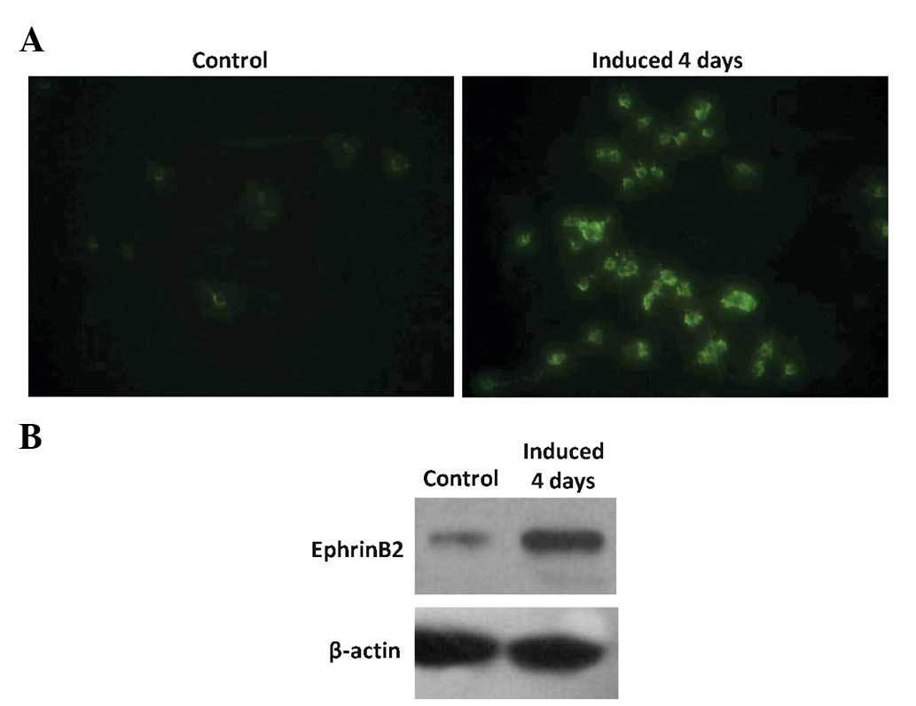
EphrinB2 expression of the induced
osteoclasts
The immunofluorescence staining (Fig. 2a) and western blot analysis
(Fig. 2b) clearly show that the
expression of EphrinB2 was higher in the induced osteoclasts than
in the control cells.
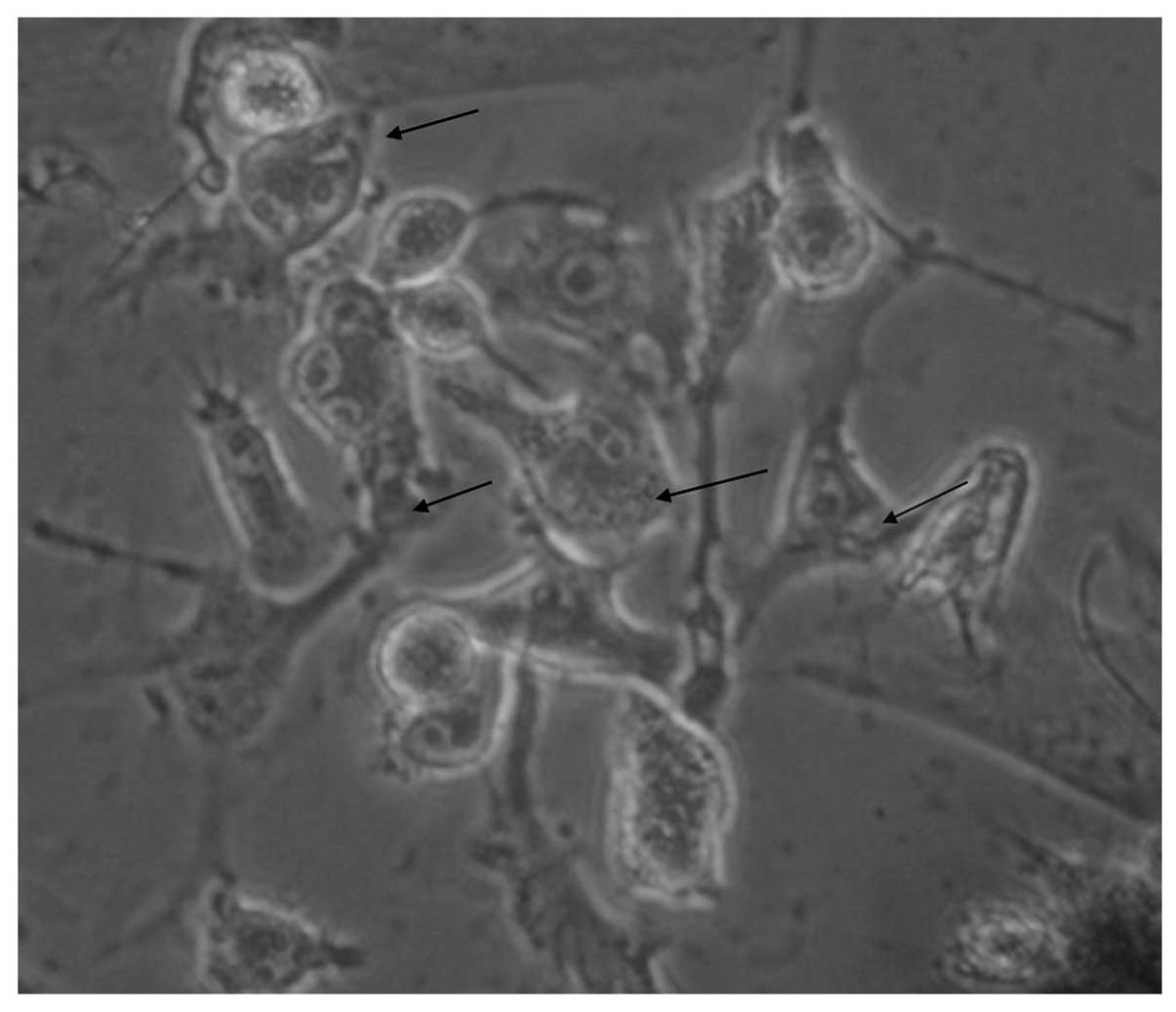
Morphological observation of the
co-cultured osteoblast-osteoclast system
Direct contact between osteoblasts and osteoclasts
was used in the present study. The results indicate that the
isolated osteoblasts and induced osteoclasts grew well when
co-cultured (Fig. 3).
Protein expression of EphB4 and
EphrinB2
As shown in Fig. 4,
immunofluorescence staining and western blot analysis were
conducted to study the changes in the expression levels of EphB4
and EphrinB2 proteins in the osteoblast-osteoblast co-culture.
After being treated with Pg-LPS at a concentration of 75 ng/ml for
24 h, the expression of EphB4 increased, while that of EphrinB2
decreased.
Gene expression of EphB4 and
EphrinB2
The gene expression of EphB4 and EphrinB2 was
detected. The results show that the relative EphB4 mRNA expression
level was significantly increased in the Pg-LPS-treated
osteoclast-osteoblast co-culture compared with that in the control
(Fig. 5a; P<0.05). However,
EphrinB2 mRNA expression was significantly decreased in the
Pg-LPS-treated co-culture compared with that in the control
(Fig. 5b; P<0.05). Therefore,
the gene studies are in line with those on protein expression.
Discussion
In the present study, the effects of Pg-LPS on
osteoblast-osteoclast bidirectional EphB4-EphrinB2 signaling were
investigated. The results show that Pg-LPS increased the expression
of EphB4 while inhibiting the expression of EphrinB2.
Our results show that many reddish-brown particles
following ALP staining were visible in the cells. A large number of
high-density black nodular aggregates of varying size were seen in
the observation of calcium nodes (Fig.
1c). Triangular or polygon-like cells centered on the scattered
aggregates, thus leading to the formation of calcium nodes. ALP
staining and the observation of calcium nodes confirmed the
successful isolation of osteoblasts.
RAW 264.7 cells, from Abelson murine leukemia
virus-induced tumors, are osteoclast precursor cells derived from
mice and are considered to represent the early differentiation
stages of the osteoclast precursor (21). Expression of EphrinB2 is one of the
indicators of induced mature osteoblasts. Hence, to verify the
successful induction of osteoclasts, two complementary assays
(immunofluorescence staining and western blot analysis) were
employed to monitor the changes in EphrinB2 (Fig. 2). The results showed that the
expression of EphrinB2 was significantly increased compared with
that in the control group. Thus, the osteoclasts were successfully
induced.
Direct contact between osteoblasts and osteoclasts
was employed in the present study, in order that certain receptors
which exert their impact through direct cell membrane contact were
able to function. The co-cultured osteoblast-osteoclast system made
it possible to mimic the real environment in vivo. After
being treated with Pg-LPS at a concentration of 75 ng/ml for 24 h,
the expression level of EphB4 increased, while that of EphrinB2
decreased. This result showed clear effects of Pg-LPS on
osteoblast-osteoclast bidirectional EphB4-EphrinB2 signaling.
Osteoblasts and osteoclasts are derived from precursors originating
in the bone marrow (19). The
interaction among cells mediated by the EphB4 receptor on
osteoblasts and the EphrinB2 ligand on osteoclasts generates
bidirectional anti-osteoclastogenic and pro-osteoblastogenic
signaling in respective cells, potentially facilitating the
transition from bone resorption to bone formation. The present
study is consistent with a report by Kubo et al(20).
When mediated with Pg-LPS, the gene expression of
EphB4 was significantly promoted while that of EphrinB2 was
inhibited. EphrinB2, involved in reverse signaling into osteoclast
precursors, is associated with the differentiation of osteoclasts.
Forward signaling through EphB4 into osteoblasts promotes
osteogenic differentiation. Contact between EphrinB2 and EphB4
inhibited the formation of osteoclasts, thus promoting the
formation of osteoblasts. The results of the present study indicate
that Pg-LPS regulates bidirectional EphB4-EphrinB2 signaling.
Therefore, the differentiation of osteoblasts was promoted, while
the differentiation of osteoclasts was inhibited. This regulation
is considered to be an effective therapeutic approach for the
treatment of bone-related diseases. Hence, this study may
contribute to the control of osteoblast differentiation and bone
formation at remodeling, and possibly also modeling, sites.
In conclusion, when treated with Pg-LPS, the EphB4
receptor on osteoblasts and the EphrinB2 ligand on osteoclasts may
generate bidirectional anti-osteoclastogenic and
pro-osteoblastogenic signaling into respective cells and
potentially facilitate the transition from bone resorption to bone
formation.
Acknowledgements
This study was supported by the National Natural
Science Foundation of China (NSFC81170999) and the Jilin Provincial
Science & Technology Department (201115104).
References
|
1
|
Irie N, Takada Y, Watanabe Y, et al:
Bidirectional signaling through ephrinA2-EphA2 enhances
osteoclastogenesis and suppresses osteoblastogenesis. J Biol Chem.
284:14637–14644. 2009. View Article : Google Scholar : PubMed/NCBI
|
|
2
|
Pasquale EB: Eph-ephrin bidirectional
signaling in physiology and disease. Cell. 133:38–52. 2008.
View Article : Google Scholar : PubMed/NCBI
|
|
3
|
Dempster DW, Lian JB and Goldring SR:
Anatomy and functions of the adult skeleton. Favus M: ASBMR Primer
on the Metabolic Bone Diseases and Disorders of Mineral Metabolism.
6th edition. American Society of Bone and Mineral Research;
Chicago, IL: pp. 7–11. 2006
|
|
4
|
Hirai H, Maru Y, Hagiwara K, Nishida J and
Takaku F: A novel putative tyrosine kinase receptor encoded by the
eph gene. Science. 238:1717–1720. 1987. View Article : Google Scholar : PubMed/NCBI
|
|
5
|
Martin TJ, Allan EH, Ho PW, Gooi JH, et
al: Communication between ephrinB2 and EphB4 within the osteoblast
lineage. Adv Exp Med Biol. 658:51–60. 2010. View Article : Google Scholar : PubMed/NCBI
|
|
6
|
Wang Y, Nakayama M, Pitulescu ME, et al:
Ephrin-B2 controls VEGF-induced angiogenesis and lymphangiogenesis.
Nature. 465:483–486. 2010. View Article : Google Scholar : PubMed/NCBI
|
|
7
|
Klein R: Eph/ephrin signaling in
morphogenesis, neural development and plasticity. Curr Opin Cell
Biol. 16:580–589. 2004. View Article : Google Scholar : PubMed/NCBI
|
|
8
|
Palmer A and Klein R: Multiple roles of
ephrins in morphogenesis, neuronal networking, and brain function.
Gene Dev. 17:1429–1450. 2003. View Article : Google Scholar : PubMed/NCBI
|
|
9
|
Holmberg J and Frisén J: Ephrins are not
only unattractive. Trends Neurosci. 25:239–243. 2002. View Article : Google Scholar : PubMed/NCBI
|
|
10
|
Egea J and Klein R: Bidirectional
Eph-ephrin signaling during axon guidance. Trends Cell Biol.
17:230–238. 2007. View Article : Google Scholar : PubMed/NCBI
|
|
11
|
Mundy GR and Elefteriou F: Boning up on
ephrin signaling. Cell. 126:441–443. 2006. View Article : Google Scholar : PubMed/NCBI
|
|
12
|
Zhao C, Irie N, Takada Y, et al:
Bidirectional ephrinB2-EphB4 signaling controls bone homeostasis.
Cell Metab. 4:111–121. 2006. View Article : Google Scholar : PubMed/NCBI
|
|
13
|
Allan EH, Häusler KD, Wei T, et al:
EphrinB2 regulation by PTH and PTHrP revealed by molecular
profiling in differentiating osteoblasts. J Bone Miner Res.
23:1170–1181. 2008. View Article : Google Scholar : PubMed/NCBI
|
|
14
|
Kojima T, Yasui S and Ishikawa I:
Distribution of Porphyromonas gingivalis in adult
periodontitis patients. J Periodontol. 64:1231–1237. 1993.
|
|
15
|
Lo Bue AM, Nicoletti G, Toscano MA,
Rossetti B, Calì G and Condorelli F: Porphyromonas
gingivalis prevalence related to other micro-organisms in adult
refractory periodontitis. New Microbiol. 22:209–218. 1999.
|
|
16
|
Wiebe SH, Hafezi M, Sandha HS, Sims SM and
Dixon SJ: Osteoclast activation in inflammatory periodontal
diseases. Oral Dis. 2:167–180. 1996. View Article : Google Scholar : PubMed/NCBI
|
|
17
|
Itoh K, Udagawa N, Kobayashi K, et al:
Lipopolysaccharide promotes the survival of osteoclasts via
Toll-like receptor 4, but cytokine production of osteoclasts in
response to lipopolysaccharide is different from that of
macrophages. J Immunol. 170:3688–3695. 2003. View Article : Google Scholar : PubMed/NCBI
|
|
18
|
Islam S, Hassan F, Tumurkhuu G, et al:
Bacterial lipopolysaccharide induces osteoclast formation in RAW
264.7 macrophage cells. Biochem Biophys Res Commun. 360:346–351.
2007. View Article : Google Scholar : PubMed/NCBI
|
|
19
|
Matsuo K and Irie N: Osteoclast-osteoblast
communication. Arch Biochem Biophys. 473:201–209. 2008. View Article : Google Scholar
|
|
20
|
Kubo T, Shiga T, Hashimoto J, et al:
Osteoporosis influences the late period of fracture healing in a
rat model prepared by ovariectomy and low calcium diet. J Steroid
Biochem Mol Biol. 68:197–202. 1999. View Article : Google Scholar : PubMed/NCBI
|
|
21
|
Kuehl WM and Bergsagel PL: Molecular
pathogenesis of multiple myeloma and its premalignant precursor. J
Clin Invest. 122:3456–3463. 2012. View
Article : Google Scholar : PubMed/NCBI
|