Introduction
Axillary lymph node dissection (ALND) provides
information for staging and prognosis that may be used to design a
therapeutic strategy (1–3). As a result of this, a large number of
patients with breast cancer in the past underwent routine ALND;
however, in many of the patients, the cancer was revealed to be
node-negative, and surgery unnecessarily exposed them to
perioperative risks and increased long-term morbidity (4). Therefore, sentinel lymph node biopsy
(SLNB) has widely replaced conventional ALND as a routine axillary
staging method in breast cancer surgery. The SLNB procedure is
accurate and safe (5–8) and results in substantially less
postoperative morbidity than ALND (9,10).
Dye-guided and γ-probe-guided methods, separately
and in combination, are used for SLNB. The dye-guided method alone
for breast cancer is considered to be inferior to the
γ-probe-guided and combined methods in terms of its accuracy and
false-negative rate. Cox et al(11) revealed the identification rates for
SLNB to be 80.3% for the dye-guided technique, 88.6% for the
γ-probe-guided method and 96.7% for the combination, based on a
large number of procedures performed in a single center. Kim et
al(12) showed that the rates
for successfully identifying the SLN were 83.1, 89.2 and 91.9%, for
the dye-guided-alone, γ-probe-guided-alone and combination methods,
respectively [with the combination method being significantly more
efficacious (P=0.007)], while the false-negative rates were 10.9,
8.8 and 7.0%, respectively (P=0.047).
The dye-guided method has the significant advantages
of being more convenient to perform, less costly and possible to
conduct in any institute with no special facilities. Although it
requires a certain training period, this method may be preferable
for all clinicians if its accuracy is demonstrated to be comparable
to that of the combination method. Morrow et al(13) reported that there were no
significant differences between the procedures with regard to the
identification rate, accuracy, time spent for identification and
number of SLNs. In the present study, the success rate in
identifying the SLN, the accuracy and the SLN occupation ratio
using the dye-guided method alone were investigated. Furthermore,
the possibility of obviating the requirement for ALND in
SLNB-positive cases was explored.
Materials and methods
Patients
From January 1999 to December 2009, 374 patients
with primary breast cancer underwent SLNB using the dye-guided
method alone followed by radical surgery at Sapporo Medical
University Hospital (Sapporo, Japan). The medical records of these
patients were analyzed retrospectively and their
clinicopathological characteristics were evaluated. This study was
approved by The Ethics Committee of Sapporo Medical University
(Sapporo, Japan). Informed consent was obtained from all patients
prior to enrollment.
Surgical procedure
Under general anesthesia, 2–4 ml blue dye (indigo
carmine) was injected at two sites surrounding the primary tumor
and areola. Approximately 5 min later, a blunt dissection was
performed through the mastectomy incision or a separate axillary
incision until a dye-stained lymphatic tract or node was
identified.
Pathological assessment
The SLN was split into three to four sections and
analyzed as frozen specimens during surgery. These specimens were
fixed in 10% formalin, stained with hematoxylin and eosin (H&E)
and then re-examined histologically following surgery. The SLN area
and the area of tumor cells in the SLN were calculated by
multiplying the longest diameter by the minor axis. The SLN
occupation ratio was defined as the proportion of area that was
occupied by tumor cells to the total SLN area. As a feasibility
study, ALND was completed for cases of at least level I or II
following SLN excision in the first 54 cases to undergo
surgery.
Statistical analysis
Continuous variables (age, maximum tumor size and
SLN occupation ratio), dichotomous variables (estrogen receptor,
progesterone receptor and human epidermal growth factor receptor
type 2 status) and categorical variables [lymph vessel invasion
(ly) status, vessel invasion (v) status and nuclear grade (NG)]
were analyzed in a univariate logistic regression model using
either the t-test or the χ2 test. All variables were
subsequently analyzed in a multivariable regression model. A
receiver operating characteristic (ROC) curve was drawn on the
basis of the SLN occupation ratio, and the area under the curve
(AUC) was calculated using SPSS version 11.5 statistical software
(SPSS, Inc., Chicago, IL, USA). GraphPad Prism version 5.0.2
(GraphPad Software, La Jolla, CA, USA) was used for all analyses.
P<0.05 was considered to indicate a statistically significant
difference.
Results
The clinicopathological characteristics of the
patients are shown in Table I. The
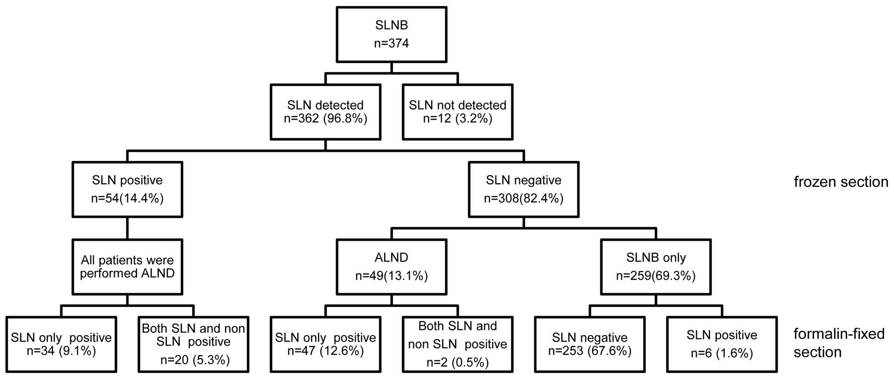
SLN was successfully identified in 362 of the 374 patients (96.8%).
The average number of resected lymph nodes was 1.9 (range, 1–10). A
total of 54 patients were identified to have SLN metastases and
ALND was performed in these patients. In 34 of the 54 patients
(63.0%) with axillary metastases, metastatic nodes were only
observed in the SLN. In the remaining 20 patients (37.0%),
metastases were observed in the SLN and the non-SLNs. Of the 308
patients without SLN metastases in the frozen sections, 49
underwent ALND. Two of these 49 had positive axillary lymph nodes
in the formalin-fixed sections. Among the 259 patients who did not
undergo ALND, six had positive formalin-fixed sections (Fig. 1).
 | Table ICharacteristics of 374 patients who
underwent SLNB. |
Table I
Characteristics of 374 patients who
underwent SLNB.
| Clinical
features | No. of patients
(%) |
|---|
| Mean age, years
(range) | 56.7 (23–87) |
| Location of
tumor |
| Medial | 103 (27.5) |
| Lateral | 264 (70.6) |
| Central | 7 (1.9) |
| Clinical tumor
stage |
| T0 | 14 (3.7) |
| T1 | 175 (46.8) |
| T2 | 161 (43.0) |
| T3 | 19 (5.1) |
| T4 | 5 (1.3) |
| Clinical nodal
stage |
| N0 | 332 (88.8) |
| N1 | 41 (11.0) |
| N2 | 1 (0.3) |
| N3 | 0 (0.0) |
| No. of SLN |
| 1 | 149 (39.8) |
| 2 | 130 (34.8) |
| 3 | 56 (15.0) |
| 4 | 18 (4.8) |
| ≥5 | 9 (2.4) |
| Not detected | 12 (3.2) |
Postoperative diagnosis of SLN
metastases
The sensitivity for the identification of a
histologically positive node was 96.4% (54/56) (Table IIA), and the specificity was 100%.
The accuracy of SLN biopsy for the detection of metastatic disease
was 98.1% (101/103). The positive predictive value was 100% (54/54)
and the negative predictive value (the correlation between the
negative SLNs and the negative axillary nodes) was 95.9% (47/49),
with a false-negative rate of 3.6% (2/56).
| Table IIDiagnosis of sentinel lymph node
metastases. |
Table II
Diagnosis of sentinel lymph node
metastases.
| A. Postoperative
diagnosis |
|---|
|
|---|
| Postoperative
diagnosis | SLN or
non-SLN-positive | SLN and
non-SLN-negative | Total |
|---|
| SLN-positive | 54 | 0 | 54 |
| SLN-negative | 2 | 47 | 49 |
| Total | 56 | 47 | 103 |
|
| B. Intraoperative
diagnosis |
|
| Intraoperative
diagnosis | SLN-positive | SLN-negative | Total |
|
| SLN-positive | 54 | 0 | 54 |
| SLN-negative | 8 | 300 | 308 |
| Total | 62 | 300 | 362 |
Intraoperative diagnosis of SLN
metastases
In 362 patients, the intraoperative diagnosis was
confirmed by the final histological examination (with
formalin-fixed specimens). However, in eight patients (12.9%) there
was a false-negative intraoperative diagnosis and a tumor was
identified on a permanent section of either the SLN or the
non-SLNs. As a result, a diagnostic accuracy of 97.8%, a
sensitivity of 87.1% and a specificity of 100% were achieved with
H&E staining of the frozen sections (Table IIB).
Clinicopathological factors correlated
with non-SLN metastasis
In 63% of the SLN-positive cases for which ALND was
performed, metastasis was limited to the SLN. This result suggested
the possibility that the resection of SLNs only may be sufficient
to treat in excess of half of the cases with positive SLNs.
Therefore, the cases were analyzed to reveal which factors were
likely to indicate the potential for omitting the following
ALND.
To investigate which factors correlated with non-SLN
metastasis, the clinicopathological characteristics of the patients
who underwent ALND and had SLN and non-SLN metastases were compared
with those who underwent ALND and just had SLN metastasis.
Univariate analyses revealed significant differences in the ly
status (P=0.0002), NG (P=0.0433), maximum tumor size (P=0.0317) and
the SLN occupation rate (P=0.0017) between the two groups. The
results of the analyses for various characteristics are summarized
in Table III.
| Table IIIClinicopathological factors correlated
with non-SLN metastasis. |
Table III
Clinicopathological factors correlated
with non-SLN metastasis.
| Clinical feature | SLN only | SLN and non-SLN | P-value |
|---|
| Mean age (years) | 59.76 | 59.85 | 0.9820 |
| Tumor size (cm) | 2.34 | 2.92 | 0.0317a |
| ly | | | 0.0002b |
| ly0 | 12 | 0 | |
| ly1–3 | 20 | 20 | |
| v | | | 0.2346 |
| v0 | 29 | 15 | |
| v1 | 3 | 5 | |
| NG | | | 0.0433a |
| 1 | 20 | 6 | |
| 2–3 | 11 | 12 | |
| Unknown | 3 | 2 | |
| ER status | | | 0.7208 |
| Negative | 7 | 3 | |
| Positive | 24 | 17 | |
| Unknown | 3 | 0 | |
| PgR status |
| Negative | 10 | 2 | 0.0949 |
| Positive | 21 | 18 | |
| Unknown | 3 | 0 | |
| HER2 status | | | 0.6958 |
| Negative | 27 | 16 | |
| Positive | 4 | 4 | |
| Unknown | 3 | 0 | |
| SLN occupation ratio
(%) | 21.47 | 46.75 | 0.0017b |
Multivariate analysis revealed that ly status was an
independent risk factor for non-SLN metastasis (Table IV). An ROC curve was generated to
assess the clinical utility of the SLN occupation ratio for the
prediction of non-SLN metastasis (sensitivity, 70.0%; specificity,
61.8%; AUC, 0.736).
| Table IVMultivariate analysis. |
Table IV
Multivariate analysis.
| Risk factor for SLN
metastasis | P-value | Relative risk | 95% CI |
|---|
| Age | 0.0938 | 1.00 | 0.93–1.07 |
| Maximum tumor
size | 0.1477 | 2.09 | 0.81–8.24 |
| ly |
| ly0 | 0.0138a | 1.00 |
1.95–5.14×10¹⁹ |
| ly1–3 | |
7.08×10⁸ | |
| v |
| v0 | 0.4950 | 1.00 | 0.27–20.47 |
| v1 | | 2.03 | |
| NG |
| 1 | 0.1258 | 1.00 | 0.68–29.32 |
| 2–3 | | 4.05 | |
| ER status |
| Negative | 0.8965 | 1.00 | 0.105–12.33 |
| Positive | | 1.16 | |
| PgR status |
| Negative | 0.0823 | 1.00 | 0.791–145.56 |
| Positive | | 7.30 | |
| HER2 status |
| Negative | 0.2760 | 1.00 | 0.43–25.07 |
| Positive | | 3.00 | |
Axillary recurrences
In this study group, 5 out of the total 374 cases
(1.3%) had an axillary recurrence. Out of all the patients, 259
patients received SLNB alone and 4/259 patients (1.5%) showed
axillary recurrence. By contrast, patients who received ALND had no
axillary recurrence. The SLN was not able to be detected in 12
cases, and axillary recurrence was observed in one of these
cases.
Discussion
In the present study, the SLN identification rate
with the dye-guided method alone was 97%, which was comparable with
previous studies (11,12). The population of patients who had
SLN metastasis was also consistent with those of previous studies.
These data indicated that the dye-guided method alone was
sufficient to detect SLN metastasis. In addition, the dye-guided
method requires no special equipment, so widespread application may
be expected in any institute.
In SLN metastasis-positive cases, subsequent ALND is
generally performed. However, in the group observed in the present
study, 63% of the patients had no axillary metastasis. These
results may indicate that in excess of half the patients receive
unnecessary additional surgery. In the management of breast cancer,
ALND may dam up the flow of lymph vessels and result in lymphedema
in the patient’s arm. Although breast cancer is a malignant
disease, survival may be relatively long subsequent to the first
surgical therapy, so it is important to support the patient’s
quality of life (QOL).
The possibility of predicting axillary lymph node
metastasis by the analysis of the SLN status was investigated. The
aim of identifying the factors that predict axillary metastasis was
to contribute to avoiding unnecessary ALND. Univariate analysis
revealed significant differences in ly status, NG, maximum tumor
size and the SLN occupation ratio between patients with axillary
metastasis and those with SLN metastasis only. In breast cancer,
lymph node metastasis may spread through the lymph nodes stepwise,
following the lymph vessel route. Thus, ly status, NG, tumor size
and the SLN occupation ratio are associated with axillary
metastasis, and the analysis of these factors is likely to enhance
our capability to predict the patients with axillary lymph node
metastasis.
The multivariate analysis of the clinicopathological
factors, including those mentioned previously, revealed a
correlation between axillary metastasis and ly status. However,
since it is necessary to assess these factors in permanent
formalin-fixed specimens, it is difficult to use them to decide on
the surgical procedure during the surgery itself. Therefore, in the
present study, the clinical usefulness of the SLN occupation ratio,
which is able to be diagnosed during surgery, was investigated. It
was demonstrated that the greater the degree of the area occupied
by tumor cells, the more invasive the tumor was to lymph vessels,
with greater proliferative ability, a larger number of malignant
cells, a more metastatic character, and the longer the time after
metastasis. Thus, this parameter was expected to be correlated with
axillary metastasis. The ROC curve analysis showed sufficient
sensitivity and specificity (70.0 and 61.8%, respectively) when the
cut-off point for deciding whether it was necessary to perform ALND
for patients who may not actually have axillary lymph node
metastasis was 12.9%. Thus, a predictive factor for axillary lymph
node metastasis in breast cancer was identified, which suggests
that it may be possible to avoid unnecessary axillary lymph node
resection, and thereby improve the patients’ QOL.
References
|
1
|
Senofsky GM, Moffat FL Jr, Davis K, et al:
Total axillary lymphadenectomy in the management of breast cancer.
Arch Surg. 126:1336–1342. 1991. View Article : Google Scholar : PubMed/NCBI
|
|
2
|
Fentiman IS and Mansel RE: The axilla: not
a no-go zone. Lancet. 337:221–223. 1991. View Article : Google Scholar : PubMed/NCBI
|
|
3
|
Noguchi M, Miwa K, Michigishi T, et al:
The role of axillary lymph node dissection in breast cancer
management. Breast Cancer. 4:143–153. 1997. View Article : Google Scholar : PubMed/NCBI
|
|
4
|
Kissin MW, Querci della Rovere G, Easton
D, et al: Risk of lymphoedema following the treatment of breast
cancer. Br J Surg. 73:580–584. 1986. View Article : Google Scholar : PubMed/NCBI
|
|
5
|
Cox C, White L, Allred N, et al: Survival
outcomes in node-negative breast cancer patients evaluated with
complete axillary node dissection versus sentinel lymph node
biopsy. Ann Surg Oncol. 13:708–711. 2006. View Article : Google Scholar : PubMed/NCBI
|
|
6
|
Kuijt GP, van de Poll-Franse LV, Voogd AC,
et al: Survival after negative sentinel lymph node biopsy in breast
cancer at least equivalent to after negative extensive axillary
dissection. Eur J Surg Oncol. 33:832–837. 2007. View Article : Google Scholar
|
|
7
|
Langer I, Guller U, Hsu-Schmitz SF, et al:
Sentinel lymph node biopsy is associated with improved survival
compared to level I & II axillary lymph node dissection in node
negative breast cancer patients. Eur J Surg Oncol. 35:805–813.
2009. View Article : Google Scholar
|
|
8
|
van der Ploeg IM, Nieweg OE, van Rijk MC,
et al: Axillary recurrence after a tumour-negative sentinel node
biopsy in breast cancer patients: a systematic review and
meta-analysis of the literature. Eur J Surg Oncol. 34:1277–1284.
2008.PubMed/NCBI
|
|
9
|
Lucci A, McCall LM, Beitsch PD, et al:
Surgical complications associated with sentinel lymph node
dissection (SLND) plus axillary lymph node dissection compared with
SLND alone in the American College of Surgeons Oncology Group Trial
Z0011. J Clin Oncol. 25:3657–3663. 2007. View Article : Google Scholar
|
|
10
|
Celebioglu F, Perbeck L, Frisell J, et al:
Lymph drainage studied by lymphoscintigraphy in the arms after
sentinel node biopsy compared with axillary lymph node dissection
following conservative breast cancer surgery. Acta Radiol.
48:488–495. 2007. View Article : Google Scholar
|
|
11
|
Cox CE, Bass SS, McCann CR, et al:
Lymphatic mapping and sentinel lymph node biopsy in patients with
breast cancer. Annu Rev Med. 51:525–542. 2000. View Article : Google Scholar : PubMed/NCBI
|
|
12
|
Kim T, Giuliano AE and Lyman GH: Lymphatic
mapping and sentinel lymph node biopsy in early-stage breast
carcinoma: a metaanalysis. Cancer. 106:4–16. 2006. View Article : Google Scholar : PubMed/NCBI
|
|
13
|
Morrow M, Rademaker AW, Bethke KP, et al:
Learning sentinel node biopsy: result of a prospective randomized
trial of two techniques. Surgery. 126:714–722. 1999. View Article : Google Scholar : PubMed/NCBI
|