Introduction
Although the inflammatory reaction is vital to
homeostatic maintenance, it may also trigger or further aggravate
various diseases, including inflammatory bowel disease (IBD), which
is a group of chronic inflammatory disorders of the
gastrointestinal tract. The hallmark of IBD is chronic,
uncontrolled inflammation involving any section of the
gastrointestinal tract in Crohn’s disease and limited to the colon
in ulcerative colitis. Both diseases are characterized by weight
loss, fever, gastric dysmotility, colonic mucosal ulceration,
shortening of the colon and diarrhea with blood and/or mucus. These
characteristics are exacerbated by immunological disorders that
activate cellular and humoral immune responses, particularly those
involving neutrophils (1–4). Although a number of studies have
indicated roles for genetic, environmental and lifestyle effects in
the pathogenesis of IBD, its etiology remains poorly understood
(5–9). Patients with IBD are typically
treated with agents that target aberrant immune responses and
inflammatory cascades, including anti-inflammatory agents
(5-aminosalicylic acid and glucocorticosteroids), immunomodulatory
therapy (azathioprine, 6-mercaptopurines and cyclosporine) and
monoclonal antibody therapy (for example, with anti-TNF-α and
leukapheresis) (10–14).
Sodium 4-phenylbutyrate (PBA) is a
phenyl-substituted short-chain fatty acid used in the treatment of
a wide range of diseases, such as urea metabolism disorders
(15), homozygous β-thalassemia
(16), spinal muscular atrophy
(17) and tumors (18). In recent studies, PBA was shown to
suppress oxidative stress by attenuating endoplasmic reticulum
stress and to exert anti-inflammatory effects by suppressing the
activity of the transcriptional factor, nuclear factor-κB (NF-κB)
(19–22). Furthermore, we previously
identified that PBA may be effective in the treatment of
neurodegenerative diseases, including Parkinson’s disease (23).
In the present study, the effects of PBA were
investigated on colonic inflammation in a mouse model of colitis
induced by dextran sulfate sodium (DSS), which is a standard animal
model of IBD.
Materials and methods
Experimental animals
Male ICR mice (weight, 28–30 g) were purchased from
Kyudo, Co., Ltd. (Saga, Japan). All mice were housed in cages (n=5
per cage) with a 12 h light:dark cycle and a constant temperature
of 20±5°C. The animals (n=45) were divided into the following
groups: The normal control group (n=5), DSS-treated control group
(n=10) and groups treated with DSS plus 50 (n=5), 100 (n=10), 150
(n=10) and 200 (n=5) mg PBA/kg body weight. Survival rates, the
development of DSS-induced colitis and cytokine levels were
analyzed in all groups. An additional 10 mice were treated with DSS
and 150 mg PBA/kg body weight, and the colon length and
histopathological changes were evaluated on day 8. The animals were
fed standard laboratory chow and had access to water ad
libitum. All animal experiments were conducted under the
University guidelines and were approved by the Ethical Committee
for Animal Care and Use of Fukuoka University (Fukuoka, Japan).
DSS and PBA treatment
Experimental colitis was induced by the addition of
3.5% (w/v) DSS (Nacalai Tesque, Inc., Kyoto, Japan) to the drinking
water of the mice. PBA (LKT Laboratories Inc., St. Paul, MN, USA)
was administered via intraperitoneal injection at doses of 50, 100,
150 or 200 mg PBA/kg body weight on days 0, 2, 4, 6, 8, 10 and
12.
Assessment of DSS-induced colitis
DSS-induced colitis was characterized by the
presence of acute colitis, bloody mucoid stools, diarrhea,
abdominal pain, weight loss, shortening of the colon and mucosal
ulceration with neutrophil infiltration (24). Mice with DSS-induced colitis were
evaluated using a disease activity index (DAI), which assigns a
score to changes in weight, a positive Hemoccult test, gross blood
in the stools and stool consistency. Body weight, stool consistency
and the condition of the peri-anal tissues were recorded daily. The
presence of fecal occult blood was tested by collecting colonic
lavage fluid immediately prior to the intraperitoneal injection of
PBA. The DAI was then scored for three categories as follows: Body
weight loss (0, none; 1, 1–5%; 2, 5–10%; 3, 10–20%; and 4,
>20%), stool consistency (0, normal; 2, loose stools; and 4,
diarrhea), and stool blood (0, negative; 2, positive Hemoccult
test; and 4, gross bleeding). Body weight loss was calculated as
the percentage difference between the body weight on day 0 and that
on the day the animal was weighed. For mortalities during this
study, the last determined DAI score was used and mice that
accidentally died during the experiment were excluded (25).
Measurement of cytokines by enzyme-linked
immunosorbent assay (ELISA)
Cytokines in the collected colonic lavage fluid were
measured by ELISA. Briefly, 96-well plates were coated with
monoclonal antibodies [2.0 μg/ml anti-mouse tumor necrosis factor-α
(TNF-α), purified; 2.0 μg/ml anti-mouse/rat interleukin-1β (IL-1β),
purified; and 1.0 μg/ml anti-mouse IL-6, purified] overnight at 4°C
and washed with phosphate-buffered saline (PBS). Blocking One
solution (Nacalai Tesque, Inc.) was diluted 5-fold with PBS and
added to the plates. After 1 h of incubation at room temperature
(RT), the collected colonic lavage fluid was added to the plates in
a 1:10 dilution. The plates were further incubated for 1 h at RT
and then washed with PBS. Biotinylated antibodies (0.8 μg/ml
biotinylated anti-mouse TNF-α; 2.0 μg/ml biotinylated
anti-mouse/rat IL-1β; and 1.0 μg/ml biotinylated anti-mouse IL-6)
were added and the plates were incubated for 1 h at RT.
Streptavidin horseradish peroxidase (SNN 1004; Biosource
International, Inc., Camarillo, CA, USA; 1:10,000) was then added
and the plates were incubated for 1 h at RT. After the plates had
been washed thoroughly, 100 μl peroxidase substrate solution
consisting of equal volumes of 3,3′,5,5′-tetramethylbenzidine (TMB)
peroxidase substrate and peroxidase substrate solution B (TMB
Microwell Peroxidase Substrate System; KPL Inc., Gaithersburg, MD,
USA) were added to each well for 10 min, followed by an equal
volume of stop solution. Absorbance was measured at 490 nm with an
ELISA reader (Bio-Rad Model 450 Microplate Reader; Bio-Rad,
Hercules, CA, USA). All anti-cytokine antibodies were purchased
from eBioscience (San Diego, CA, USA).
Measurement of colon length and
histopathological evaluation
On day 8, the 10 mice with DSS-induced colitis that
were treated with 150 mg PBA/kg body weight (as described in
Experimental animals) were sacrificed and a section of the colon
extending from the cecocolic junction to the anus was removed.
Colon length was measured between the cecocolic junction and the
anal verge. The resected tissue was fixed overnight in 10%
neutral-buffered formalin (Nacalai Tesque, Inc.) and then routinely
processed for embedding in paraffin blocks. Sections of 3-μm thick
tissues were cut and stained with hematoxylin and eosin for
examination under a microscope (OLYMPUS BX51; Olympus Corporation,
Tokyo, Japan).
Statistical analysis
The experimental data were statistically analyzed
using the log-rank test (Fig. 1),
one-way analysis of variance followed by Dunnet’s post hoc test
(Fig. 2), Student’s t-test
(Figs. 3 and 4A) and the Tukey-Kramer post hoc test
(Fig. 4B). P<0.05 was
considered to indicate a statistically significant difference. Data
are expressed as the mean ± standard error.
Results
Survival rate
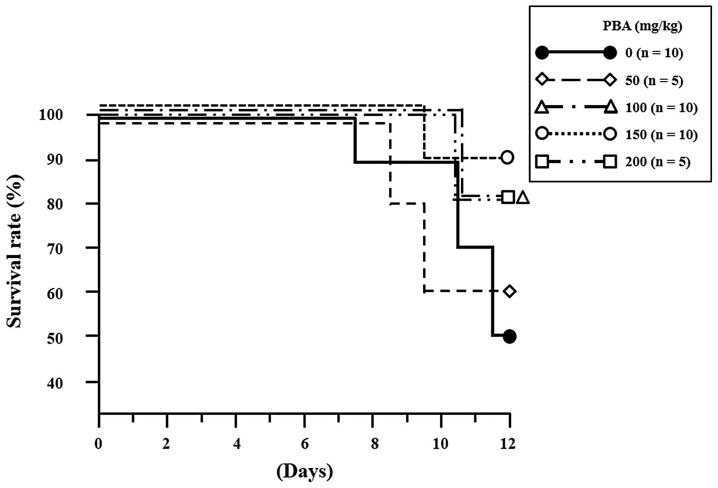
On day 12, the survival rate of DSS-treated control
mice was 50% (5/10), whereas the mice treated with 50, 100, 150 and
200 mg PBA/kg body weight showed survival rates of 60% (3/5), 80%
(8/10), 90% (9/10) and 80% (4/5), respectively (Fig. 1). The larger number of animals in
the 100 and 150 mg PBA/kg body weight groups reflects an initial
test group of five animals and then the testing of the effects of
these drug doses in five additional animals. The differences in
survival rates from those in the untreated control group were not
identified to be significant, possibly due to the small sample
size. However, there was a positive trend in the survival rates of
the PBA-treated animals. The following results in this study refer
to those obtained from the mice treated with 150 mg PBA/kg body
weight as this dose yielded the highest survival rate.
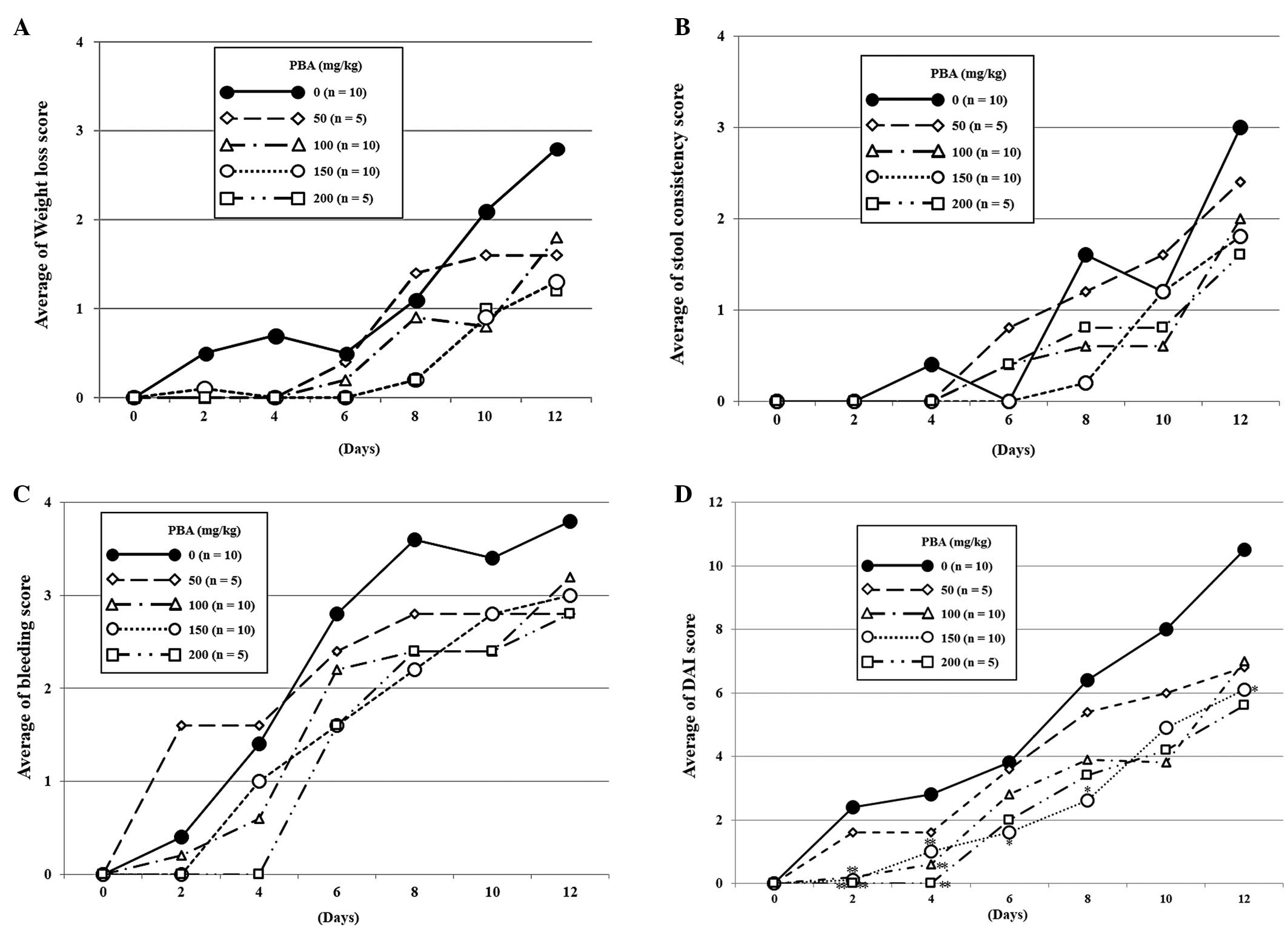
DAI score
In the DSS control group, occult blood positivity
was observed on day 2 in two of the 10 mice and on day 4 in the
remaining eight mice. Gross bleeding was observed in four mice on
day 6. In the mice treated with 150 mg PBA/kg body weight, occult
blood positivity was detected on days 4 (5/10) and 6 (8/10), which
was later than that of the DSS control group. Only one mouse showed
gross bleeding, which was observed on day 6 (Fig. 2C). By the end of the experiment,
there were seven cases of severe weight loss (DAI score of ≥3) in
the DSS control group, but only one case in the PBA group, which
was detected on day 12 (Fig. 2B).
Overall, the DAI score of the PBA-treated group was significantly
lower than that of the DSS control group on days 2, 4, 6, 8 and 12
(Fig. 2D).
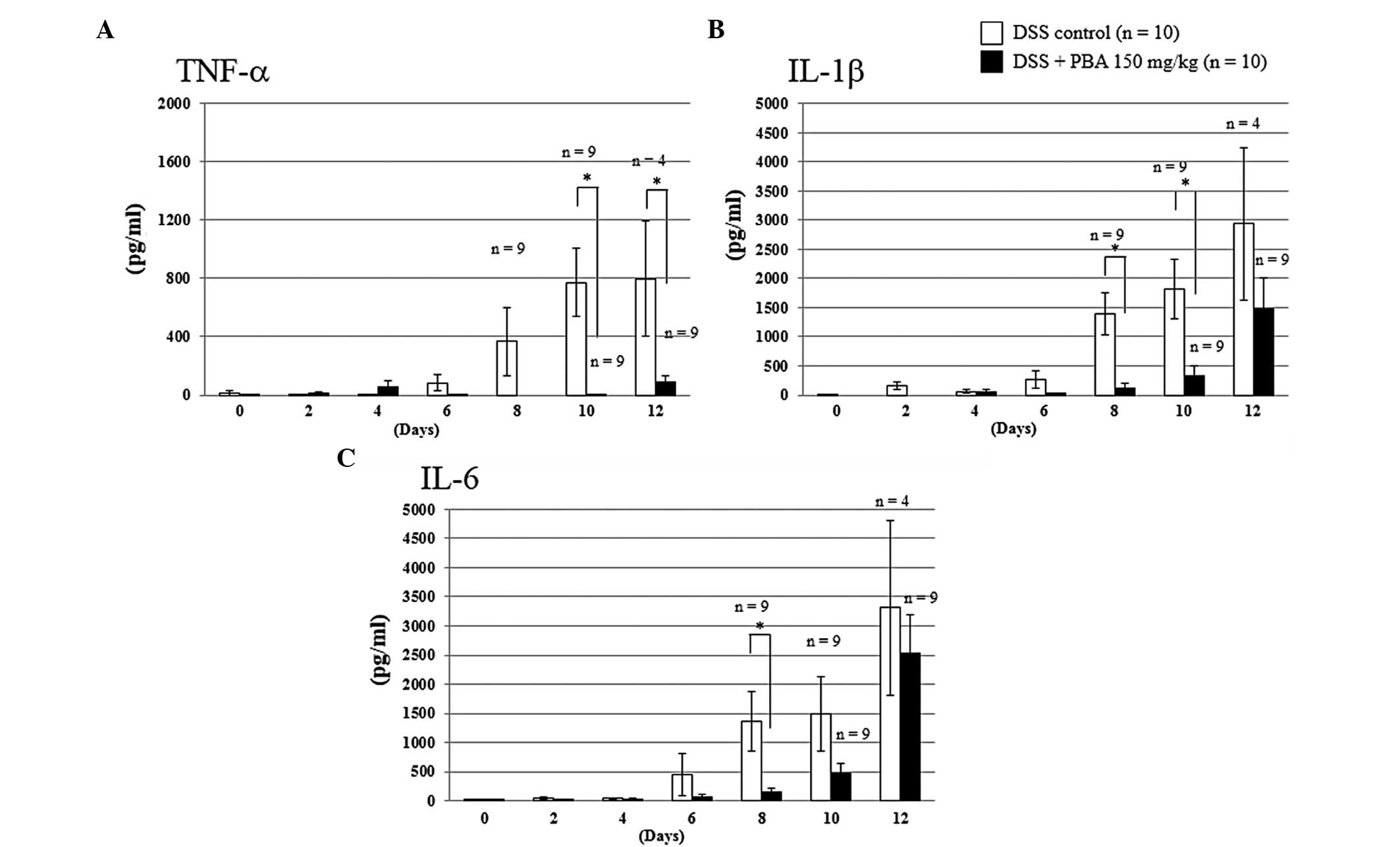
Inflammatory cytokine production
(ELISA)
The concentrations of cytokines in colonic lavage
fluid were determined from a standard curve prepared for each
plate. In the DSS-treated control group, the level of TNF-α
production appeared to increase from day 6, reaching 365.7±232.6
pg/ml by day 8. However, in the PBA group, TNF-α production was
suppressed completely until day 8 and only low levels were detected
on day 10. Similarly, on day 8 the levels of IL-1β and IL-6
production in the PBA-treated group were suppressed by 91.3%
(121.9±72.4 pg/ml) and 88.9% (151.3±68.8 pg/ml), respectively,
compared with those of the DSS control group (1,398.0±358.5 and
1,362.8±502.8 pg/ml, respectively). The differences between the
DSS-treated control and PBA groups were significant (P<0.01) for
all three cytokines (Fig. 3).
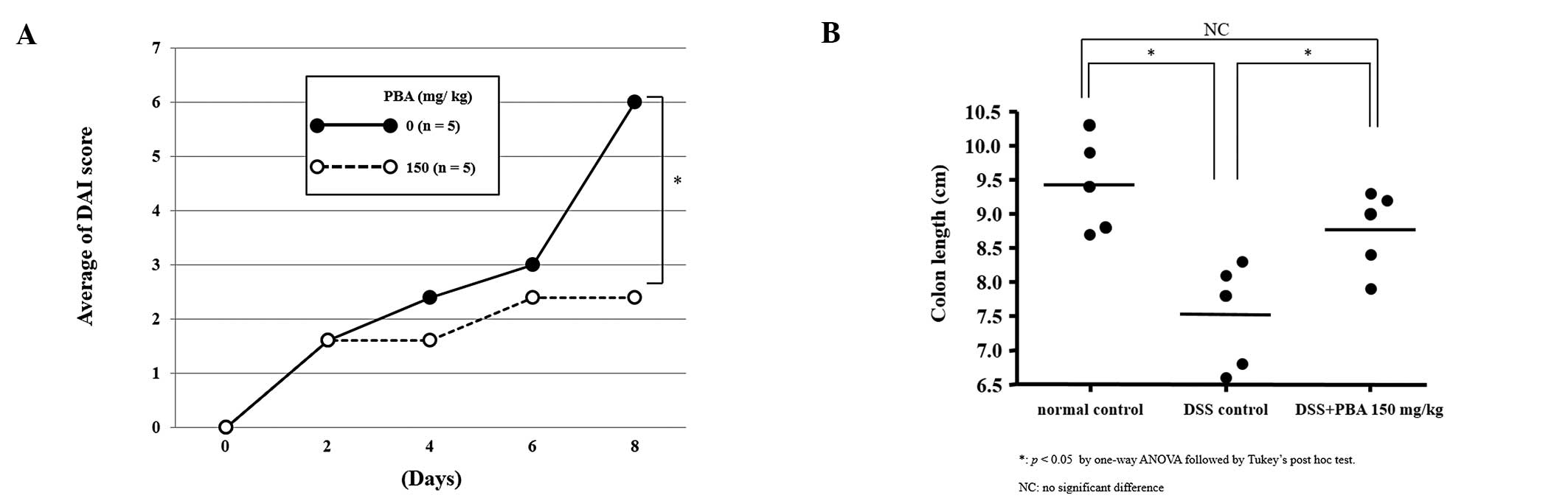
Colon length
The protective effects of PBA against DSS-induced
shortening of the colon were examined in five mice on day 8, when
the DAI score of the PBA-treated group was significantly lower than
that of the DSS control group (Fig.
4A). The animals were sacrificed and the length of the colon
from the cecocolic junction to the anal verge was measured. The
tissues were then processed for histology. The length of the colon
was 9.4±0.3 cm in healthy control animals, 7.5±0.3 cm in the DSS
control group and 8.8±0.3 cm in the PBA-treated group. The
difference between the latter two groups was statistically
significant, indicating marked suppression of the DSS-induced
shortening of the colon in mice treated with 150 mg PBA/kg body
weight (Fig. 4B).
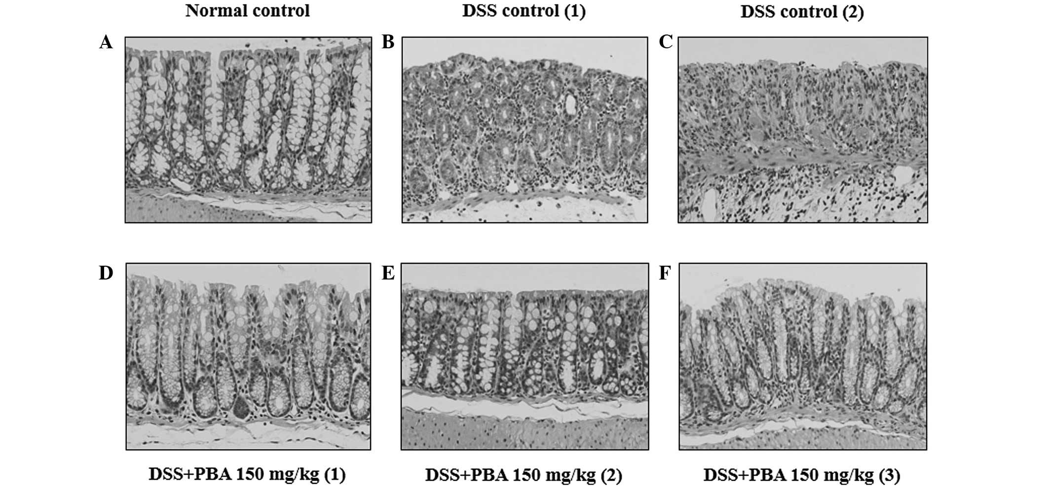
Histological findings
Microscopic examination of the excised colon
segments in the DSS control group showed chronic inflammatory cells
infiltrating the lamina propria around the crypts. The surface
epithelia were partially exfoliated, revealing the underlying
intestinal mucosa. The epithelium did not contain mucin of a mature
goblet cell and thus indicated mucin depletion (Fig. 5B and C). By contrast, in the
PBA-treated group the mucosa showed a normal appearance and
resembled that of healthy animals (Fig. 5A, D–F).
Discussion
In the present study, the effects of PBA were
investigated on DSS-induced colitis in mice as a model of IBD.
DSS-treated mice tested positive for occult blood and loose stools
were observed as early as day 2. Evident weight loss developed by
day 6, and by day 10, five of the 10 mice had died. However, in DSS
mice treated with 150 mg PBA/kg body weight, positive occult blood
was first detected on day 4 and loose stools and weight loss did
not occur until day 8. By day 10 there was only one mortality
(Fig. 2A–D). Thus, by day 12, mice
in the PBA-treated group had a 40% better survival rate than those
of the DSS control group (Fig. 1),
which is consistent with the lower DAI score (an index of clinical
state) in the PBA-treated group. PBA also significantly restricted
DSS-induced shortening of the colon (Fig. 4B) and maintained mucosal integrity
to the extent of that observed in the healthy control group
(Fig. 5A,D–F). These results
suggest that PBA treatment suppressed or limited the development of
DSS-induced colitis, histological inflammation and shortening of
the colon.
Previous studies have demonstrated the pivotal role
of abnormally high levels of pro-inflammatory cytokines, such as
TNF-α, IL-1β and IL-6, within colonic tissues in the pathogenesis
of IBD. Thus, blocking the production of these inflammatory
mediators may be of therapeutic potential in patients with IBD
(26–28). Notably, promising results have been
obtained in patients with IBD who were treated with anti-TNF-α
therapy (12,29,30).
Agents targeting interferon-γ or the IL-6 receptor are currently
undergoing clinical trials (31).
However, as with most biological therapeutics, the risk of
developing immunogenic side-effects, such as infusion reactions and
serum sickness-like reactions, are of major concern, particularly
in patients treated for chronic diseases. Small-molecule
therapeutics, including PBA, are devoid of these side-effects and
have the additional benefit of an oral route of administration
(32,33). In the present study, the
concentrations of TNF-α, IL-1β and IL-6, which were measured in the
colonic lavage fluids, increased in parallel with worsening of the
disease state in the DSS control group, whereas this was not the
case in the PBA-treated group. PBA treatment almost completely
suppressed TNF-α production until the end of the experiment.
However, while IL-1β and IL-6 production were suppressed by PBA
during the early disease phase, by the end of the experiment the
levels of both cytokines had increased and did not differ
significantly from those of the DSS-induced colitis group. However,
this early suppression was sufficient to inhibit the onset of
DSS-induced colitis, as indicated by the DAI, the histopathological
findings and the improved survival rate of the PBA-treated
mice.
The etiology of IBD remains poorly understood and
affected patients are only treated symptomatically. Moreover, the
few medications that have been demonstrated to be effective are
often associated with considerable adverse side-effects, which
further complicates the medical therapy of IBD. In the present
study, PBA treatment was shown to suppress the onset of DSS-induced
colitis. While the efficacy of PBA appears to involve the
suppression of pro-inflammatory cytokines, further studies are
required to elucidate the precise mechanisms of action. The
findings of these studies may ultimately allow the clinical use of
PBA to improve the pathological conditions and disease remission
rates of patients with IBD.
References
|
1
|
Fiocchi C: Inflammatory bowel disease:
etiology and pathogenesis. Gastroenterology. 115:182–205. 1998.
View Article : Google Scholar : PubMed/NCBI
|
|
2
|
Podolsky DK: Inflammatory bowel disease. N
Engl J Med. 347:417–429. 2002. View Article : Google Scholar
|
|
3
|
Kaser A, Zeissig S and Blumberg RS:
Inflammatory bowel disease. Annu Rev Immunol. 28:573–621. 2010.
View Article : Google Scholar
|
|
4
|
Pullman WE, Elsbury S, Kobayashi M, Hapel
AJ and Doe WF: Enhanced mucosal cytokine production in inflammatory
bowel disease. Gastroenterology. 102:529–537. 1992.PubMed/NCBI
|
|
5
|
Bouma G and Strober W: The immunological
and genetic basis of inflammatory bowel disease. Nat Rev Immunol.
3:521–533. 2003. View
Article : Google Scholar : PubMed/NCBI
|
|
6
|
Strober W, Fuss I and Mannon P: The
fundamental basis of inflammatory bowel disease. J Clin Invest.
117:514–521. 2007. View
Article : Google Scholar : PubMed/NCBI
|
|
7
|
Sartor RB: Mechanisms of disease:
pathogenesis of Crohn’s disease and ulcerative colitis. Nat Clin
Pract Gastroenterol Hepatol. 3:390–407. 2006.
|
|
8
|
Maloy KJ and Powrie F: Intestinal
homeostasis and its breakdown in inflammatory bowel disease.
Nature. 474:298–306. 2011. View Article : Google Scholar : PubMed/NCBI
|
|
9
|
Scharl M and Rogler G: Inflammatory bowel
disease pathogenesis: what is new? Curr Opin Gastroenterol.
28:301–309. 2012. View Article : Google Scholar : PubMed/NCBI
|
|
10
|
Sands BE: Therapy of inflammatory bowel
disease. Gastroenterology. 118:S68–S82. 2000. View Article : Google Scholar
|
|
11
|
Shteyer E and Wilschanski M: Novel
therapeutic modalities in pediatric inflammatory bowel disease. Isr
Med Assoc J. 10:816–820. 2008.PubMed/NCBI
|
|
12
|
Reddy JG and Loftus EV Jr: Safety of
infliximab and other biologic agents in the inflammatory bowel
disease. Gastroenterol Clin North Am. 35:837–855. 2006. View Article : Google Scholar : PubMed/NCBI
|
|
13
|
Katz JA: Management of inflammatory bowel
disease in adults. J Dig Dis. 8:65–71. 2007. View Article : Google Scholar : PubMed/NCBI
|
|
14
|
Yamamoto T, Umegae S and Matsumoto K:
Mucosal healing in patients with ulcerative colitis during a course
of selective leukocytapheresis therapy: a prospective cohort study.
Inflamm Bowel Dis. 16:1905–1911. 2010. View Article : Google Scholar
|
|
15
|
Maestri NE, Brusilow SW, Clissold DB and
Bassett SS: Long-term treatment of girls with ornithine
transcarbamylase deficiency. N Engl J Med. 335:855–859. 1996.
View Article : Google Scholar : PubMed/NCBI
|
|
16
|
Collins AF, Pearson HA, Giardina P,
McDonagh KT, Brusilow SW and Dover GJ: Oral sodium phenylbutyrate
therapy in homozygous beta thalassemia: a clinical trial. Blood.
85:43–49. 1995.PubMed/NCBI
|
|
17
|
Mercuri E, Bertini E, Messina S,
Pelliccioni M, D’Amico A, Colitto F, Mirabella M, Tiziano FD,
Vitali T, Angelozzi C, et al: Pilot trial of phenylbutyrate in
spinal muscular atrophy. Neuromuscul Disord. 14:130–135. 2004.
View Article : Google Scholar : PubMed/NCBI
|
|
18
|
Camacho LH, Olson J, Tong WP, Young CW,
Spriggs DR and Malkin MG: Phase I dose escalation clinical trial of
phenylbutyrate sodium administered twice daily to patients with
advanced solid tumors. Invest New Drugs. 25:131–138. 2007.
View Article : Google Scholar : PubMed/NCBI
|
|
19
|
Roy A, Ghosh A, Jana A, Liu X, Brahmachari
S, Gendelman HE and Pahan K: Sodium phenylbutyrate controls
neuroinflammatory and antioxidant activities and protects
dopaminergic neurons in mouse models of Parkinson’s disease. PLoS
One. 7:e381132012.PubMed/NCBI
|
|
20
|
Luo ZF, Feng B, Mu J, Qi W, Zeng W, Guo
YH, Pang Q, Ye ZL, Liu L and Yuan FH: Effects of 4-phenylbutyric
acid on the process and development of diabetic nephropathy induced
in rats by streptozotocin: regulation of endoplasmic reticulum
stress-oxidative activation. Toxicol Appl Pharmacol. 246:49–57.
2010. View Article : Google Scholar
|
|
21
|
Park JS, Lee EJ, Lee JC, Kim WK and Kim
HS: Anti-inflammatory effects of short chain fatty acids in
IFN-gamma-stimulated RAW 264.7 murine macrophage cells: involvement
of NF-kappaB and ERK signaling pathways. Int Immunopharmacol.
7:70–77. 2007. View Article : Google Scholar : PubMed/NCBI
|
|
22
|
Vij N, Fang S and Zeitlin PL: Selective
inhibition of endoplasmic reticulum-associated degradation rescues
DeltaF508-cystic fibrosis transmembrane regulator and suppresses
interleukin-8 levels: therapeutic implications. J Biol Chem.
281:17369–17378. 2006. View Article : Google Scholar
|
|
23
|
Ono K, Ikemoto M, Kawarabayashi T, Ikeda
M, Nishinakagawa T, Hosokawa M, Shoji M, Takahashi M and Nakashima
M: A chemical chaperone, sodium 4-phenylbutyric acid, attenuates
the pathogenic potency in human alpha-synuclein A30P + A53T
transgenic mice. Parkinsonism Relat Disord. 15:649–654.
2009.PubMed/NCBI
|
|
24
|
Wirtz S, Neufert C, Weigmann B and Neurath
MF: Chemically induced mouse models of intestinal inflammation. Nat
Protoc. 2:541–546. 2007. View Article : Google Scholar : PubMed/NCBI
|
|
25
|
Cooper HS, Murthy SN, Shah RS and
Sedergran DJ: Clinicopathologic study of dextran sulfate sodium
experimental murine colitis. Lab Invest. 69:238–249.
1993.PubMed/NCBI
|
|
26
|
Dionne S, D’Agata ID, Hiscott J, Vanounou
T and Seidman EG: Colonic explant production of IL-1 and its
receptor antagonist is imbalanced in inflammatory bowel disease
(IBD). Clin Exp Immunol. 112:435–442. 1998. View Article : Google Scholar : PubMed/NCBI
|
|
27
|
Tountas NA, Casini-Raggi V, Yang H, Di
Giovine FS, Vecchi M, Kam L, Melani L, Pizarro TT, Rotter JI and
Cominelli F: Functional and ethnic association of allele 2 of the
interleukin-1 receptor antagonist gene in ulcerative colitis.
Gastroenterology. 117:806–813. 1999. View Article : Google Scholar : PubMed/NCBI
|
|
28
|
Kwon KH, Murakami A, Hayashi R and
Ohigashi H: Interleukin-1beta targets interleukin-6 in progressing
dextran sulfate sodium-induced experimental colitis. Biochem
Biophys Res Commun. 337:647–654. 2005. View Article : Google Scholar : PubMed/NCBI
|
|
29
|
Rutgeerts P, Van Assche G and Vermeire S:
Optimizing anti-TNF treatment in inflammatory bowel disease.
Gastroenterology. 126:1593–1610. 2004. View Article : Google Scholar : PubMed/NCBI
|
|
30
|
Yun L and Hanauer S: Selecting appropriate
anti-TNF agents in inflammatory bowel disease. Expert Rev
Gastroenterol Hepatol. 3:235–248. 2009. View Article : Google Scholar : PubMed/NCBI
|
|
31
|
Bosani M, Ardizzone S and Porro GB:
Biologic targeting in the treatment of inflammatory bowel diseases.
Biologics. 3:77–97. 2009.PubMed/NCBI
|
|
32
|
Baert F, Noman M, Vermeire S, Van Assche
G, D’Haens G, Carbonez A and Rutgeerts P: Influence of
immunogenicity on the long-term efficacy of infliximab in Crohn’s
disease. N Engl J Med. 348:601–608. 2003.
|
|
33
|
Farrell RJ, Alsahli M, Jeen YT, Falchuk
KR, Peppercorn MA and Michetti P: Intravenous hydrocortisone
premedication reduces antibodies to infliximab in Crohn’s disease:
a randomized controlled trial. Gastroenterology. 124:917–924.
2003.PubMed/NCBI
|