Introduction
Spinal cord injury (SCI) is a common type of severe
trauma. Maximum recovery of spinal cord function has become
increasingly studied for the treatment of SCI. It was previously
hypothesized that following injury, the central nerve is not able
to regenerate; however, recent studies have identified that by
altering the local environment following SCI, the injured nerve
axons are able to regenerate and partial functioning of the spinal
cord can be restored (1,2). The following methods alter the local
environment of the spinal cord following injury and aid with the
regeneration of injured nerve axons (3): Transplantation of fetal spinal cord
tissue, peripheral nerves, neural stem cells, Schwann cells,
macrophages, olfactory ensheathing cells and umbilical cord blood
stem cells (4–13), transplantation of fibroblasts with
a neurotrophic factor injection or secretion (14,15)
and removal of myelin-related nerve growth inhibitory factors.
Previous studies have identified that marrow
mesenchymal stem cells (MSCs) can be differentiated into neural
cells in vitro and in vivo via induction. This
provides a novel method for the treatment of SCI and has been shown
to be effective in certain clinical applications (16–18).
Previously, MSCs were found to exist in the cord blood and be
induced to differentiate into bone, fat or neuron-like cells in
certain conditions or when cultured in vitro; indicating
that cord blood-derived MSCs have the potential to differentiate
into a variety of cells, including nerve cells (19–22).
Clinical trials of stem cell transplantation for the treatment of a
number of nervous system disorders have been conducted for several
years, achieving positive results (23) and providing confidence in current
stem cell research and its clinical applications for the treatment
of SCI. Several previous studies have indicated that stem cell
transplantation effectively promotes the repair of the spinal cord
in animals, thus, reducing SCI (24–26).
Although numerous studies have been performed on cord blood-derived
MSCs (6,27,28),
few have focused on the applications of MSCs. In the present study,
human umbilical cord blood (HUCB)-MSC transplantation was used for
the treatment of a rat SCI model. The behavior and histological
changes exhibited by the rats were investigated to evaluate the
therapeutic effect of HUCB-MSC transplantation. The aim of the
current study was to identify a type of cell that is suitable for
the treatment of SCI and to provide further experimental evidence
for its clinical application.
Materials and methods
HUCB
HUCB was obtained from the Departments of Gynecology
and Obstetrics at the First and Third Affiliated Hospitals of
Zhengzhou University and Zhengzhou People’s Hospital (Zhengzhou,
China). HUCB was obtained from healthy full-term cesarean or
full-term eutocia puerpera patients who tested negative for the
hepatitis B virus. The present study was conducted in accordance
with the Declaration of Helsinki and with approval from the Ethics
Committee of Zhengzhou University. Written informed consent was
obtained from all the participants.
Animals
In total, 46 healthy adult female Wistar rats
(weight, 250–280 g) were obtained from the Experimental Animal
Center of Henan (Zhengzhou, China). The rats were housed in a
specific pathogen-free room at a constant temperature of 25°C and
humidity of 45%. The present study was conducted in strict
accordance with the recommendations in the Guide for the Care and
Use of Laboratory Animals of the National Institutes of Health. The
animal use protocol was reviewed and approved by the Institutional
Animal Care and Use Committee of The First Affiliated Hospital of
Zhengzhou University.
Model establishment and grouping
The SCI model was established in accordance with the
modified Allen’s method. The rats were anesthetized with 400 mg/kg
chloral hydrate via intraperitoneal injection. A laminectomy was
subsequently performed on the entire spinous process, the vertebral
plate of T9 and part of the vertebral plate of T8 and T10, to
expose the dorsal (posterior) surface of the spinal cord. The
exposed spinal cord at the T9 level was vulnerated with a 10 g
weight dropped from a height of 2.5 cm (vulnerating energy, 25
g/cm). Following injury, the rats were randomly divided into three
groups. The injury group (n=15) received no treatment following
injury, the control group (n=15) were treated with physiological
saline and the transplantation group (n=16) were treated with the
HUCB-MSC suspension.
Cell isolation, culture and
identification
HUCB 50–80-ml samples were collected in a blood
collection bag containing composite citrate phosphate dextrose
adenine-1 anticoagulant under sterile conditions and stored at 4°C.
The samples were isolated within 12 h of collection using
Ficoll-Hypaque, according to the manufacturer’s instructions. HUCB
was diluted with physiological saline (1:1), placed in the
Ficoll-Hypaque and centrifuged at 626 × g for 20 min (the
centrifuge was provided by Hunan Xingke Medical Scientific
Instrument Co., Ltd., Changsha, China). Following centrifugation,
the cord blood mononuclear cells, which contained MSCs, were
collected. The HUCB was removed, placed in a second tube and washed
three times in physiological saline. The cells were stained with
trypan blue and the viable cells were counted under an optical
microscope (Nikon Corp., Tokyo, Japan). HUCB mononuclear cells were
diluted to a cell density of 1.0×10⁶/ml with a
specialized MSC medium (Stemcell Technologic Inc., Vancouver,
Canada) and placed in an incubator with a 5% CO2
atmosphere and saturated humidity of 37°C. After six days, the
culture media was removed and changed, then subsequently changed
every three to four days. When cell growth was exponential, the
cells were passaged at a 2:1 ratio for the two initial passages and
then at a 1:1 ratio for the third passage.
Preparation and implantation
The third passage of the HUCB-MSCs cultured in
vitro was collected and diluted to a density of
1.0×10⁷/ml. A 5-μl cell suspension was implanted into
the wounded site of the rats with SCI. The control group underwent
the same procedure using physiological saline.
Behavior and histological changes
At week one, two and four following transplantation,
an inclined plane test was conducted and Basso, Beattie, Bresnahan
(BBB) locomotor rating scale (29,30)
values were obtained for the rats in the control and
transplantation groups. Samples collected from the rats at week one
and four were stained with hematoxylin and eosin (HE) or by
immunohistochemistry (IHC), to examine the histological changes
(the related kits and reagents were provided by Beijing Zhongshan
Biotechnology Co., Ltd., Beijing, China).
Statistical analysis
Statistical analysis was performed using SPSS
software 10.0 (SPSS, Inc., Chicago, IL, USA). Data are expressed as
the mean ± SD. Differences among the groups and different time
periods were compared using the t-test and P<0.05 was considered
to indicate a statistically significant difference.
Results
Isolation of HUCB-MSCs and culture
The mononuclear cells that were isolated from the
HUCB consisted of two types of cell; a small number of spindle-like
cells and a large number of osteoclast-like cells. Osteoclast-like
cells were large, round or oval-shaped and possessed multiple
nuclei. The majority of the spindle-like cells were HUCB-MSCs,
which were successfully isolated from 18 of the 32 samples of HUCB,
however, only four were amplified and cultured in vitro.
In the early stages of culture, the HUCB-MSCs were
round. However, when the quantity of adherent cells increased, the
cell body gradually became spindle-like. The HUCB-MSCs were
mononuclear and predominantly distributed in a diffuse pattern,
with a small number growing in colonies. When cultured in
vitro, a number of the MSCs developed into heterogeneous
adherent cells. The cells varied in shape, exhibiting small and
round structures or irregular shapes; a number of the cells were
shaped like a poached egg or a star and certain cells were large
with multiple nuclei. Approximately three weeks after culturing,
with the rapid proliferation of the cells, the HUCB-MSCs appeared
to be relatively uniform, exhibiting long spindle-like structures
and colony distribution. Once the cells had grown to 80–90%
confluence, they were harvested and inoculated in passage culture
flasks. After 15 days, the cells were subcultured and amplified to
the third passage; the HUCB-MSCs were implanted in the rats with
SCI, according to the methods described previously by Wang et
al (31).
Animal behavior
Normal rats were graded on a 21-point scale,
according to the BBB ratings, prior to surgery (30,31).
Following surgery and transplantation, the rats in the three groups
were graded at various time points. At day one after the induction
of SCI, the rats scored zero points. After one week, the scores
improved, although no significant differences were identified among
the three groups (P>0.05). At week two following treatment, the
BBB ratings of the rats in the transplantation group were greater
than that of the injury and control groups (P<0.05). In
addition, at week four following treatment, the BBB ratings of the
rats in the transplantation group exhibited improved recovery when
compared with those in the other groups (P<0.05). The rats were
able to stand on their hind limbs and exhibited concordant
movements with their fore and hind limbs (Table I).
 | Table IBBB locomotor ratings of the rats in
the three groups. |
Table I
BBB locomotor ratings of the rats in
the three groups.
| BBB locomotor
ratings |
|---|
|
|
|---|
| Group | Day 1 | Week 1 | Week 2 | Week 3 | Week 4 |
|---|
| A | 0 | 6.6±0.7 | 9.6±1.2 | 10.1±1.3 | 11.0±1.5 |
| B | 0 | 6.4±0.5 | 9.4±1.3 | 9.8±1.6 | 10.9±1.8 |
| C | 0 | 7.3±0.6 | 12.8±1.6a | 13.5±2.1a | 14.2±2.3a |
Neuron-specific enolase (NSE) and glial
fibrillary acidic protein (GFAP) expression
At week one, two and four following treatment, the
HE and IHC staining were performed on the spinal cord tissue of
rats. HE staining results identified that the SCI had resulted in a
marked inflammatory reaction at week one; there was a large
quantity of inflammatory cell infiltration. At week four following
treatment, there was a degree of inflammatory cell infiltration and
a marginal difference was observed in the HE sections between the
control and transplantation groups.
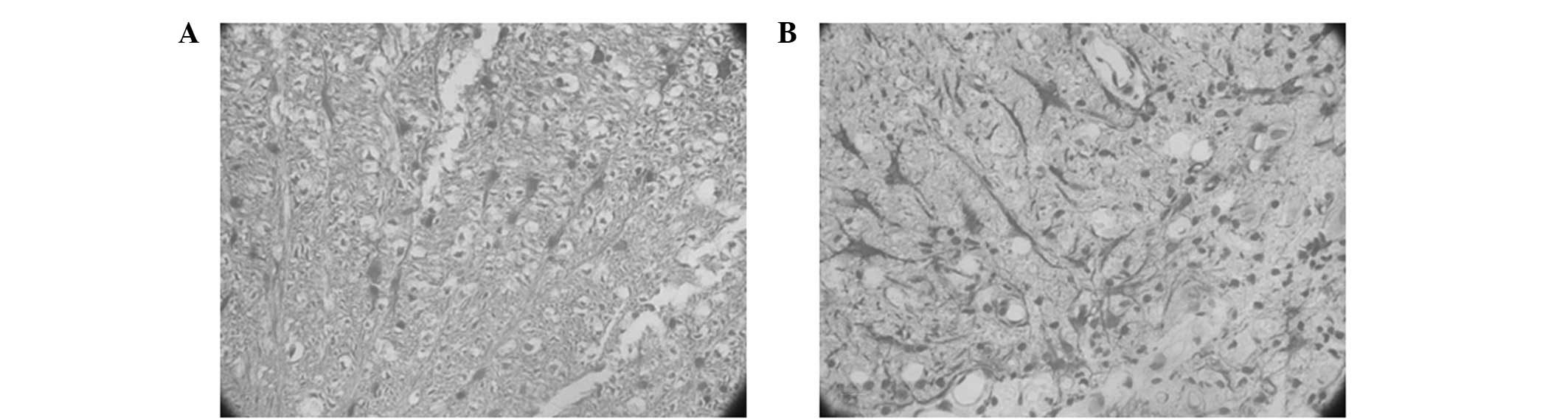
The expression of NSE and GFAP in the spinal cord
tissue was detected by IHC. There were virtually no NSE+
or GFAP+ cells identified in the injury and control
groups at week four following the treatment; however, there was a
small number of NSE+ cells and a large number of
GFAP+ cells observed in the transplantation group. The
NSE+ cells appeared in a streak or group-like manner and
there were small protrusions around the NSE+ cells. The
GFAP+ cells were stained a dark color and exhibited
morphological diversity. The processes of the GFAP+
cells increased in length and a number of the cells were fibrous
and dendritic cell-like, becoming interlaced into a network within
the spinal cord (Fig. 1).
Discussion
In the present study, HUCB-MSCs were effectively
isolated and amplified in vitro, and growth was observed in
the specialized MSC medium. Redundant MSCs were obtained through
passaging, which may provide the experimental foundation for future
investigations and clinical applications.
The results of the present study demonstrated that
HUCB-MSC transplantation for the treatment of rat SCI significantly
improved the neurological function of the damaged spinal cord at
week two following treatment. In addition, at week four after
treatment, further improvement was observed, which was consistent
with the results of previous studies regarding the treatment of SCI
by transplantation of MSCs (32,33).
Thus, the results indicate that the damaged spinal cord may recover
and regenerate following the implantation of HUCB-MSCs. However,
the underlying mechanisms behind how the transplanted MSCs survive,
concentrate and migrate to the damaged spinal cord, remain unclear.
The present study indicated that at week two following HUCB-MSC
transplantation into the damaged spinal cord, the MSCs adapted to
the microenvironment of the spinal cord and spontaneously secreted
or induced other cells to produce nerve repair factors. This
promoted local nerve repair and stimulated the surviving nerve
axons to extend their lateral branches towards the damaged axons,
resulting in improved function of the spinal cord nerves (34–36).
In addition, the mechanism may be associated with the integration
of the transplanted cells into the neural circuits (37,38).
The IHC results at week four identified that a number of the
implanted MSCs transformed into nerve cells and were able to
survive for long periods of time. The MSCs adapted to the
environment of the body and continued to promote the recovery of
the injured spinal cord. The HE results at week one and four
revealed that immune rejection of the MSC transplantation was not
significantly different from the group with non-implanted cells.
Therefore, HUCB-MSC transplantation for the treatment of rats with
SCI is considered to be a safe method.
In conclusion, the present study identified that
MSCs can be isolated from HUCB, cultured and passaged in
vitro. Following transplantation of the passaged MSCs into rats
with SCI, the isolated MSCs were able to survive in the bodies of
the rats without experiencing immune rejection. The implanted MSCs
were able to differentiate into nerve cells, which was involved in
the recovery of the damaged spinal cord. Thus, the BBB locomotor
rating scale scores were improved and the recovery of motor
function following SCI was promoted. Therefore, the results provide
a theoretical and experimental basis for HUCB-MSC transplantation
for the treatment of SCI. Following further investigation, it may
be possible to apply HUCB-MSC transplantation to the clinical
treatment of SCI. Compared with other stem cells, HUCB-MSCs exhibit
certain advantages, including favorable primitiveness, strong
amplification ability, simple acquisition and weak in vivo
rejection, without any damage to the donor. Therefore, HUCB-MSCs
may provide a novel cell source and method for the treatment of
SCI.
References
|
1
|
Akiyama Y, Honmou O, Kato T, Uede T, Hashi
K and Kocsis JD: Transplantation of clonal neural precursor cells
derived from adult human brain establishes functional peripheral
myelin in the rat spinal cord. Exp Neurol. 167:27–39. 2001.
View Article : Google Scholar : PubMed/NCBI
|
|
2
|
Blesch A, Lu P and Tuszynski MH:
Neurotrophic factors, gene therapy, and neural stem cells for
spinal cord repair. Brain Res Bull. 57:833–838. 2002. View Article : Google Scholar : PubMed/NCBI
|
|
3
|
Einstein O and Ben-Hur T: The changing
face of neural stem cell therapy in neurological diseases. Arch
Neurol. 65:452–456. 2008. View Article : Google Scholar : PubMed/NCBI
|
|
4
|
Girard C, Bemelmans AP, Dufour N, et al:
Grafts of brain-derived neurotrophic factor and neurotrophin
3-transduced primate Schwann cells lead to functional recovery of
the demyelinated mouse spinal cord. J Neurosci. 25:7924–7933. 2005.
View Article : Google Scholar : PubMed/NCBI
|
|
5
|
Goldman S: Stem and progenitor cell-based
therapy of the human central nervous system. Nat Biotechnol.
23:862–871. 2005. View
Article : Google Scholar : PubMed/NCBI
|
|
6
|
Kuh SU, Cho YE, Yoon DH, Kim KN and Ha Y:
Functional recovery after human umbilical cord blood cells
transplantation with brain-derived neutrophic factor into the
spinal cord injured rat. Acta Neurochir (Wien). 147:985–992. 2005.
View Article : Google Scholar : PubMed/NCBI
|
|
7
|
Kwon BK, Fisher CG, Dvorak MF and Tetzlaff
W: Strategies to promote neural repair and regeneration after
spinal cord injury. Spine (Phila Pa 1976). 30(17 Suppl): S3–S13.
2005. View Article : Google Scholar : PubMed/NCBI
|
|
8
|
López-Vales R, Forés J, Verdú E and
Navarro X: Acute and delayed transplantation of olfactory
ensheathing cells promote partial recovery after complete
transaction of the spinal cord. Neurobiol Dis. 21:57–68.
2006.PubMed/NCBI
|
|
9
|
Nakamura M, Toyama Y and Okano H:
Transplantation of neural stem cells for spinal cord injury. Rinsho
Shinkeigaku. 45:874–876. 2005.(In Japanese).
|
|
10
|
Rizek PN and Kawaja MD: Cultures of rat
olfactory ensheathing cells are contaminated with Schwann cells.
Neuroreport. 17:459–462. 2006. View Article : Google Scholar : PubMed/NCBI
|
|
11
|
Deumens R, Koopmans GC, Lemmens M, et al:
Neurite outgrowth promoting effects of enriched and mixed OEC/ONF
cultures. Neurosci Lett. 397:20–24. 2006. View Article : Google Scholar : PubMed/NCBI
|
|
12
|
Zhao ZM, Li HJ, Liu HY, et al: Intraspinal
transplantation of CD34+ human umbilical cord blood
cells after spinal cord hemisection injury improves functional
recovery in adult rats. Cell Transplant. 13:113–122. 2004.
|
|
13
|
Bregman BS, Kunkel-Bagden E, Schnell L,
Dai HN, Gao D and Schwab ME: Recovery from spinal cord injury
mediated by antibodies to neurite growth inhibitors. Nature.
378:498–501. 1995. View
Article : Google Scholar : PubMed/NCBI
|
|
14
|
Bregman BS, McAtee M, Dai HN and Kuhn PL:
Neurotrophic factors increase axonal growth after spinal cord
injury and transplantation in the adult rat. Exp Neurol.
148:475–494. 1997. View Article : Google Scholar : PubMed/NCBI
|
|
15
|
Popovich PG, Guan Z, Wei P, Huitinga I,
van Rooijen N and Stokes BT: Depletion of hematogenous macrophages
promotes partial hindlimb recovery and neuroanatomical repair after
exprimental spinal cord injury. Exp Neurol. 158:351–365. 1999.
View Article : Google Scholar
|
|
16
|
Kishk NA, Gabr H, Hamdy S, et al: Case
control series of intrathecal autologous bone marrow mesenchymal
stem cell therapy for chronic spinal cord injury. Neurorehabil
Neural Repair. 24:702–708. 2010. View Article : Google Scholar : PubMed/NCBI
|
|
17
|
Pal R, Venkataramana NK, Bansal A, et al:
Ex vivo-expanded autologous bone marrow-derived mesenchymal stromal
cells in human spinal cord injury/paraplegia: a pilot clinical
study. Cytotherapy. 11:897–911. 2009. View Article : Google Scholar : PubMed/NCBI
|
|
18
|
Saito F, Nakatani T, Iwase M, et al:
Spinal cord injury treatment with intrathecal autologous bone
marrow stromal cell transplantation: the first clinical trial case
report. J Trauma. 64:53–59. 2008. View Article : Google Scholar : PubMed/NCBI
|
|
19
|
Chiu B, Wan JZ, Abley D and Akabutu J:
Induction of vascular endothelial phenotype and cellular
proliferation from human cord blood stem cells cultured in
simulated microgravity. Acta Astronaut. 56:918–922. 2005.
View Article : Google Scholar : PubMed/NCBI
|
|
20
|
Hong SH, Gang EJ, Jeong JA, et al: In
vitro differentiation of human umbilical cord blood-derived
mesenchymal stem cells into hepatocyte-like cells. Biochem Biophys
Res Commun. 330:1153–1161. 2005. View Article : Google Scholar : PubMed/NCBI
|
|
21
|
Jurga M, Markiewicz I, Sarnowska A, et al:
Neurogenic potential of human umbilical cord blood: neural-like
stem cells depend on previous long-term culture conditions. J
Neurosci Res. 83:627–637. 2006. View Article : Google Scholar : PubMed/NCBI
|
|
22
|
Walczak P, Chen N, Eve D, et al: Long term
cultured human umbilical cord neural-like cells transplanted into
the striatum of NOD SCID mice. Brain Res Bull. 74:155–163. 2007.
View Article : Google Scholar : PubMed/NCBI
|
|
23
|
El-Badri NS, Hakki A, Saporta S, et al:
Cord blood mesenchymal stem cells: Potential use in neurological
disorders. Stem Cells Dev. 15:497–506. 2006. View Article : Google Scholar : PubMed/NCBI
|
|
24
|
Ankeny DP, McTigue DM and Jakeman LB: Bone
marrow transplants provide tissue protection and directional
guidance for axons after contusive spinal cord injury in rats. Exp
Neurol. 190:17–31. 2004. View Article : Google Scholar : PubMed/NCBI
|
|
25
|
Mansilla E, Marin GH, Sturla F, et al:
Human mesenchymal stem cells are tolerized by mice and improve skin
and spinal cord injuries. Transplant Proc. 37:292–294. 2005.
View Article : Google Scholar : PubMed/NCBI
|
|
26
|
Schultz SS: Adult stem cell application in
spinal cord injury. Curr Drug Targets. 6:63–73. 2005. View Article : Google Scholar : PubMed/NCBI
|
|
27
|
Harris DT: Non-haematological uses of cord
blood stem cells. Br J Haematol. 147:177–184. 2009. View Article : Google Scholar : PubMed/NCBI
|
|
28
|
Kim SU and de Vellis J: Stem cell-based
cell therapy in neurological diseases: a review. J Neurosci Res.
87:2183–2200. 2009. View Article : Google Scholar : PubMed/NCBI
|
|
29
|
Basso DM, Beattie MS and Bresnahan JC: A
sensitive and reliable locomotor rating scale for open field
testing in rats. J Neurotrauma. 12:1–21. 1995. View Article : Google Scholar : PubMed/NCBI
|
|
30
|
Tomita S, Mickle DA, Weisel RD, et al:
Improved heart function with myogenesis and angiogenesis after
autologous porcine bone marrow stromal cell transplantation. J
Thorac Cardiovasc Surg. 123:1132–1140. 2002. View Article : Google Scholar : PubMed/NCBI
|
|
31
|
Wang TT, Tio M, Lee W, Beerheide W and
Udolph G: Neural differentiation of mesenchymal-like stem cells
from cord blood is mediated by PKA. Biochem Biophys Res Commun.
357:1021–1027. 2007. View Article : Google Scholar : PubMed/NCBI
|
|
32
|
Pal R, Gopinath C, Rao NM, et al:
Functional recovery after transplantation of bone marrow-derived
human mesenchymal stromal cells in a rat model of spinal cord
injury. Cytotherapy. 12:792–806. 2010. View Article : Google Scholar : PubMed/NCBI
|
|
33
|
Bossolasco P, Cova L, Calzarossa C, et al:
Neuro-glial differentiation of human bone marrow stem cells in
vitro. Exp Neurol. 193:312–325. 2005. View Article : Google Scholar : PubMed/NCBI
|
|
34
|
Bjugstad KB, Redmond DE Jr, Teng YD, et
al: Neural stem cells implanted into MPTP-treated monkeys increase
the size of endogenous tyrosine hydroxylase-positive cells found in
the striatum: a return to control measures. Cell Transplant.
14:183–192. 2005. View Article : Google Scholar
|
|
35
|
Garbuzova-Davis S, Willing AE, Saporta S,
et al: Novel cell therapy approaches for brain repair. Prog Brain
Res. 157:207–222. 2006. View Article : Google Scholar
|
|
36
|
Leu S, Lin YC, Yuen CM, et al:
Adipose-derived mesenchymal stem cells markedly attenuate brain
infarct size and improve neurological function in rats. J Transl
Med. 8:632010. View Article : Google Scholar : PubMed/NCBI
|
|
37
|
Djouad F, Delorme B, Maurice M, et al:
Microenvironmental changes during differentiation of mesenchymal
stem cells towards chondrocytes. Arthritis Res Ther. 9:R332007.
View Article : Google Scholar : PubMed/NCBI
|
|
38
|
Crigler L, Robey RC, Asawachaicharn A,
Gaupp D and Phinney DG: Human mesenchymal stem cell subpopulations
express a variety of neuro-regulatory molecules and promote
neuronal cell survival and neuritogenesis. Exp Neurol. 198:54–64.
2006. View Article : Google Scholar : PubMed/NCBI
|