Introduction
Rheumatoid arthritis (RA) is the most common type of
inflammatory arthritis, affecting between 0.5 and 1% of the
population worldwide, regardless of geographical location and
ethnicity (1,2). Although the etiology of RA remains to
be fully elucidated, numerous studies have suggested that a
combination of environmental and genetic factors are responsible.
However, although environmental and genetic factors have been
demonstrated to be important, they are insufficient for full
expression of the disease. The primary inflammatory site in RA is
the synovium. The proliferation of synovial fibroblasts is one of
the main characteristics of RA and is considered to be necessary
for the initiation and long-term progression of joint destruction
in RA (3–6). In the long-term, joint destruction
may lead to limited functions, decreased work ability and, more
importantly, a decrease in the quality of life for patients with
RA. In addition, RA is associated with an increased risk of
cardiovascular disease and, thus, the life expectancy of patients
with RA may be reduced by 3–18 years (7). Therefore, the inhibition of synovial
hyperplasia during the early stages of disease progression may
provide a potential therapeutic approach for the treatment of
RA.
Houttuynia cordata Thunb (HCT) is a perennial
herbaceous plant that grows in the wild in moist, shady areas in
numerous Asian countries, including India. HCT has been widely used
in China, Japan and other Asian countries as a medicine due to its
anti-inflammatory properties (8).
Previous studies have demonstrated that HCT has anti-inflammatory
effects in a wide range of diseases (8–11).
During the outbreak of hand, foot and mouth disease (HFMD) in 2008
in China, HCT was used as a therapeutic drug (12). Several studies demonstrated that
HCT inhibited enterovirus 71 and coxsackievirus A16, which are the
two main causative agents of HFMD (13,14).
It has also been demonstrated that HCT water extract was able to
treat severe acute respiratory syndrome (8,15,16)
and herpes simplex virus infection (17). Sodium houttuyfonate (SH), an
addition compound of sodium bisulfite and houttuynia, is the stable
form of houttuynia and exhibits the same effect as HCT. Certain
studies have demonstrated that SH has an antibacterial effect
against 21 strains of Staphylococcus aureus (18) and it has previously been used for
the treatment of cationic bovine serum albumin-induced membranous
glomerulonephritis in BALB/c mice (19). Previous studies have also
demonstrated that SH exerts an anti-inflammatory effect by
inhibiting the tumor necrosis factor-α (TNF-α) pathway (20), which led to the hypothesis that SH
may also be effective for the treatment of RA.
Materials and methods
Materials
Synovial tissue was obtained from a patient at the
Department of Orthopedics and Pathology, Shanghai Third People’s
Hospital (Shanghai, China). SH was purchased from Shanghai Qingping
Pharmaceutical Co., Ltd. (Shanghai, China; batch number 0701-3).
D-Hank’s solution and RPMI-1640 nutrient medium were provided by
the laboratory of the Shanghai Third People’s Hospital, School of
Medicine, Shanghai Jiao Tong University (Shanghai, China). Fetal
bovine serum (FBS) was purchased from Beijing Ruizekang Biotech
Co., Ltd. (Beijing, China). Type II collagenase and trypsin were
obtained from Shanghai Qifa Experimental Reagent Co., Ltd.
(Shanghai, China). The MTT kit was purchased from Sigma (St. Louis,
MO, USA). Informed consent was obtained from the patient.
Cultivation of primary cells from a
patient with RA
The fat and fibrous tissue was removed from the
synovial tissue. The tissue was then washed three times with
D-Hank’s solution and cut into two sections (1–2 mm in size). The
tissue was placed in 25 cm2 culture bottles containing 2
ml RPMI-1640 nutrient solution in 10% FBS and 2 ml 0.4% type II
collagenase. The culture bottles were incubated at 37°C and 5%
CO2 for 2 h. Unattached cells were then transferred into
centrifuge tubes and centrifuged for 10 min. A total of 4 ml 0.25%
trypsin was added and the cells were incubated for 30 min. The
solution was then filtered using a 200-mesh nylon net. Following
centrifugation for 10 min, the cells were incubated as
aforementioned for 24 h. The unattached cells were discarded,
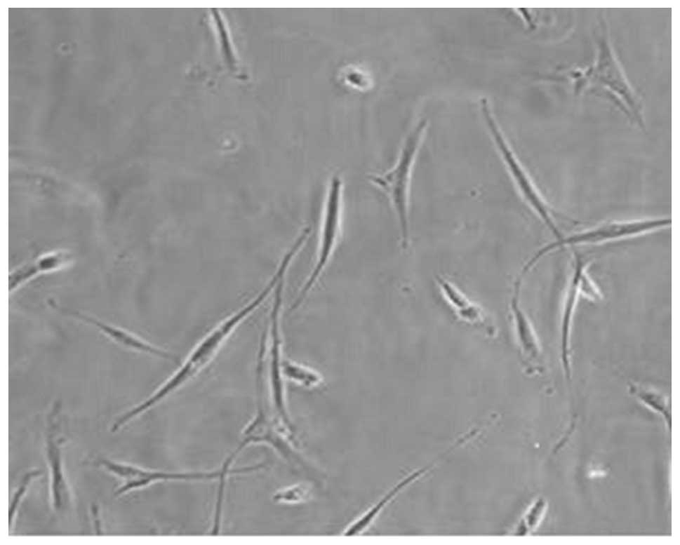
leaving primary cells from a patient with RA (Fig. 1).
Experimental groups and
administration
The primary cells were equally divided into five
groups as follows: the control group (group 1), cells treated with
25 μg/ml SH (group 2), 50 μg/ml SH (group 3), 100 μg/ml SH (group
4) and 200 μg/ml SH (group 5). Group 1 was administered an
equivalent amount of normal saline (NS), whilst groups 2 to 5 were
treated with corresponding amounts of SH. NS and SH were
administered daily for 7 days by transfer pipette. Following the
final administration, the five groups of synovial cells were
measured using an MTT assay for analysis of the growth inhibitory
rate of SH on synovial proliferation.
MTT assay
Sequential dilutions of cells in the culture medium
between 106 and 103 cells/ml were prepared. A
total of 100 μl each dilution was analyzed in triplicate, using a
microplate reader (Bio-Rad, Hercules, CA, USA) and three control
wells containing medium only were used as an absorbance reference.
The cells were then incubated for 24 h. A total of 10 μl MTT
reagent (0.25% MTT) was added to each well and the cells were
further incubated for 4 h until a purple precipitate was observed.
A total of 100 μl detergent reagent was added to each well and
swirled gently, and the plate was then incubated in the dark
overnight at room temperature. The absorbance in each well at 570
nm was measured using a microplate reader. Finally, the data were
recorded and the results were analyzed.
Data interpretation and statistical
analysis
If the absorbance rate was lower than the control,
this was considered to indicate a reduction in cell proliferation.
By contrast, if the absorbance rate was higher, this indicated an
increase in cell proliferation. The difference in the inhibition
rate between the groups was analyzed using one-way analysis of
covariance. P<0.05 was considered to indicate a statistically
significant difference.
Results
Inhibition rate of SH on synovial
proliferation in cells from a patient with RA
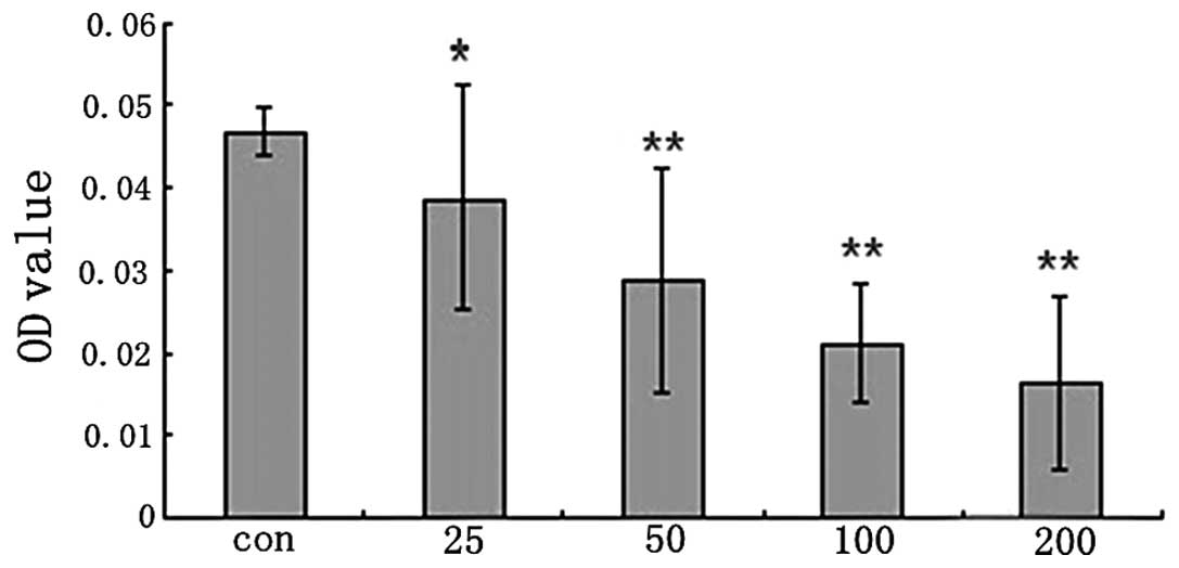
As shown in Fig. 2,
the proliferation rate of synovial cells was markedly higher in the
control group compared with the other groups (P<0.05). In the
SH-treated groups, the proliferation rates of synovial cells were
significantly decreased compared with those in group 1 (P<0.05
for group 2; P<0.01 for groups 3–5). These results indicated
that SH decreased the level of synovial proliferation in cells from
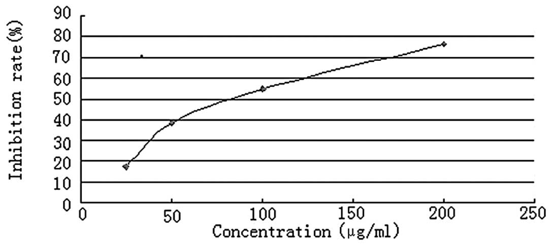
a patient with RA. Fig. 3 shows
the inhibition rate of different concentrations of SH on synovial
proliferation. The inhibition rates of different concentrations of
SH are shown numerically in Table
I and the correlation coefficient was found to be 0.961718.
 | Table IAssociation between the inhibition
rate of sodium houttuyfonate and its concentration. The correlative
coefficient is 0.961718. |
Table I
Association between the inhibition
rate of sodium houttuyfonate and its concentration. The correlative
coefficient is 0.961718.
| Inhibition rate
(%) | Concentration
(μg/ml) |
|---|
| 17.02 | 25 |
| 38.30 | 50 |
| 54.79 | 100 |
| 76.60 | 200 |
Discussion
The proliferation of synovial fibroblasts leads to
the development of RA and initiates joint destruction in the long
term (3–5). Previous studies have demonstrated
that TNF-α, interleukin (IL)-6 and IL-8 are important
proinflammatory cytokines in the pathogenesis of RA (21–24),
and that the inhibition of TNF-α and IL-6 is effective in the
treatment of patients with RA (25–27).
Notably, several studies have indicated that HCT was able to
efficiently inhibit IL-6, IL-8 and TNF-α (24,28).
In addition, nonsteroidal anti-inflammatory drugs (NSAIDs) have
been demonstrated to be efficacious in the treatment of RA by
binding to cyclooxygenase (COX) enzymes and therefore inhibiting
the production of prostaglandins. Previous studies also
demonstrated that HCT supercritical extract exerted an
anti-inflammatory effect by inhibiting the COX-2/prostaglandin E2
pathway. Owing to the gastrointestinal side-effects of NSAIDs, HCT
may be a better drug candidate for the alleviation of symptoms
caused by RA (20). The present
study demonstrated that the synovial proliferation rate
significantly decreased following treatment with SH. Furthermore,
the inhibition rate of SH was found to be dose dependent.
Therefore, these results suggest that SH is able to inhibit the
proliferation of synovial cells from a patient with RA.
The results from the present study provide a
potential theoretical basis for the treatment of RA. Furthermore,
due to the dose-dependent reaction of SH observed in the present
study, a suitable dose of SH may almost completely inhibit synovial
proliferation and therefore be highly effective in the clinical
treatment of patients with RA.
Acknowledgements
The authors would like to thank the doctors at the
Department of Orthopedics and Pathology, as well as the technicians
at the laboratory of Shanghai Third People’s Hospital, School of
Medicine, Shanghai Jiao Tong University for their technical
assistance.
References
|
1
|
Gabriel SE and Michaud K: Epidemiological
studies in incidence, prevalence, mortality, and comorbidity of the
rheumatic diseases. Arthritis Res Ther. 11:2292009. View Article : Google Scholar : PubMed/NCBI
|
|
2
|
Yilmaz S and Simek I: Early intervention
in the treatment of rheumatoid arthritis: focus on tocilizumab.
Ther Clin Risk Manag. 9:403–408. 2013.PubMed/NCBI
|
|
3
|
Mor A, Abramson SB and Pillinger MH: The
fibroblast-like synovial cell in rheumatoid arthritis: a key player
in inflammation and joint destruction. Clin Immunol. 115:118–128.
2005. View Article : Google Scholar : PubMed/NCBI
|
|
4
|
Huber LC, Distler O, Tarner I, Gay RE, Gay
S and Pap T: Synovial fibroblasts: key players in rheumatoid
arthritis. Rheumatology (Oxford). 45:669–675. 2006. View Article : Google Scholar : PubMed/NCBI
|
|
5
|
Bartok B and Firestein GS: Fibroblast-like
synoviocytes: key effector cells in rheumatoid arthritis. Immunol
Rev. 233:233–255. 2010. View Article : Google Scholar : PubMed/NCBI
|
|
6
|
Parada-Turska J, Nowicka-Stążka P, Majdan
M, Jabłoński M, Turski WA and Rzeski W: Anti-epileptic drugs
inhibit viability of synoviocytes in vitro. Ann Agric Environ Med.
20:571–574. 2013.PubMed/NCBI
|
|
7
|
Pincus T, Kavanaugh A and Sokka T:
Benefit/risk of therapies for rheumatoid arthritis: underestimation
of the ‘side effects’ or risks of RA leads to underestimation of
the benefit/risk of therapies. Clin Exp Rheumatol. 22(Suppl 35):
S2–S11. 2004.PubMed/NCBI
|
|
8
|
Lu HM, Liang YZ, Yi LZ and Wu XJ:
Anti-inflammatory effect of Houttuynia cordata injection. J
Ethnopharmacol. 104:245–249. 2006. View Article : Google Scholar
|
|
9
|
Miyata M, Koyama T and Yazawa K: Water
extract of Houttuynia cordata Thunb. leaves exerts
anti-obesity effects by inhibiting fatty acid and glycerol
absorption. J Nutr Sci Vitaminol (Tokyo). 56:150–156. 2010.
|
|
10
|
Hayashi K, Kamiya M and Hayashi T:
Virucidal effects of the steam distillate from Houttuynia
cordata and its components on HSV-1, influenza virus, and HIV.
Plant Med. 61:237–241. 1995. View Article : Google Scholar : PubMed/NCBI
|
|
11
|
Chang JS, Chiang LC, Chen CC, Liu LT, Wang
KC and Lin CC: Antileukemic activity of Bidens pilosa L.
var. minor (Blume) Sherff and Houttuynia cordata Thunb. Am J
Chin Med. 29:303–312. 2001.
|
|
12
|
Yang F, Ren L, Xiong Z, et al: Enterovirus
71 outbreak in the People’s Republic of China in 2008. J Clin
Microbiol. 47:2351–2352. 2009.
|
|
13
|
Chen X, Wang C, Xu L, et al: A laboratory
evaluation of medicinal herbs used in China for the treatment of
hand, foot, and mouth disease. Evid Based Complement Alternat Med.
2013:5045632013.PubMed/NCBI
|
|
14
|
Lin TY, Liu YC, Jheng JR, Tsai HP, Jan JT,
Wong WR and Horng JT: Anti-enterovirus 71 activity screening of
Chinese herbs with anti-infection and inflammation activities. Am J
Chin Med. 37:143–158. 2009. View Article : Google Scholar : PubMed/NCBI
|
|
15
|
Lau KM, Lee KM, Koon CM, et al:
Immunomodulatory and anti-SARS activities of Houttuynia
cordata. J Ethnopharmacology. 118:79–85. 2008. View Article : Google Scholar : PubMed/NCBI
|
|
16
|
Li S, Wang R, Zhang Y, Zhang X, Layon AJ,
Li Y and Chen M: Symptom combinations associated with outcome and
therapeutic effects in a cohort of cases with SARS. Am J Chin Med.
34:937–947. 2006. View Article : Google Scholar : PubMed/NCBI
|
|
17
|
Chen X, Wang Z, Yang Z, Wang J, Xu Y, Tan
RX and Li E: Houttuynia cordata blocks HSV infection through
inhibition of NF-κB activation. Antiviral Res. 92:341–345. 2011.
View Article : Google Scholar
|
|
18
|
Liu G, Xiang H, Tang X, et al:
Transcriptional and functional analysis shows sodium
houttuyfonate-mediated inhibition of autolysis in Staphylococcus
aureus. Molecules. 16:8848–8865. 2011. View Article : Google Scholar : PubMed/NCBI
|
|
19
|
Pan P, Wang YJ, Han L, Liu X, Zhao M and
Yuan YF: Effects of sodium houttuyfonate on expression of NF-κB and
MCP-1 in membranous glomerulonephritis. J Ethnopharmacology.
131:203–209. 2010.PubMed/NCBI
|
|
20
|
Shin S, Joo SS, Jeon JH, et al:
Anti-inflammatory effects of a Houttuynia cordata
supercritical extract. J Vet Sci. 11:273–275. 2010. View Article : Google Scholar
|
|
21
|
Choy EH and Panayi GS: Cytokine pathways
and joint inflammation in rheumatoid arthritis. N Engl J Med.
344:907–916. 2001. View Article : Google Scholar : PubMed/NCBI
|
|
22
|
Feldmann M: Development of anti-TNF
therapy for rheumatoid arthritis. Nat Rev Immunol. 2:364–371. 2002.
View Article : Google Scholar : PubMed/NCBI
|
|
23
|
Maini RN: Anti-TNF therapy from the bench
to the clinic: a paradigm of translational research. Clin Med.
10:161–162. 2010. View Article : Google Scholar : PubMed/NCBI
|
|
24
|
Lee HJ, Seo HS, Kim GJ, et al:
Houttuynia cordata Thunb inhibits the production of
pro-inflammatory cytokines through inhibition of the NFκB signaling
pathway in HMC-1 human mast cells. Mol Med Rep. 8:731–736.
2013.
|
|
25
|
Maini RN and Taylor PC: Anti-cytokine
therapy for rheumatoid arthritis. Annu Rev Med. 51:207–229. 2000.
View Article : Google Scholar : PubMed/NCBI
|
|
26
|
Chu K, Zheng H, Li H, Zhang Y, Zhang X, Xu
W and Chen L: Shuangtengbitong tincture treatment of
collagen-induced arthritis via downregulation of the expression of
IL-6, IL-8, TNF-α and NF-κB. Exp Ther Med. 5:423–428.
2013.PubMed/NCBI
|
|
27
|
Weinblatt ME, Keystone EC, Furst DE, et
al: Adalimumab, a fully human anti-tumor necrosis factor alpha
monoclonal antibody, for the treatment of rheumatoid arthritis in
patients taking concomitant methotrexate: the ARMADA trial.
Arthritis Rheum. 48:35–45. 2003. View Article : Google Scholar
|
|
28
|
Park E, Kum S, Wang C, Park SY, Kim BS and
Schuller-Levis G: Anti-inflammatory activity of herbal medicines:
inhibition of nitric oxide production and tumor necrosis
factor-alpha secretion in an activated macrophage-like cell line.
Am J Chin Med. 33:415–424. 2005. View Article : Google Scholar
|