Introduction
Colorectal cancer (CRC) is the third most common
cancer in humans worldwide (1). It
has recently been reported that CRC mortality accounts for ~9% of
all cancer mortalities (2). The
survival rate for CRC is higher at early stages following surgical
resection; however, the long-term survival rate and prognosis for
patients with CRC remain poor (3).
Currently available cancer markers are not suitable for current
clinical practice and require further investigation. Therefore,
there is an urgent requirement for the discovery of novel targets
and useful biomarkers for CRC prognosis.
Protein phosphatase, Mg2+/Mn2+
dependent, 1D (PPM1D) is a member of the protein phosphatase 2C
family and is involved in a wide range of physiological functions,
including cell signaling, apoptosis and the cell cycle (4). A growing body of evidence suggests
that PPM1D is involved in tumorigenesis. PPM1D has been reported to
be upregulated in human primary breast, ovarian and neuroblastoma
tumors (5–7). Of note, PPM1D has been found to
complement oncogenes during cellular transformation (8). However, the role of PPM1D in CRC is
yet to be elucidated. The present study investigated the expression
pattern and clinical significance of PPM1D in CRC, including the
correlation of PPM1D with clinical outcome.
Materials and methods
Patients and samples
Based on tissue data availability, 368 cases of CRC,
between 2003 and 2007, were included in the present study.
Clinicopathological features of the patients with CRC are shown in
Table I. Formalin-fixed
paraffin-embedded tissues were collected from the Second and First
Affiliated Hospitals of Hunan University of Chinese Medicine
(Changsha, China). This study was approved by the Ethics Committees
of Hunan University of Chinese Medicine. Informed consent was
obtained from all participants and the study was performed in
accordance with the Declaration of Helsinki. Follow-up data were
obtained from medical records and direct communication with the
patients or their relatives. The follow-up period was defined as
the time from the date of surgery to the date of patient mortality
or the final follow-up in January 2012.
 | Table IClinicopathological characteristics
and PPM1D expression in patients with colorectal cancer. |
Table I
Clinicopathological characteristics
and PPM1D expression in patients with colorectal cancer.
| | PPM1D expression | |
|---|
| |
| |
|---|
| Characteristic | n | Low | High | P-value |
|---|
| Total | 368 | 116 | 252 | |
| Age (years) | | | | 0.371 |
| <60 | 144 | 52 | 92 | |
| ≥60 | 224 | 64 | 160 | |
| Gender | | | | 0.49 |
| Male | 208 | 72 | 136 | |
| Female | 160 | 44 | 116 | |
| Tumor location | | | | 0.875 |
| Right colon | 80 | 24 | 56 | |
| Left colon | 92 | 32 | 60 | |
| Rectum | 196 | 60 | 136 | |
| Histology
(differentiation) | | | | 0.098 |
| Well | 172 | 48 | 124 | |
| Moderate | 136 | 56 | 80 | |
| Poor | 60 | 12 | 48 | |
| Node metastasis | | | | 0.0024 |
| N0 | 160 | 72 | 88 | |
| N1–3 | 208 | 44 | 164 | |
| Distant
metastasis | | | | <0.001 |
| No | 288 | 112 | 176 | |
| Yes | 80 | 4 | 76 | |
| TNM stage | | | | 0.0016 |
| I | 28 | 12 | 16 | |
| II | 112 | 24 | 88 | |
| III | 148 | 72 | 76 | |
| IV | 80 | 8 | 72 | |
Histopathological evaluation and
scoring
Paraffin-embedded serial sections of CRC specimens
were analyzed for PPM1D protein expression using an anti-PPM1D
antibody (Abcam, Cambridge, MA, USA), as described previously
(9,10). Negative control sections were
incubated with pre-immunized rabbit serum (Abcam).
Immunostaining was assessed by two independent,
blinded pathologists and scored by multiplying the intensity of the
staining by the percentage of stained cells. The staining intensity
was graded on a scale of 0 to 3. The percentage of stained tumor
cells was graded as follows: 0 (<5%), 1 (5–25%), 2 (26–50%), 3
(51–75%) and 4 (>75%). The final scores ranged from 0 to 12. For
any subsequent analysis, scores between 0 and 4 were defined as low
expression and those between 5 and 12 were defined as high
expression (11). Inconsistent
scores were re-evaluated by two pathologists until a consensus
score was established.
Quantitative polymerase chain reaction
(qPCR) analysis
Total RNA was extracted from cells using an RNA
extraction kit (Qiagen Co., Ltd., Shanghai, China) according to the
manufacturer’s instructions. Total RNA was used for quantification
using the One-Step RT-PCR kit (Invitrogen Life Technologies,
Carslbad, CA, USA) in a Roche Light Cycler (Roche Diagnostics,
Basel, Switzerland). The primer sequences used in the study were as
follows: PPM1D, 5′-CAATTGGCCTTG TGCCTACT-3′ (forward) and
5′-TCTTTCGCTGTGAGGTTGTG-3′ (reverse); and β-actin,
5′-CCTGTACGCCAACACAGTGC-3′ (forward) and 5′-ATACTCCTGCTTGCTGATCC-3′
(reverse). The PCR cycling conditions consisted of a denaturation
step at 95°C for 3 min, followed by 40 cycles of 95°C for 10 sec
and 60°C for 1 min. Samples were normalized using β-actin mRNA
expression and relative expression was calculated using the
ΔΔCt method.
Western blot analysis
Cells were lysed in lysis buffer and protein
concentrations were measured using a BCA protein assay kit (Qiagen
Co., Ltd.). Total protein was separated using SDS-PAGE on a 12.5%
gel and electroblotted onto polyvinylidene fluoride membranes
(Millipore Corporation, Billerica, MA, USA). Membranes were
immunoblotted overnight at 4°C with primary antibodies against
human PPM1D (1:1,000 dilution; Sigma-Aldrich, St. Louis, MO, USA)
or β-actin (1:2,000 dilution; Sigma-Aldrich). Following three
washes with Tris-buffered saline containing Tween 20, membranes
were incubated with horseradish peroxidase-conjugated IgG secondary
antibodies (1:2,000 dilution; Santa Cruz Biotechnology, Inc., Santa
Cruz, CA, USA) for 2 h. Immunoreactive signals were detected using
an enhanced chemiluminescence detection reagent (Pierce
Biotechnology, Rockford, IL, USA). Expression levels were
quantified from the images using Quantity One® (Bio-Rad,
Hercules, CA, USA). All experiments were performed in
triplicate.
Cell culture and transfection
HCT-116, RKO and COLO-320 CRC cell lines were
purchased from American Type Culture Collection (Rockville, MD,
USA). Cells were maintained in RPMI-1640 in 5% CO2 at
37°C. PPM1D siRNA and scrambled siRNA were purchased from
Invitrogen Life Technologies and transfected using
Lipofectamine® 2000 (Invitrogen Life Technologies)
according to the manufacturer’s instructions.
MTT assay
Cell viability was assessed using an MTT assay.
Following transfection, cells were plated in 96-well plates and
incubated for 24, 48 and 72 h. A total of 20 μl 5 mg/ml MTT
(Sigma-Aldrich) was added to each corresponding test well and
incubated for 4 h at 37°C. The supernatant was then discarded and
200 μl dimethyl sulfoxide was added to each well to dissolve the
formazan. Optical density was assessed by measuring the absorbance
of each well at 490 nm using a spectrophotometer (SpectraMax
Plus384; Molecular Devices, Sunnyvale, CA, USA). All experiments
were performed in triplicate.
Cell invasion assay
At 12 h after transfection, the mixed medium
containing Lipofectamine 2000 was discarded and cells were
maintained in serum-free RPMI-1640 medium overnight. Cell invasion
was then assessed using a 24-well Cell Invasion assay (BD
Biosciences, San Jose, CA, USA) with 8 μm pores according to the
manufacturer’s instructions. Cells were incubated for 72 h. The
upper compartment was coated with 50 μg Matrigel™ (BD Biosciences)
and the lower compartment was filled with medium containing 10%
fetal calf serum as a chemoattractant. Spectrophotometry was
conducted using a microtiter plate reader (SpectraMax Plus384;
Molecular Devices) at 540 nm. All assays were performed in
triplicate.
Statistical analysis
All statistical analyses were performed using SPSS
16.0 software (SPSS, Inc, Chicago, IL, USA). The χ2 test
was performed for categorical data. Kaplan-Meier estimates,
log-rank tests and multivariate Cox proportional hazard regression
models were performed for survival analyses. A value of P<0.05
was considered to indicate statistical significance.
Results
Correlation between PPM1D and
clinicopathological variables
PPM1D expression was analyzed in 368 CRC tissue
samples and paired non-cancerous colorectal tissue samples. Among
the 368 CRC tissues, 68.48% (252/368) of cases demonstrated high
PPM1D expression, while only 9.24% (34/368) of the matched,
non-cancerous colorectal tissue samples showed high PPM1D
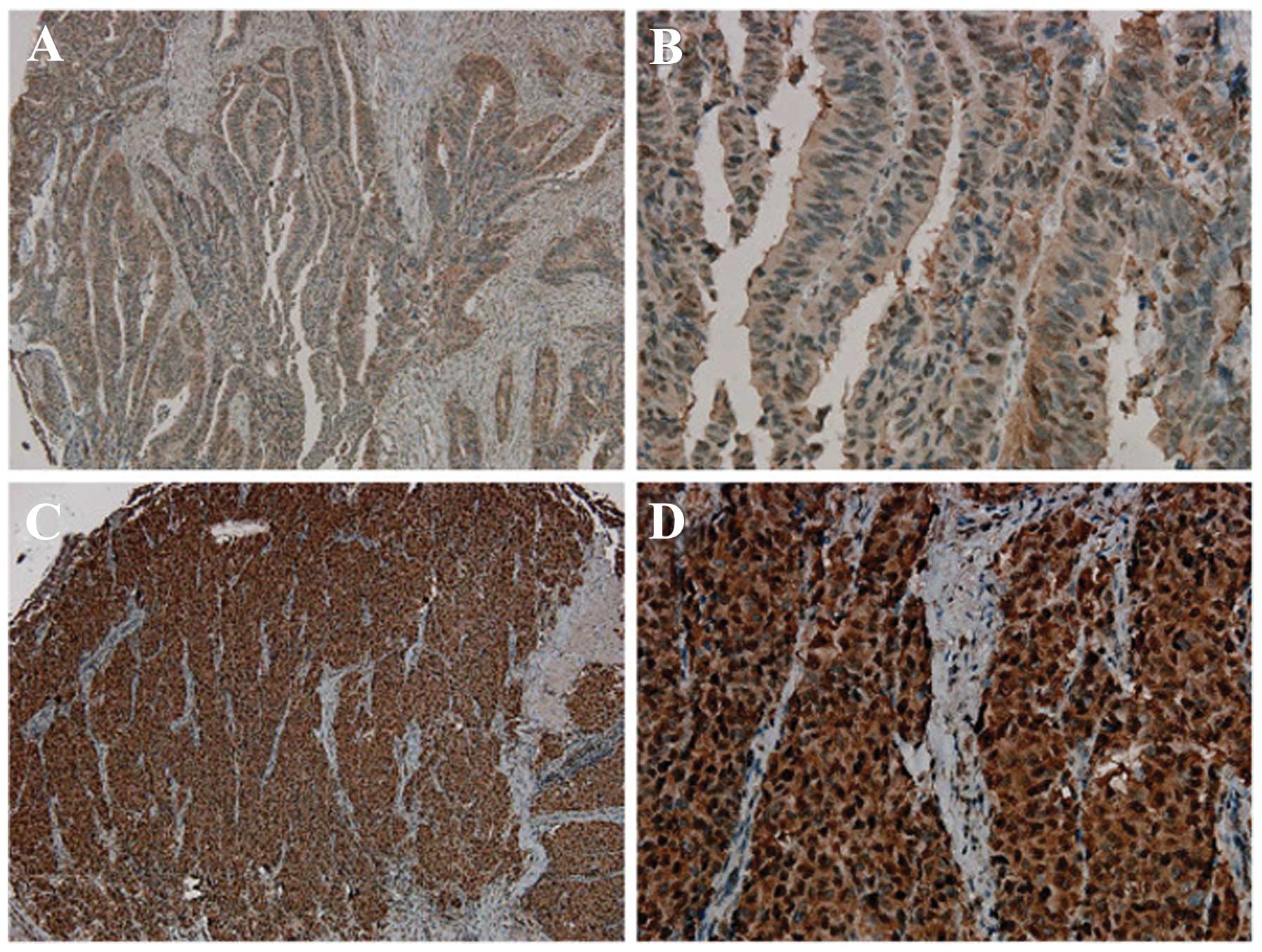
expression (P<0.001). Immunohistochemistry revealed a
predominantly nuclear localization of PPM1D (Fig. 1) and showed that PPM1D expression
was significantly higher in CRC tissue than in non-cancerous,
normal colorectal tissue. In addition, significant differences in
PPM1D expression were observed between tumors with node metastasis
(P=0.0024), distant metastasis (P<0.001) and different TNM
stages (P=0.0016; Table I). No
significant correlation was observed between PPM1D expression and
patient age or gender or tumor location or histology (Table I).
PPM1D expression and survival in patients
with CRC
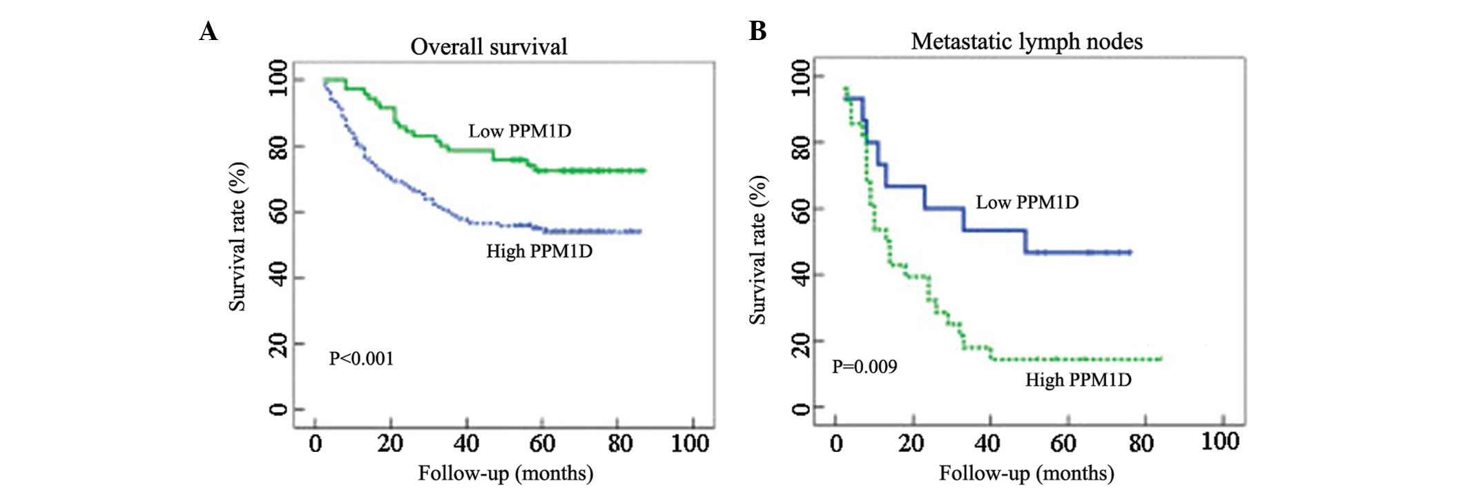
Kaplan-Meier survival analysis revealed that the
patients with low PPM1D expression had significantly longer
survival than those with high PPM1D expression (log-rank,
P<0.001; Fig. 2A). The
correlation between metastatic lymph node PPM1D expression and
patient survival was also assessed. Kaplan-Meier analysis revealed
that patients with low PPM1D expression in the metastatic lymph
nodes had significantly longer overall survival than patients with
high metastatic lymph node PPM1D expression (Fig. 2B, log-rank P=0.009).
Furthermore, multivariate analysis was performed
using the Cox proportional hazards model. High PPM1D expression was
identified to be a significant independent prognostic factor for OS
(hazard ratio=0.24; 95% confidence interval, 0.13–0.86; P=0.004) as
shown in Table II.
| Table IIUni- and multivariate analyses of
survival in patients with colorectal cancer. |
Table II
Uni- and multivariate analyses of
survival in patients with colorectal cancer.
| Overall survival |
|---|
|
|
|---|
| Univariate | Multivariate |
|---|
|
|
|
|---|
| Variable | HR (95% CI) | P-value | HR (95% CI) | P-value |
|---|
| Age (years) |
| <65 | 1 | | | |
| ≥65 | 1.003 (0.61,
1.58) | 0.73 | | |
| Gender |
| Male | 1 | | | |
| Female | 0.74 (0.43,
1.19) | 0.46 | | |
| TNM stage |
| I | 1 | | 1 | |
| II | 0.36 (0.07,
1.51) | 0.18 | 0.96 (0.21,
4.14) | 0.96 |
| III | 0.09 (0.02,
0.41) | 0.001 | 0.47 (0.06,
1.18) | 0.08 |
| IV | 0.34 (0.19,
0.56) | <0.001 | 0.19 (0.10,
0.43) | <0.001 |
|
Differentiation |
| Well | 1 | | | |
| Moderate/poor | 0.14 (0.07,
0.235) | 0.098 | | |
| PPM1D status |
| Low
expression | 1 | | 1 | |
| High
expression | 0.04 (0.01,
0.09) | <0.001 | 0.24 (0.13,
0.86) | 0.004 |
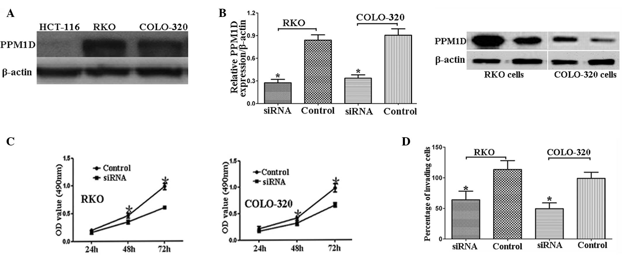
PPM1D siRNA significantly reduces CRC
cell proliferation and invasion
PPM1D expression was analyzed in three CRC cell
lines: HCT-116, RKO and COLO-320. PPM1D expression was observed to
be higher in RKO and COLO-320 cells than in HCT-116 cells (Fig. 3A). Based on this finding, RKO and
COLO-320 cells were used for the subsequent functional
analysis.
siRNA-induced PPM1D knockdown was confirmed using
qPCR and western blot analyses (Fig.
3B). PPM1D siRNA was observed to significantly reduce
proliferation and invasion in RKO and COLO-320 CRC cells (Fig. 3C and D).
Discussion
The present study investigated the correlation
between PPM1D expression, survival and clinical and pathological
features in patients with CRC. Significant correlations were
observed between PPM1D expression, metastasis, TNM stage and
mortality. Furthermore, these correlations were found to be
independent of other patient characteristics. These findings
indicate that high PPM1D expression may be a useful prognostic
marker for CRC.
PPM1D has been reported to be upregulated in
neuroblastoma, as well as pancreatic, lung, bladder, liver, ovarian
and breast cancer (6,7,12,13).
However, the role of PPM1D in CRC is yet to be elucidated. In the
present study, immunohistochemistry revealed that PPM1D was
upregulated in CRC tissues compared with the levels in paired
non-cancerous tissues. In human breast tissue, PPM1D overexpression
has been found to contribute to malignant progression through
inactivating wild-type p53 and p38 mitogen-activated protein
kinase, as well as through decreasing p16 protein expression
(14). PPM1D has also been shown
to be a prognostic marker in patients with lung adenocarcinoma
(15). Furthermore, high PPM1D
expression has been reported to be correlated with poor prognosis
in patients with pancreatic neuroendocrine tumors and
medulloblastoma (16,17). In the present study, patients with
CRC with high PPM1D expression were observed to have a worse
outcome than those with low PPM1D expression. Furthermore,
multivariate analysis suggested that high PPM1D expression was an
independent prognostic factor for patients with CRC. In
vitro experiments in CRC cells were also performed and revealed
that PPM1D siRNA significantly inhibited CRC cell proliferation and
invasion. These findings indicate that PPM1D may not only be a
prognostic marker, but also a potential therapeutic target.
In conclusion, PPM1D may be a prognostic biomarker
for CRC and its high expression is associated with poorer
prognosis. Further investigations are required to validate the
findings of the present study and to elucidate the underlying
mechanisms through which PPM1D affects CRC.
References
|
1
|
Jemal A, Siegel R, Xu J and Ward E: Cancer
statistics, 2010. CA Cancer J Clin. 60:277–300. 2010. View Article : Google Scholar
|
|
2
|
Siegel R, Naishadham D and Jemal A: Cancer
statistics, 2013. CA Cancer J Clin. 63:11–30. 2013. View Article : Google Scholar
|
|
3
|
Speetjens FM, Zeestraten EC, Kuppen PJ,
Melief CJ and van der Burg SH: Colorectal cancer vaccines in
clinical trials. Expert Rev Vaccines. 10:899–921. 2011. View Article : Google Scholar : PubMed/NCBI
|
|
4
|
Lu G and Wang Y: Functional diversity of
mammalian type 2C protein phosphatase isoforms: new tales from an
old family. Clin Exp Pharmacol Physiol. 35:107–112. 2008.
View Article : Google Scholar : PubMed/NCBI
|
|
5
|
Li J, Yang Y, Peng Y, Austin RJ, van
Eyndhoven WG, Nguyen KC, Gabriele T, McCurrach ME, Marks JR, Hoey
T, Lowe SW and Powers S: Oncogenic properties of PPM1D located
within a breast cancer amplification epicenter at 17q23. Nat Genet.
31:133–134. 2002. View
Article : Google Scholar : PubMed/NCBI
|
|
6
|
Hirasawa A, Saito-Ohara F, Inoue J, Aoki
D, Susumu N, Yokoyama T, Nozawa S, Inazawa J and Imoto I:
Association of 17q21–q24 gain in ovarian clear cell adenocarcinomas
with poor prognosis and identification of PPM1D and APPBP2 as
likely amplification targets. Clin Cancer Res. 9:1995–2004.
2003.
|
|
7
|
Saito-Ohara F, Imoto I, Inoue J, Hosoi H,
Nakagawara A, Sugimoto T and Inazawa J: PPM1D is a potential target
for 17q gain in neuroblastoma. Cancer Res. 63:1876–1883.
2003.PubMed/NCBI
|
|
8
|
Demidov ON, Kek C, Shreeram S, Timofeev O,
Fornace AJ, Appella E and Bulavin DV: The role of the MKK6/p38 MAPK
pathway in Wip1-dependent regulation of ErbB2-driven mammary gland
tumorigenesis. Oncogene. 26:2502–2506. 2007. View Article : Google Scholar : PubMed/NCBI
|
|
9
|
Morikawa T, Kuchiba A, Qian ZR,
Mino-Kenudson M, Hornick JL, Yamauchi M, Imamura Y, Liao X,
Nishihara R, Meyerhardt JA, Fuchs CS and Ogino S: Prognostic
significance and molecular associations of tumor growth pattern in
colorectal cancer. Ann Surg Oncol. 19:1944–1953. 2012. View Article : Google Scholar : PubMed/NCBI
|
|
10
|
Morikawa T, Kuchiba A, Yamauchi M,
Meyerhardt JA, Shima K, Nosho K, Chan AT, Giovannucci E, Fuchs CS
and Ogino S: Association of CTNNB1 (beta-catenin) alterations, body
mass index, and physical activity with survival in patients with
colorectal cancer. JAMA. 305:1685–1694. 2011. View Article : Google Scholar : PubMed/NCBI
|
|
11
|
Weichert W, Röske A, Gekeler V, Beckers T,
Ebert MP, Pross M, Dietel M, Denkert C and Röcken C: Association of
patterns of class I histone deacetylase expression with patient
prognosis in gastric cancer: a retrospective analysis. Lancet
Oncol. 9:139–148. 2008. View Article : Google Scholar : PubMed/NCBI
|
|
12
|
Loukopoulos P, Shibata T, Katoh H, Kokubu
A, Sakamoto M, Yamazaki K, Kosuge T, Kanai Y, Hosoda F, Imoto I,
Ohki M, Inazawa J and Hirohashi S: Genome-wide array-based
comparative genomic hybridization analysis of pancreatic
adenocarcinoma: identification of genetic indicators that predict
patient outcome. Cancer Sci. 98:392–400. 2007. View Article : Google Scholar
|
|
13
|
Wang P, Rao J, Yang H, Zhao H and Yang L:
Wip1 over-expression correlated with TP53/p14(ARF) pathway
disruption in human astrocytomas. J Surg Oncol. 104:679–684. 2011.
View Article : Google Scholar : PubMed/NCBI
|
|
14
|
Yu E, Ahn YS, Jang SJ, Kim MJ, Yoon HS,
Gong G and Choi J: Overexpression of the wip1 gene abrogates the
p38 MAPK/p53/Wip1 pathway and silences p16 expression in human
breast cancers. Breast Cancer Res Treat. 101:269–278. 2007.
View Article : Google Scholar : PubMed/NCBI
|
|
15
|
Satoh N, Maniwa Y, Bermudez VP, Nishimura
K, Nishio W, Yoshimura M, Okita Y, Ohbayashi C, Hurwitz J and
Hayashi Y: Oncogenic phosphatase Wip1 is a novel prognostic marker
for lung adenocarcinoma patient survival. Cancer Sci.
102:1101–1106. 2011. View Article : Google Scholar : PubMed/NCBI
|
|
16
|
Hu W, Feng Z, Modica I, Klimstra DS, Song
L, Allen PJ, Brennan MF, Levine AJ and Tang LH: Gene amplifications
in well-differentiated pancreatic neuroendocrine tumors inactivate
the p53 pathway. Genes Cancer. 1:360–368. 2010. View Article : Google Scholar : PubMed/NCBI
|
|
17
|
Castellino RC, De Bortoli M, Lu X, Moon
SH, Nguyen TA, Shepard MA, Rao PH, Donehower LA and Kim JY:
Medulloblastomas overexpress the p53-inactivating oncogene
WIP1/PPM1D. J Neurooncol. 86:245–256. 2008. View Article : Google Scholar : PubMed/NCBI
|