Introduction
Paraquat (PQ) is a widely-used herbicide that has
been shown to cause severe and often fatal pulmonary fibrosis in
humans and laboratory animals in developing countries, such as
China and Sri Lanka. Following the ingestion of large amounts of
concentrated formulation, the rapid development of multi-organ
failure and cardiogenic shock is almost universally fatal (1,2).
Even with the ingestion of smaller amounts, PQ is actively taken up
into pulmonary epithelial cells where redox cycling and free
radical generation triggers a fibrotic process that may lead to
mortality. Management of PQ poisoning has remained predominantly
supportive and the results for the treatment of PQ poisoning,
including immunosuppressive therapy (3,4),
prolonged extracorporeal elimination (5) and lung transplantation, have been
disappointing (6,7).
A reliable predictor of prognosis may guide
treatment and future clinical research on antidotes and other
therapies (8). The measurement of
plasma PQ concentration has been considered as a marker of severity
and prognosis (1). Proudfoot et
al produced a nomogram in 1979 that indicated a correlation
between the outcome to the plasma PQ concentration on admission
with the time from ingestion to blood collection. Patients with PQ
levels lower than a line connecting concentrations of 2.0, 0.6,
0.3, 0.16 and 0.1 mg/ml at 4, 6, 10, 16 and 24 h, respectively,
have been shown to survive (9).
Scherrmann et al identified that 30 survivors following PQ
poisoning had plasma PQ levels of >C mg/ml, where C = 1/[0.471 ×
time (h) since ingestion × 1.302] (10). The studies by Hart et al
(11) and Gil et al
(12) demonstrated a very strong
correlation between measured concentrations and survival rates. In
addition, the correlation between the urine PQ concentration and
the time following ingestion has also been used to evaluate the
prognosis of patients with PQ intoxication (13). However, a major constraint of these
methods is the inability of numerous hospitals in the developing
world, where the majority of patients present with this condition,
to perform the required assay. An additional important predictor of
mortality is the quantity of PQ consumed. However, estimates on the
amount ingested are often unobtainable or unreliable in a number of
intoxicated patients.
Recently, the serum lactate level has been used as a
prognostic marker in patients with acute PQ poisoning, and higher
arterial lactate levels have been shown to be associated with a
higher risk of mortality through multiple logistic regression [odds
ratio (OR), 7.02; 95% confidence interval (CI), 2.06–23.91;
P=0.002] (14). In an additional
study, receiver operating characteristic (ROC) curve analysis
revealed that the initial arterial lactate concentration had an
area of 0.749 (95% CI, 0.714–0.856) and a cut-off level of 2.5
mmol/l for the prediction of prognosis in patients with acute PQ
poisoning (15).
However, initial arterial lactate concentrations may
vary, even in patients that have ingested the same amount of PQ,
since blood samples are collected at different times. Therefore, it
has been hypothesized that the initial arterial lactate
concentration alone is not sufficiently predictive to infer
effectiveness. However, initial arterial lactate concentration-time
data may have a better predictive power compared with initial
arterial lactate concentration alone. Thus, the aim of the present
study was to assess initial arterial lactate concentration-time
data as a prognostic marker of mortality and morbidity in patients
with PQ intoxication.
Materials and methods
Patients and setting
The study protocol was reviewed and approved by the
Clinical Trial Committee of Pingjin Hospital (Tiajin, China).
Informed consent was provided by the patients or by their next of
kin prior to therapy.
The study included 170 patients with PQ poisoning.
The time lag following PQ ingestion and the arterial lactate and
plasma PQ concentrations were measured at the same time following
admission. However, not all the patients had their urine PQ
concentration measured quantitatively, as a dithionite method was
applied, which provided a qualitative determination. Demographic
variables, including age and gender, were recorded in all the
patients, and in particular, whether mortality occurred during the
stay at hospital. All the patients who survived to the time of
discharge were visited after six months to determine if there had
been any delayed mortalities.
This retrospective observational study occurred in
an 18-bed poisoning Emergency Treatment Center of the University
affiliated Pingjin Hospital between June 2008 and June 2012. Blood
samples were collected on admission and the arterial lactate levels
were measured with a blood gas analyzer (GEM premier 3000;
Instrumentation Laboratory, Bedford, IL, USA) immediately following
admission. Quantitative analysis of the plasma samples from all the
PQ-poisoned patients were conducted in the hospital laboratory
using a gas chromatography method (16). Patients who met any of the
following criteria were excluded from the study: Non-oral ingestion
poisoning, admission to a different hospital or PQ exposure of
>24 h previous to presentation.
Treatment
To prevent the absorption of PQ by the
gastrointestinal tract, gastric lavage was performed via a
nasogastric tube using 1 g/kg activated charcoal in 500 ml saline
(0.9%) once every 4 h. In addition, SMECTA (Beaufour Ipsen Pharmacy
Co., Ltd., Tianjin, China) and magnesium sulfate powder (Tianjin
Huairen Pharmacy Co., Ltd., Tianjin, China) were placed into 20%
mannitol (Shuanghe Pharmaceutical Co., Ltd., Tianjin, China), which
was administered rectally. All patients received activated charcoal
hemoperfusion therapy (Braun Diapact CRRT machine; B Braun Medical,
Ltd., Hesse, Germany), followed by 12 h continuous veno-venous
hemofiltration (CVVH) therapy with a 4 h interval following
hemoperfusion. During the 4 h interval and following CVVH, the
patients received high-dose therapy comprising an intravenous
infusion of 15 mg/kg/day cyclophosphamide (Guangdong Qingping
Pharmacy Co., Ltd., Guangzhou, China) in 250 ml glucose saline (5%)
for 1 h for two days and 1 g/day methylprednisolone sodium
succinate injection (Pfizer, Inc., New York, NY, USA) iin 250 ml
glucose saline (5%) for 2 h for three days. Starting on day 4,
patients also received intravenous injections of 5 mg dexamethasone
(Jilin Extrawell Changbaishan Pharmaceutical Co., Ltd., Jilin,
China) every 6 h. In addition, vitamin E capsules (Xinyi
Pharmaceutical Co., Ltd., Shanghai, China), metoprolol
(AstraZeneca, London, UK) and vitamin E injections (Zhongjing
Biotechnology Co. Ltd., Harbin, China) were administered.
Statistical analysis
SPSS statistical software package 20.0 (IBM, Armonk,
NY, USA), MedCalc 12.4 (MedCalc Software, Ostend, Belgium) and
GraphPad Prism v 4.0 (GraphPad Software, Inc., La Jolla, CA, USA)
were used to perform statistical analysis. Data are presented as
the mean ± standard deviation or as the median with the range.
Statistically significant differences between the two groups were
analyzed using the independent two-sample t-test or the
Mann-Whitney U-test. Arterial lactate concentration, arterial
lactate-time and PQ concentration-time data were compared using ROC
curve analysis to analyze the statistical significance of the
differences between the areas under the ROC curve, according to the
method by DeLong et al (17). The cut-off values were determined
by analyzing the Youden’s index and the maximized area under the
ROC curve.
In addition, multiple logistic regression analysis
was performed with log transformed data to derive a formula that
had the best predicted survival rates of the study population.
Multiple logistic regression analysis of the initial parameters
focused on mortality using a backward elimination method.
Results
Baseline characteristics
A total of 170 patients were enrolled in the study
and the baseline characteristics of the patients are described in
Table I. Of the 170 subjects,
there were 97 females and 73 males, and 93 survivors and 77
patients that succumbed to acute PQ intoxication. In all the
patients, the average time interval between PQ ingestion and the
first sample collection was 6.46 h (range, 2.5–19 h). However, the
median times for the survivors and non-survivors were 5.00 (range,
4.00–8.00 h) and 6.00 h (range, 4.00–8.75 h), respectively.
Non-survivors exhibited a higher average arterial lactate
concentration of 5.00 mmol/l (range, 2.00–10.00 mmol/l) and a
plasma PQ concentration of 10.0 mg/l (range, 6.00–15.00 mg/l) when
compared with the survivors that had arterial lactate and PQ
concentrations of 2.00 mmol/l (range, 1.00–2.50 mmol/l) and 5.00
mg/l (3.00–7.00 mg/l), respectively.
 | Table IDemographic and laboratory
observations of the survivors and non-survivors among the 170
patients with acute PQ poisoning. |
Table I
Demographic and laboratory
observations of the survivors and non-survivors among the 170
patients with acute PQ poisoning.
| Parameter | Survivors (n=93) | Non-survivors
(n=77) | P-value |
|---|
| Age (years) | 30.00 (23.00,
45.5) | 29.00 (24.00,
46.50) | 0.518 |
| Time lag after PQ
ingestion (h) | 5.00 (4.00,
8.00) | 6.00 (4.00,
8.75) | 0.117 |
| Arterial lactate
(mmol/l) | 2.00 (1.00,
2.50) | 5.00 (2.00,
10.00) | <0.001 |
| Arterial lactate-time
(mmol/l.h) | 10.00 (5.00,
17.50) | 26.10 (15.00,
53.55) | <0.001 |
| PQ concentration
(mg/l) | 5.00 (3.00,
7.00) | 10.00 (6.00,
15.00) | <0.001 |
| PQ concentration-time
(mg/l.h) | 26.50 (13.50,
47.50) | 59.00 (35.50,
104.50) | <0.001 |
ROC curve analysis
With regard to the ROC curve analysis (Table II), the arterial lactate
concentration had an area of 0.774 and the cut-off value was 4.2
mmol/l (sensitivity, 82.80%; specificity, 63.64%; Youden’s index,
0.464). The arterial lactate-time data had an area of 0.782 with a
cut-off value of 11.95 mmol/l h (sensitivity, 64.52%; specificity,
84.42%; Youden’s index, 0.490). Positive correlations were observed
between initial arterial lactate and plasma PQ concentrations
(ρ=0.414), as well as between arterial lactate-time and PQ
concentration-time (ρ=0.485; Table
III). Results from the multiple logistic regression analysis of
the initial parameters on mortality using a backward elimination
method are described in Table IV.
The results indicated that increased lactate concentrations (OR,
0.838; 95% CI, 0.755–0.930; P<0.001) were associated with a
significantly higher risk of mortality when the time lag following
PQ ingestion and PQ concentration for the two patients was
equal.
| Table IIPrediction of the mortality rate in
acute PQ poisoning. |
Table II
Prediction of the mortality rate in
acute PQ poisoning.
| Parameter | Cut-off point | Sensitivity (%) | Specificity (%) | AUC (95% CI) | Youden’s index |
|---|
| Arterial lactate
(mmol/l) | 4.20 | 82.80 (73.57,
89.83) | 63.64 (51.89,
74.30) | 0.774 (0.703,
0.834) | 0.464 |
| Arterial lactate-time
(mmol/l.h) | 11.95 | 64.52 (53.91,
74.17) | 84.42 (74.36,
91.68) | 0.782 (0.712,
0.841) | 0.490 |
| PQ concentration
(mg/l) | 9.35 | 86.02 (77.28,
92.34) | 59.74 (47.94,
70.77) | 0.765 (0.694,
0.826) | 0.462 |
| PQ concentration-time
(mg/l.h) | 22.70 | 44.01 (32.79,
53.69) | 92.21 (83.81,
97.09) | 0.768 (0.697,
0.829) | 0.362 |
| Time lag after PQ
ingestion (h) | 6.50 | 66.67 (56.31,
75.96) | 48.05 (36.52,
59.74) | 0.568 (0.490,
0.644) | 0.145 |
| Table IIICorrelation analysis between arterial
lactate and PQ concentrations, measured at the same time after
acute PQ poisoning. |
Table III
Correlation analysis between arterial
lactate and PQ concentrations, measured at the same time after
acute PQ poisoning.
| Parameter | Statistical
parameter | PQ concentration | PQ
concentration-time |
|---|
| Arterial lactate
(n=170) | Correlation
coefficient (ρ) | 0.414 | |
| P-value | <0.001 | |
| Arterial lactate-time
(n=170) | Correlation
coefficient (ρ) | | 0.485 |
| P-value | | <0.001 |
| Table IVMultiple logistic regression analysis
of the initial parameters on mortality following PQ ingestion. |
Table IV
Multiple logistic regression analysis
of the initial parameters on mortality following PQ ingestion.
| Parameter | OR | 95% CI | P-value |
|---|
| Time lag after PQ
ingestion | 0.869 | 0.774, 0.978 | 0.002 |
| Arterial lactate | 0.838 | 0.755, 0.930 | <0.001 |
| PQ concentration | 0.856 | 0.794, 0.923 | <0.001 |
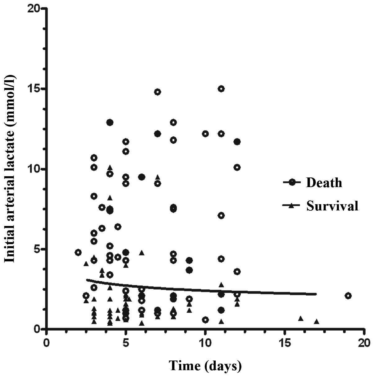
A logarithmic plot of the initial arterial lactate
concentration against the time since ingestion is show in Fig. 1. To calculate the predicted
probability of survival for any specified time and initial arterial
lactate concentration, the following formula was derived based on
the logistic regression coefficients: Logit(p) = 3.066 − 0.139 ×
(time lag after PQ ingestion) − 0.177 × (initial arterial lactate
concentration); where the probability of survival = 1/1 +
e−logit(p).
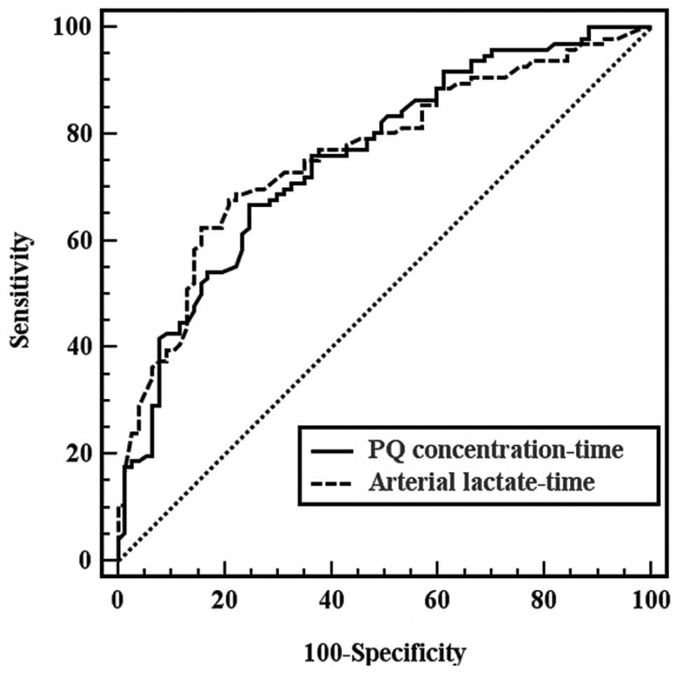
Pairwise comparison of the ROC curves in Fig. 2 demonstrated that there was no
statistically significant difference between the areas for arterial
lactate-time and PQ concentration-time (z=0.712; P=0.864).
Discussion
Increased blood lactate levels have been associated
with significant morbidity and mortality since their first
description in 1843 by Scherer (18). A number of studies have emphasized
the prognostic importance of measuring a single lactate level
during treatment (19). Despite
this strong and already long-lasting predictive power of lactate
levels, little evidence exists on the prognosis of acute PQ
poisoned patients. Recently, a number of studies found that the
arterial lactate concentration had predictive power for the
prognosis of acute PQ poisoned patients (14,15).
The results of the present study revealed that the arterial lactate
concentration was not only higher in non-survivors (average, 5.00
mmol/l; range, 2.00–10.00 mmol/l) compared with survivors (average,
2.00; range, 1.00–2.50 mmol/l; P<0.001), but also exhibited a
positive correlation with the PQ concentration (ρ=0.414). These
observations may validate the hypothesis that the arterial lactate
level possesses a good predictive power in evaluating the prognosis
of patients with acute PQ poisoning.
However, initial arterial lactate concentrations may
vary, even in patients that have ingested the same amount of PQ,
since blood samples are collected at different times. The time
following PQ ingestion should be considered as a marker of severity
and prognosis. As shown in Table
IV, logistic regression analysis, using time and arterial
lactate concentration as variables, produced very significant
independent effects for each variable as a predictor of survival.
Therefore, we hypothesized that the initial arterial lactate
concentration-time data may have a better predictive power compared
with the initial arterial lactate concentration alone.
Plasma PQ concentration-time data have been used as
a practical tool to predict the prognosis of acute PQ poisoned
patients. The present study demonstrated that there was a positive
correlation between the arterial lactate-time and PQ
concentration-time (ρ=0.485). The arterial lactate-time data
exhibited a similar discriminative power to the plasma PQ
concentration-time data (z=0.712; P=0.864), thus, arterial
lactate-time data may have a discriminative power as a practical
tool in predicting the prognosis of acute PQ poisoned patients. To
calculate the predicted probability of survival for any specified
time and initial arterial lactate concentration, the following
formula was derived based on the logistic regression coefficients:
Logit(p) = 3.066 − 0.139 × (time lag after PQ ingestion) − 0.177 ×
(initial arterial lactate concentration); where the probability of
survivors = 1/1 + e−logit(p). The use of the logistic
regression equation allows the prediction of the probability of
survival for any specified time and initial arterial lactate
concentration following the ingestion of PQ for ≤19 h.
The present study reports a novel correlation
between the initial arterial lactate concentration and time, which
may aid the prediction of patient survival following the ingestion
of PQ for ≤19 h (Fig. 1). The
logistic regression equation together with the survival curve
(Fig. 1) may produce highly
significant independent effects for each variable to predict the
probability of survival. However, the new survival curve requires
prospective validation to determine the sensitivity and specificity
for predicting the outcome in patients with acute PQ poisoning.
There were several limitations in the present study.
Firstly, this was a retrospective study; thus, a limited amount of
quality data were collected. Secondly, since the times of mortality
for the non-survivors were not collected, survival analysis of the
arterial lactate concentration and arterial lactate-time was unable
to be performed. Finally, the results may have been more
informative if the urine PQ concentrations had been measured.
In conclusion, the arterial lactate-time data had a
better predictive power compared with the arterial lactate
concetration alone for evaluating the prognosis of patients with
acute PQ poisoning. Therefore, measuring the initial arterial
lactate concentration and the time of poisoning may be a simple and
practical tool for assessing the severity of PQ poisoning.
Acknowledgements
The authors thank the research doctors, nursing and
other medical staff in the study hospitals for their assistance
during the study.
References
|
1
|
Jones AL, Elton R and Flanagan R: Multiple
logistic regression analysis of plasma paraquat concentrations as a
predictor of outcome in 375 cases of paraquat poisoning. QJM.
92:573–578. 1999. View Article : Google Scholar : PubMed/NCBI
|
|
2
|
Rebello G and Mason JK: Pulmonary
histological appearances in fatal paraquat poisoning.
Histopathology. 2:53–66. 1978. View Article : Google Scholar : PubMed/NCBI
|
|
3
|
Lin JL, Lin-Tan DT, Chen KH, et al:
Improved survival in severe paraquat poisoning with repeated pulse
therapy of cyclophosphamide and steroids. Intensive Care Med.
37:1006–1013. 2011. View Article : Google Scholar : PubMed/NCBI
|
|
4
|
Addo E and Poon-King T: Leucocyte
suppression in treatment of 72 patients with paraquat poisoning.
Lancet. 1:1117–1120. 1986. View Article : Google Scholar : PubMed/NCBI
|
|
5
|
Shi Y, Bai Y, Zou Y, et al: The value of
plasma paraquat concentration in predicting therapeutic effects of
haemoperfusion in patients with acute paraquat poisoning. PLoS One.
7:e409112012. View Article : Google Scholar : PubMed/NCBI
|
|
6
|
Matthew H, Logan A, Woodruff MF and Heard
B: Paraquat poisoning - lung transplantation. Br Med J. 3:759–763.
1968. View Article : Google Scholar : PubMed/NCBI
|
|
7
|
Licker M, Schweizer A, Hohn L, Morel DR
and Spiliopoulos A: Single lung transplantation for adult
respiratory distress syndrome after paraquat poisoning. Thorax.
53:620–621. 1998. View Article : Google Scholar : PubMed/NCBI
|
|
8
|
Senarathna L, Eddleston M, Wilks MF, et
al: Prediction of outcome after paraquat poisoning by measurement
of the plasma paraquat concentration. QJM. 102:251–259. 2009.
View Article : Google Scholar : PubMed/NCBI
|
|
9
|
Proudfoot AT, Stewart MS, Levitt T and
Widdop B: Paraquat poisoning: significance of plasma paraquat
concentrations. Lancet. 2:330–332. 1979. View Article : Google Scholar : PubMed/NCBI
|
|
10
|
Scherrmann JM, Houze P, Bismuth C and
Bourdon R: Prognostic value of plasma and urine paraquat
concentration. Hum Toxicol. 6:91–93. 1987. View Article : Google Scholar : PubMed/NCBI
|
|
11
|
Hart TB, Nevitt A and Whitehead A: A new
statistical approach to the prognostic significance of plasma
paraquat concentrations. Lancet. 2:1222–1223. 1984. View Article : Google Scholar : PubMed/NCBI
|
|
12
|
Gil HW, Kang MS, Yang JO, et al:
Association between plasma paraquat level and outcome of paraquat
poisoning in 375 paraquat poisoning patients. Clin Toxicol (Phila).
46:515–518. 2008. View Article : Google Scholar : PubMed/NCBI
|
|
13
|
Fock KM: Clinical features and prognosis
of paraquat poisoning: a review of 27 cases. Singapore Med J.
28:53–56. 1987.PubMed/NCBI
|
|
14
|
Lee Y, Lee JH, Seong AJ, et al: Arterial
lactate as a predictor of mortality in emergency department
patients with paraquat intoxication. Clin Toxicol (Phila).
50:52–56. 2012. View Article : Google Scholar : PubMed/NCBI
|
|
15
|
Liu XW, Ma T, Qu B, et al: Prognostic
value of initial arterial lactate level and lactate metabolic
clearance rate in patients with acute paraquat poisoning. Am J
Emerg Med. 31:1230–1235. 2013. View Article : Google Scholar : PubMed/NCBI
|
|
16
|
Posecion NC, Ostrea EM and Bielawski DM:
Quantitative determination of paraquat in meconium by sodium
borohydride-nickel chloride chemical reduction and gas
chromatography/mass spectrometry (GC/MS). J Chromatogr B Analyt
Technol Biomed Life Sci. 862:93–99. 2008. View Article : Google Scholar
|
|
17
|
DeLong ER, DeLong DM and Clarke-Pearson
DL: Comparing the areas under two or more correlated receiver
operating characteristic curves: a nonparametric approach.
Biometrics. 44:837–845. 1988. View
Article : Google Scholar : PubMed/NCBI
|
|
18
|
Kompanje EJ, Jansen TC, van der Hoven B
and Bakker J: The first demonstration of lactic acid in human blood
in shock by Johann Joseph Scherer (1814–1869) in January 1843.
Intensive Care Med. 33:1967–1971. 2007.PubMed/NCBI
|
|
19
|
Aduen J, Bernstein WK, Khastgir T, et al:
The use and clinical importance of a substrate-specific electrode
for rapid determination of blood lactate concentrations. JAMA.
272:1678–1685. 1994. View Article : Google Scholar : PubMed/NCBI
|