Introduction
Diabetes mellitus (DM) could lead to fibrosis of
multiple organs, including atrial structural remodeling (1). Atrial fibrosis, as a hallmark of
atrial structural remodeling, plays a critical role in the
occurrence and maintenance of atrial arrhythmogenicity. Fibrosis
isolates groups of atrial and individual myocytes and thus impairs
cell-to-cell coupling, leading to inhomogeneities in intra- and
interatrial conduction and retarded conduction velocity. The
mechanisms responsible for atrial fibrosis remain to be clarified.
Currently, there are no specific drugs available for the prevention
or treatment of atrial fibrosis in DM.
The Ras homolog gene family, member A (RhoA)/Rho
associated coiled-coil forming protein kinase (ROCK) is a member of
the serine/threonine kinase family. ROCK is the most studied Rho
downstream effector. It has two known isoforms (ROCK1 and ROCK2)
and regulates cytoskeletal reorganization by phosphorylating myosin
phosphatase, which results in an increase in myosin light chain
(MLC) phosphorylation (2). Through
this, the RhoA/ROCK pathway is involved in regulating endothelial
migration, platelet activation, thrombosis, and oxidative stress as
well as smooth muscle contraction (3,4). A
number of abnormal activations of the RhoA/ROCK pathway are present
in diseases (5). It is activated
by multiple cytokines and inflammatory mediators, including
platelet-derived growth factor, transforming growth factor-β,
endothelin-1 and angiotensin II (6). Previous studies have suggested that
the RhoA/Rock pathway also plays a significant role in bowel
(7), liver (8), renal (9–10),
pulmonary (11), and myocardial
fibrosis (12–13). Thus inhibition of this signaling
pathway is effective for treating a wide range of cardiovascular
and non-cardiovascular disease. Fasudil is a Rho-kinase inhibitor,
and has previously been shown to attenuate fibrosis in multiple
organs (9,11–12).
We hypothesized that the RhoA/ROCK pathway is
involved in atrial fibrosis, and that fasudil, which is a highly
selective inhibitor of the two ROCK isoforms, may inhibit the
development of atrial fibrosis. Therefore, the present study
focused on the expression and function of the RhoA/ROCK pathway in
a rat model of type 2 diabetes.
Materials and methods
The experimental protocol was approved by the Ethics
Review Committee for Animal Experimentation of the Beijing
Friendship Hospital (Beijing, China).
Animal model
Eight-week-old male Sprague-Dawley rats, weighing
240–260 g, were purchased from Vital River Laboratories (Beijing,
China). After two weeks of acclimatization, the rats were randomly
assigned to receive a standard diet as the normal control group or
a high-fat diet (HFD; 73% standard diet, 25% lard, and 2% yolk
powder) for four weeks. At the end of the sixth week, control rats
were injected intraperitoneally (i.p.) with vehicle (0.1 mol/l
citric acid buffer) once and given a standard diet sequentially.
HFD rats were injected i.p. with a low-dose (30 mg/kg) of
streptozotocin (STZ; Sigma-Aldrich, St. Louis, MO, USA) and given
an HFD sequentially. Blood glucose (BG) was measured in whole blood
collected from the tail vein by a portable glucometer and BG levels
were recorded every week. Rats with BG levels of >16.7 mmol/l
and that were stable for four weeks were considered to be diabetic.
From the 10th week, control rats remained on a standard diet and
were treated with i.p. injection of sterile vehicle every day.
Diabetic rats were randomly divided into two groups: i) Treated
diabetic rats were maintained on a HFD and received fasudil
hydrochloride hydrate (10 mg/kg/day; i.p.) (Tianjin Hongri Company,
Tianjin, China) every day for 14 weeks; and ii) untreated diabetic
rats were treated with i.p. injection of sterile vehicle every day
for 14 weeks. At week 24, fasting plasma was collected for further
measurement of fasting insulin (FINS) and fasting BG (FBG).
Homeostasis model assessment for insulin resistance (HOMA-IR) was
calculated as FBG x FINS/22.5 and was used in assessing insulin
resistance. The rats were sacrificed and the hearts were harvested.
A section of tissue from the left atrium was fixed in 4%
paraformaldehyde and embedded in paraffin for histological
examination. The remainder of the left and right atria were
snap-frozen and stored at −80°C until processing for the extraction
of mRNA and protein.
Masson staining
The tissue samples were washed with water and then
stained using the following steps: i) Fixated in Bouin’s solution,
microwaved for 1 min, left to stand for 15 min; ii) washed under a
running tap water to remove the picric acid for 5 min; iii) stained
in Weigert’s hematoxylin working solution for 10 min; iv) waited
for it to turn blue under running tap water for 5 min and then
rinsed with distilled water; v) added Biebrich scarlet solution for
5 min; vi) rinsed with distilled water; vii) differentiated in
phosphotungstic/phosphomolybdic acid solution for 10 min; viii)
transferred directly into aniline blue solution for 5 min; ix)
rinsed with distilled water; x) differentiated in 1% acetic acid
for 1 min, rinsed with distilled water and xi) dehydrated, cleared
and mounted on a coverslip.
Isolation of RNA and quantitative
polymerase chain reaction (qPCR)
Small sections of the atria were sampled. Total RNA
was prepared from the right and left atrial free walls using
TRIzol® reagent according to the manufacturer’s
instructions. The purity of isolated RNA was identified by
ultraviolet spectrometry. cDNA was synthesized by reverse
transcription using oligo dT (deoxythymine) primer and M-MLV
(moloney murine leukemia virus) reverse transcriptase (Promega
Corp., Fitchburg, WI, USA), which was used as a template in the
subsequent PCR analysis. The mRNA levels of RhoA, ROCK1, ROCK2,
collagen type-I and collagen type-III were evaluated by qPCR. The
level of GAPDH was also evaluated as the internal control. qPCR was
performed with a BioEasy SYBR-Green I Real Time PCR kit manual
(Hangzhou Bioer Technology Co., Ltd., Hangzhou, China). The primers
were as follows: RhoA, forward: 5′-CATCCCAGAAAAGTGGACTCCA-3′ and
reverse 5′-CCT TGTGTGCTCATCATTCCG-3′, 103 bp; ROCK1, forward:
5′-GAATGACATGCAAGCGCAAT-3′ and reverse: 5′-GTC
CAAAAGTTTTGCACGCA-3′, 113 bp; ROCK2, forward:
5′-GAAACAACTGGATGAAGCTAATGC-3′ and reverse:
5′-GTTTCAAGCAGGCAGTTTTTATCTT-3′, 150 bp; type-I procollagen,
forward: 5′-TTCACCTACAGCACGCTTGT-3′, reverse:
5′-TTGGGATGGAGGGAGTTTAC-3′, 196 bp; type-III procollagen, forward:
5′-TTGAATATCAAACAC GCAAGGC-3′ and reverse: 5′-GGTCACTTTCACTGGTTG
ACGA-3′, 201 bp; GAPDH, forward: 5′-GATGGGTGT GAACCACGAGAAA-3′ and
reverse: 5′-ACGGATACATTG GGGGTAGGAA-3′, 330 bp. The RhoA, ROCK1,
ROCK2, collagen type I and III amplification conditions were:
Pre-denaturation at 95°C for 2 min, denaturation at 95°C for 20
sec, annealing at 58°C for 25 sec, and extension at 72°C for 30
sec, for a total of 45 cycles. The PCR products underwent
electrophoresis and were scanned with a gel image analysis system
[UV-2000; UNICO (Shanghai) Instruments Co., Ltd., Shanghai, China].
The intensity of RhoA, ROCK1, ROCK2, collagen type I and III was
standardized to that of the GAPDH mRNA levels.
Western blot analysis
Small sections of the atria of the rats were lysed.
Protein was extracted and measured using a bicinchoninic acid (BCA)
protein assay kit (Pierce Biotechnology, Inc., Rockford, IL, USA).
Protein (~50 μg) was separated by 10% SDS-PAGE and transferred to
polyvinylidene fluoride (PVDF) membranes. The membranes were
blocked with 5% fat-free milk in Tris-buffered saline with Tween
(TBST) buffer (20 mmol/l Tris-HCl, pH 7.5, 150 mmol/l NaCl and
0.05% Tween 20), and subsequently incubated with the following
primary antibodies: polyclonal rabbit anti-rat RhoA (Santa Cruz
Biotechnology, Inc., Santa Cruz, CA, USA), polyclonal goat anti-rat
ROCK1 (Santa Cruz Biotechnology, Inc.), and polyclonal goat
anti-rat ROCK2 (Santa Cruz Biotechnology, Inc.) at 4°C overnight.
The mixture was washed and then incubated for 1 h with horseradish
peroxidase (HRP)-conjugated secondary antibodies (Kirkegaard &
Perry Laboratories, Inc., Gaithersburg, MA, USA). The membranes
were developed using an enhanced chemiluminescence kit (Pierce
Biotechnology, Inc.). Quantification of bands was performed by gel
densitometry with a gel image analysis system (UVP, LLC, Upland,
CA, USA). The phosphorylation level was normalized by total
protein-band densitometry individually.
Statistical analysis
The SPSS 17.0 statistical software package (SPSS,
Chicago, IL, USA) was used for analysis. Data are presented as the
mean ± standard deviation. All the groups were tested for normal
distribution and equal variance. Differences among the three groups
(at week 24) were assessed using one-way analysis of variance.
P<0.05 was considered statistically significant.
Results
At week 24, compared with the control rats, the
levels of FBG, FINS, and HOMA-IR were significantly increased in
untreated and fasudil-treated diabetic rats. No significant
differences were identified in these indices between untreated and
treated diabetic rats, suggesting that the 10 mg/kg/day fasudil
administered had no effect on glucose metabolism and insulin
resistance (Table I).
 | Table IThe effects of fasudil on glucose
metabolism parameters in rats at week 24. |
Table I
The effects of fasudil on glucose
metabolism parameters in rats at week 24.
| Group | Control | Diabetic | DM+fas |
|---|
| FBG (mmol/l) | 5.65±0.23 | 19.57±0.71a | 20.10±0.61a,b |
| FINS (mU/l) | 33.95±2.51 | 52.53±5.47a | 47.08±2.90a,b |
| HOMA-IR | 8.50±0.64 | 44.13±3.25a | 41.93±2.51a,b |
Myocardial fibrosis
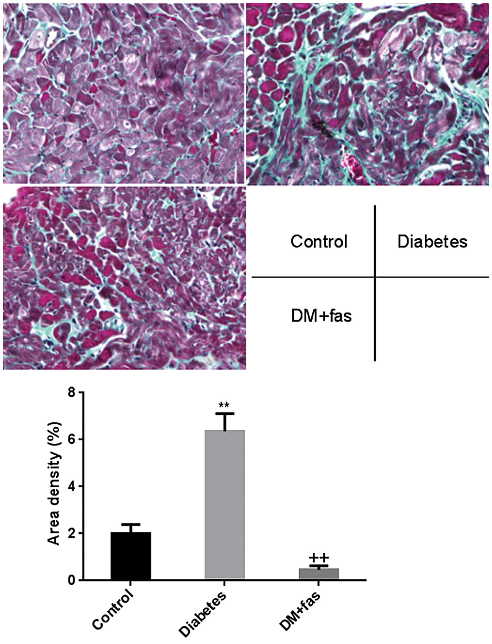
Representative patterns of Masson staining
demonstrating atrial fibrosis are shown in Fig. 1. The deposition of collagen in the
atrial interstitium from untreated diabetic hearts was
significantly increased compared with that observed in the control
rats. However, treatment with fasudil significantly reduced the
deposition of collagen.
Gene expression
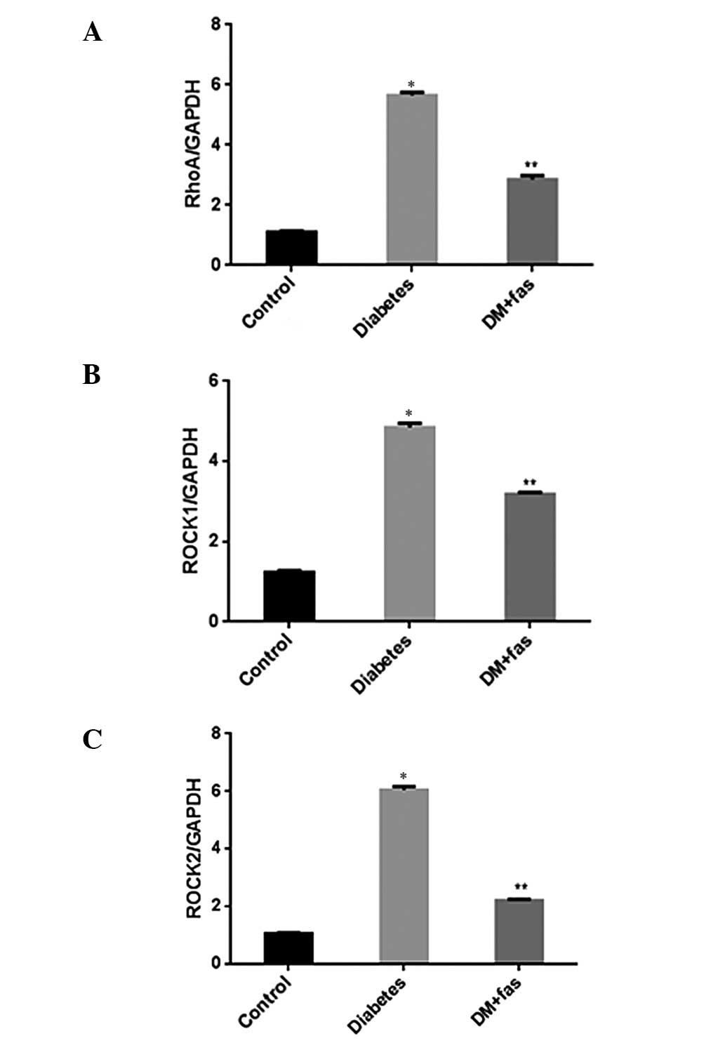
Compared with the control rats, mRNA expression
levels of RhoA, ROCK1 and ROCK2 (Fig.
2A–C) were significantly increased in the hearts of untreated
diabetic rats. The upregulation of mRNA expression of RhoA, ROCK1
and ROCK2 was blocked by treatment with fasudil. mRNA expression of
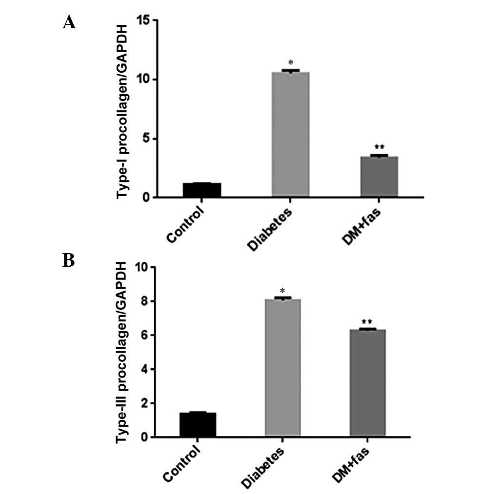
type I and III procollagen (Fig. 3A
and B) was significantly upregulated in the hearts of the
untreated diabetic rats, however, this expression was inhibited by
treatment with fasudil. These results suggest that fasudil
exhibited inhibitory effects on diabetes-induced gene upregulation
of type I and III procollagen, RhoA, ROCK1 and ROCK2.
Protein expression
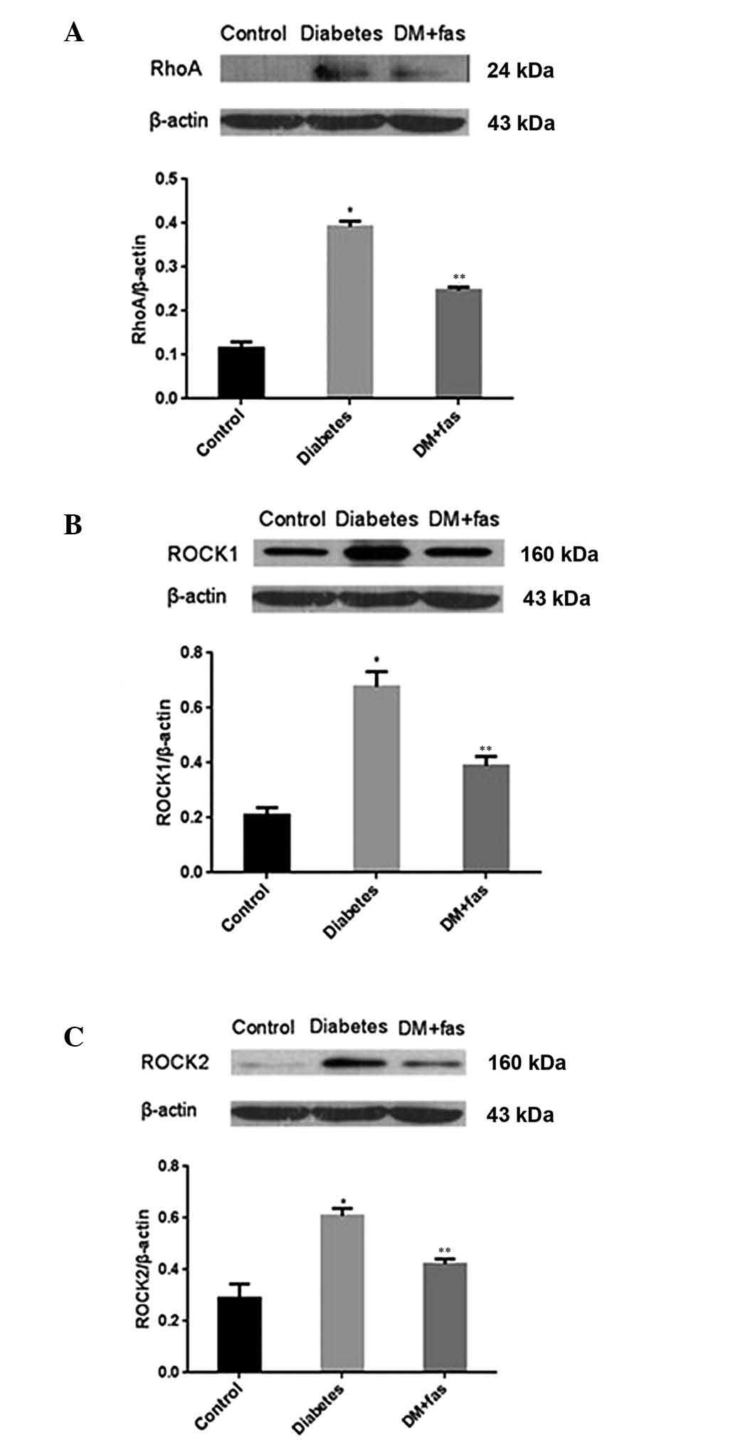
The protein levels of RhoA (Fig. 4A), ROCK1 (Fig. 4B) and ROCK2 (Fig. 4C) were measured, and all three
proteins were significantly enhanced in the hearts of diabetic rats
compared with those of control rats. In diabetic rats, RhoA, ROCK1
and ROCK2 were attenuated by treatment with fasudil compared to the
untreated diabetic rats. These results suggested that fasudil
inhibited activation of the Rho/ROCK pathways.
Discussion
Previous studies have shown that rats fed a HFD
develop insulin resistance (14–15),
and low-dose STZ is known to induce a mild impairment of insulin
secretion (16). In the present
study, the experimental rats fed HFD combined with low-dose STZ (30
mg/kg, once, i.p.) exhibited hyperglycemia and hyperinsulinemia.
The results suggested insulin resistance in diabetic rats.
Therefore, the present study successfully established a model of
type 2 DM in the rat, and this model closely mimics the natural
history and metabolic characteristics of type 2 DM in humans.
In the present study, the atria of untreated
diabetic rats showed notable increases in RhoA/ROCK activity and
marked atrial fibrosis as compared to that in the control rats.
Treatment with fasudil hydrochloride hydrate significantly reduced
RhoA/ROCK activity and atrial fibrosis. The inhibitory effect of
fasudil on collagen fibers was regulated at least as far upstream
as the transcriptional level, since treatment with fasudil
suppressed the mRNA expression of type I and III procollagen. The
findings are in agreement with the results of other previous
studies (9–12,17).
The Rho family of proteins is a group of small
guanosine triphosphate-binding proteins. ROCK is a downstream
signaling molecule of RhoA that has been widely investigated. ROCK
is activated by RhoA and phosphorylates the cytoplasmic MLC.
Through this pathway, ROCK regulates several biological processes,
including cell adhesion, chemotaxis and contraction. Inhibitors can
inhibit ROCK activity through competitive binding to the adenosine
triphosphate-binding site of the ROCK catalytic domain (18).
Fasudil and Y-27632 are non-isoform-selective ROCK
inhibitors and equivalently inhibit ROCK1 and ROCK2. Furthermore,
at higher concentrations, these ROCK inhibitors also inhibit other
serine-threonine kinases, including protein kinase (PK) A and PKCU.
Nevertheless, fasudil and its active metabolite, hydroxyfasudil,
are more selective for ROCKs than other kinases, with
hydroxyfasudil being slightly more selective than fasudil and
Y-27632 (19). In the present
study, the effect of restraining atrial fibrosis was associated
with the RhoA/ROCK pathway.
The RhoA/ROCK pathway controls a wide variety of
signal transduction pathways. ROCK1 has been shown to be a
regulator of glucose homeostasis and insulin sensitivity in
vivo (20). However, previous
studies on the effects of ROCK inhibitors on glucose and lipid
metabolism in vivo have yielded conflicting results. The
different conclusions obtained among these studies may be due to
the use of different inhibitors, doses, treatment times and animal
models. Kikuchi et al (9)
found that a low-dose of fasudil (30 mg/kg/day) did not affect
glucose and lipid metabolism. However, high-dose fasudil (100
mg/kg/day) ameliorated the metabolic disorder. Chronic treatment of
obese db/db mice with fasudil (10 mg/kg/day) has no effect on BG
levels and blood pressure (10).
Komers et al (21) found
that the fibrosis inhibitory effect of fasudil was without
reduction of blood pressure. In the present study, a low-dose of
fasudil (10 mg/kg/day) exhibited no effects on BG, insulin
resistance nor blood pressure in diabetic rats. Thus it appears
that the effect of fasudil was independent of blood pressure and
glycemic control.
The present study showed that RhoA/ROCK was involved
in atrial fibrosis, and fasudil hydrochloride hydrate ameliorated
atrial fibrosis through the RhoA/ROCK pathway in rats with type 2
diabetes. Inhibition of the RhoA/ROCK pathway may therefore be a
novel therapeutic target for the prevention of atrial fibrosis.
Additionally, the RhoA/ROCK pathway is involved in atrial fibrosis
in rats with type 2 diabetes. However, there were certain
experimental limitations in this study. First, to improve
understanding of the mechanism of action involved, cell culture
studies are required to verify the cause and effect associations
with regards to the role of these signaling molecules in diabetic
cardiac fibrosis. Second, it is known that atrial fibrosis is
associated with significantly increased oxidative stress and
inflammation. Therefore, the characteristics of oxidative stress
and inflammation in the heart should be described. Third, the small
number of rats in the present study, may limit the statistical
power and results. Fourth, as the conclusion of fasudil exhibiting
no effect on blood pressure was obtained from the preceding
studies, measures should be taken to further verify the result. The
treatments used to prevent the development and progression of
atrial fibrosis are not fully confirmed, and further clinical
trials are required to corroborate the conclusions of the present
study.
Acknowledgements
This study was supported by the Basic Clinical
Scientific Research Cooperation Fund of Capital Medical University
(no. 10JL32). The authors would like to express their gratitude to
Professors Yongquan Wu and Zhuowei Hu for their constant
encouragement and guidance.
References
|
1
|
Kato T, Yamashita T, Sekiguchi A, et al:
What are arrhythmogenic substrates in diabetic rat atria? J
Cardiovasc Electrophysiol. 17:890–894. 2006. View Article : Google Scholar : PubMed/NCBI
|
|
2
|
Shimokawa H and Takeshita A: Rho-kinase is
an important therapeutic target in cardiovascular medicine.
Arterioscler Thromb Vasc Biol. 25:1767–1775. 2005. View Article : Google Scholar : PubMed/NCBI
|
|
3
|
Shimokawa H: Rho-kinase as a novel
therapeutic target in treatment of cardiovascular diseases. J
Cardiovasc Pharmacol. 39:319–327. 2002. View Article : Google Scholar : PubMed/NCBI
|
|
4
|
Rolfe BE, Worth NF, World CJ, et al: Rho
and vascular disease. Atherosclerosis. 183:1–16. 2005. View Article : Google Scholar : PubMed/NCBI
|
|
5
|
Tapia PC: RhoA, Rho kinase, JAK2, and
STAT3 may be the intracellular determinants of longevity implicated
in the progeric influence of obesity: Insulin, IGF-1, and leptin
may all conspire to promote stem cell exhaustion. Med Hypotheses.
66:570–576. 2006. View Article : Google Scholar : PubMed/NCBI
|
|
6
|
Burch ML, Zheng W and Little PJ: Smad
linker region phosphorylation in the regulation of extracellular
matrix synthesis. Cell Mol Life Sci. 68:97–107. 2011. View Article : Google Scholar : PubMed/NCBI
|
|
7
|
Hudson VM: Rethinking cystic fibrosis
pathology: the critical role of abnormal reduced glutathione (GSH)
transport caused by CFTR mutation. Free Radic Biol Med.
30:1440–1461. 2001. View Article : Google Scholar : PubMed/NCBI
|
|
8
|
Yang L, Dan D, Zhu R, et al: Beta-elemene
inhibits expression of ANG II and RhoA/ROCK signaling in hepatic
stellate cells. Zhongguo Zhong Yao Za Zhi. 34:458–463. 2009.(In
Chinese).
|
|
9
|
Kikuchi Y, Yamada M, Imakiire T, et al: A
Rho-kinase inhibitor, fasudil, prevents development of diabetes and
nephropathy in insulin-resistant diabetic rats. J Endocrinol.
192:595–603. 2007. View Article : Google Scholar : PubMed/NCBI
|
|
10
|
Kolavennu V, Zeng L, Peng H, et al:
Targeting of RhoA/ROCK signaling ameliorates progression of
diabetic nephropathy independent of glucose control. Diabetes.
57:714–723. 2008. View Article : Google Scholar : PubMed/NCBI
|
|
11
|
Jiang C, Huang H, Liu J, et al: Fasudil, a
rho-kinase inhibitor, attenuates bleomycin-induced pulmonary
fibrosis in mice. Int J Mol Sci. 13:8293–8307. 2012. View Article : Google Scholar : PubMed/NCBI
|
|
12
|
Zhou H, Li YJ, Wang M, et al: Involvement
of RhoA/ROCK in myocardial fibrosis in a rat model of type 2
diabetes. Acta Pharmacol Sin. 32:999–1008. 2011. View Article : Google Scholar : PubMed/NCBI
|
|
13
|
Gao HC, Zhao H, Zhang WQ, et al: The role
of the Rho/Rock signaling pathway in the pathogenesis of acute
ischemic myocardial fibrosis in rat models. Exp Ther Med.
5:1123–1128. 2013.PubMed/NCBI
|
|
14
|
Tanaka S, Hayashi T, Toyoda T, et al:
High-fat diet impairs the effects of a single bout of endurance
exercise on glucose transport and insulin sensitivity in rat
skeletal muscle. Metabolism. 56:1719–1728. 2007. View Article : Google Scholar : PubMed/NCBI
|
|
15
|
Flanagan AM, Brown JL, Santiago CA, et al:
High-fat diets promote insulin resistance through cytokine gene
expression in growing female rats. J Nutr Biochem. 19:505–513.
2008. View Article : Google Scholar : PubMed/NCBI
|
|
16
|
Reed MJ, Meszaros K, Entes LJ, et al: A
new rat model of type 2 diabetes: the fat-fed,
streptozotocin-treated rat. Metabolism. 49:1390–1394. 2000.
View Article : Google Scholar : PubMed/NCBI
|
|
17
|
Guan SJ, Ma ZH, Wu YL, et al: Long-term
administration of fasudil improves cardiomyopathy in
streptozotocin-induced diabetic rats. Food Chem Toxicol.
50:1874–1882. 2012. View Article : Google Scholar : PubMed/NCBI
|
|
18
|
Wettschureck N and Offermanns S:
Rho/Rho-kinase mediated signaling in physiology and
pathophysiology. J Mol Med (Berl). 80:629–638. 2002. View Article : Google Scholar : PubMed/NCBI
|
|
19
|
Rikitake Y, Kim HH, Huang Z, et al:
Inhibition of Rho kinase (ROCK) leads to increased cerebral blood
flow and stroke protection. Stroke. 36:2251–2257. 2005. View Article : Google Scholar : PubMed/NCBI
|
|
20
|
Lee DH, Shi J, Jeoung NH, et al: Targeted
disruption of ROCK1 causes insulin resistance in vivo. J Biol Chem.
284:11776–11780. 2009. View Article : Google Scholar : PubMed/NCBI
|
|
21
|
Komers R, Oyama TT, Beard DR, et al: Rho
kinase inhibition protects kidneys from diabetic nephropathy
without reducing blood pressure. Kidney Int. 79:432–442. 2011.
View Article : Google Scholar : PubMed/NCBI
|