Introduction
Corrosive substances are commonly used in daily
life, and accidents due to these substances continue to be a
frequent reason for the emergency hospital admission of children
throughout the world. This hazard remains despite aggressive
accident prevention education programs aimed at children and
adults, preventive labeling and packaging and even legislation
limiting the strength and availability of caustic substances. In
rural areas and in developing countries, such as Turkey, caustic
soda in crystal and liquid form is used in home industries for soap
making, fruit drying and cleaning containers on farms. In addition,
the high number of over-the-counter caustic cleaning agents means
that children are likely to continue to accidentally ingest these
agents. The natural curiosity of children and their tendency to
taste everything, coupled with the availability of certain
chemicals around the house, create the setting for corrosive
esophageal injury. Although the mortality rate is not high,
accidental ingestion of corrosive substances is usually harmful and
results in lifelong damage (1). It
is reported that 20–40% of the cases with corrosive substance
ingestion result in esophageal damage (2,3). The
main strategy for corrosive esophageal burn treatment involves the
inhibition of inflammation, bacterial colonization and the
subsequent narrowing that may occur (4,5).
When a corrosive agent has been ingested, the presence or absence
of an esophageal burn must be established immediately, and if a
burn is diagnosed, the aim is to prevent stricture formation.
Treatment methods for the prevention of stricture formation include
antibiotics and steroids alone or in combination with bouginage,
total parenteral nutrition, nasogastric tube and intraluminal
stents. However, in previously reported studies, the stricture
ratios have remained high (3,4).
None of the treatment strategies have yielded uniform success, and
they have failed to receive universal acceptance. This has resulted
in an increasing number of studies investigating novel therapeutic
strategies for the prevention of stricture formation in the
esophagus following burns. However, despite experimental evidence
indicating that the majority of agents (heparin, pentoxifylline,
estradiol, antioxidants and epidermal growth factor) are beneficial
in preventing esophageal strictures, the agents have not yet gained
clinical application, possibly due to inconsistencies resulting
from the requirement for systemic administration of these drugs. In
addition, a number of the drugs have not yet been shown to be
suitable for human use (6).
Therefore, further investigation is required to determine the
efficacy of different treatments in eliminating stricture
formation.
Keratinocyte growth factor (KGF), a member of the
growth factor family, exerts its effect by inducing epithelial
proliferation, migration, and in certain tissues, epithelial
differentiation. KGF suppresses inflammation by modulating the
cytokine profile. The growth factor induces the upregulation of
enzyme expression, which detoxifies free oxygen radicals and exerts
a cytoprotective effect by inhibiting DNA breakage and epithelial
cell apoptosis. KGF has a trophic effect in the oral and intestinal
mucosa and is able to prevent atrophic alterations in these tissues
(7,8).
Palifermin (Kepivance®) is a recombinant
form of human KGF produced in Escherichia coli. In
palifermin, the first 23 N-terminal amino acids found in endogenous
KGF were deleted in order to obtain a more stable protein product.
Palifermin was approved by the US Food and Drug Administration
(FDA) in December 2004 to decrease the incidence and duration of
severe oral mucositis in patients with hematological malignancies
receiving myelotoxic therapy and requiring hematopoietic stem cell
support. Although the FDA has only approved palifermin for use in
bone marrow transplantation in the treatment of hematological
malignancies, it is currently under investigation for use in
additional therapies targeting mucosal injury, including colorectal
cancer chemotherapy regimens and head and neck radiation (9). In the present study, the therapeutic
effect of palifermin for the prevention of esophageal strictures
was investigated in a rat alkaline esophageal injury model.
Materials and methods
Ethics
Experimental protocols were approved by the
Experimental Ethics Committee of Bülent Ecevit University School of
Medicine (Kozlu, Turkey). All the rats were housed at 24±1°C under
controlled lighting (12-h light/dark cycle), humidity and human
activity. Animals were allowed to acclimatize to these conditions
for 10 days prior to the start of the experiment. All the study
groups rats were housed in identical wire-bottomed cages to prevent
coprophagy, and all animals received human care. Following ethical
approval, a preliminary study was performed to standardize the
experimental model and surgical techniques.
Experimental model
A total of 32 female Wistar albino rats, weighing
190–240 g, were divided into four groups, which included the
control (C), burn (B), steroid (S) and steroid plus palifermin
(S/P) groups. An experimental corrosive esophageal burn model was
established in the B, S and S/P groups.
Following overnight fasting, rats were anesthetized
with 10 mg/kg xylazine (2%) (Rompun®, Bayer Healthcare
AG, Leverkusen, Germany) and 100 mg/kg ketamine
(Ketalar®, Pfizer, New York, USA) subcutaneously. The
experimental model of caustic esophageal burn was established as
previously described by Gehanno and Guedon (10), with modifications as in the study
by Liu and Richardson (11).
Following a median laparotomy, a 2-cm segment of the abdominal
esophagus at the gastroesophageal junction was dissected and
isolated. A catheter (1 mm internal diameter and 2.2 mm external
diameter) (Bıçakçılar, Istanbul, Turkey) was advanced to the upper
region of the isolated segment, while a venous catheter of 24 G
size was introduced into the lower part of the segment via the
stomach for drainage. The two ends of the segments were then
secured. A total of 10 ml sodium hydroxide at 30% concentration was
infused through the upper catheter for 90 sec in the B, S and S/P
groups, whilst isotonic saline was used in the control group.
Subsequently, distilled water was used to irrigate the burned
segment for ≥15 sec. The catheters were then withdrawn and the
gastric insertion site was repaired. Following closure of the
laparotomy, 10 ml saline and 5% dextrose solutions (İE Ulagay,
Istanbul, Turkey) were injected intraperitoneally. The rats were
subsequently fasted for 24 h following surgery. At postoperative
day one, intraperitoneal injections of the study group’s
medications were initiated and rats were provided with food ad
libitum. All the animals were housed in identical cages that
provided food and water during the study period.
Study design
In the control group, the esophagus of the rats was
uninjured and untreated. For the rats in group B, a standard
esophageal burn was produced, and the group received
intraperitoneal injections of 150 mg/kg/day ampicillin
(Ampisina®; Mustafa Nevzat Pharmaceuticals, Istanbul,
Turkey). With regard to the rats in group S, the esophagus was
injured and treated with 150 mg/kg/day ampicillin and 1 mg/kg/day
dexametazon (Dekort®; Deva, Istanbul, Turkey)
intraperitoneally. In the S/P group, the esophagus was injured and
the rats were intraperitoneally administered 150 mg/kg/day
ampicillin, 1 mg/kg/day dexametazon and 60 μg/kg/day palifermin
(Kepivance®; Amgen, Thousand Oaks, CA, USA). The drugs
were administered daily throughout the seven day treatment period.
During the observation period, animals were weighed daily. The rats
were sacrificed after 21 days. The efficacy of the treatment was
subsequently assessed by analyzing the weight gain, stenosis index
and histopathological evaluation of the burned segments in the rats
in each group.
At postoperative day 21, the animals were sacrificed
and the esophageal burn segment was removed, fixed in 10%
formaldehyde and embedded in paraffin. Next, 5-μm sections were
stained with hematoxylin-eosin and Masson’s trichrome connective
tissue dye (Merck Millipore, Billerica, MA, USA). Esophageal wall
thickness and luminal diameters were assessed using a millimetric
ocular microscope. The thickness of the esophageal wall and the
lumen diameter were measured in order to calculate the stenosis
index (12). The stenosis index
was calculated as follows: Stenosis index = [wall thickness (A1 +
A2)/2]/[lumen diameter (B1 + B2)/2]. Submucosal collagen
accumulation, muscularis mucosa injury, tunica muscularis injury
and collagen accumulation were evaluated semi-quantitatively and
scored as shown in Table I
(1).
 | Table IHistopathological analysis
criteria. |
Table I
Histopathological analysis
criteria.
| Histopathological
parameters | Score |
|---|
| Submucosal collagen
accumulation |
| Absent | 0 |
| 2-fold increase in
muscularis mucosa thickness | +1 |
| >2-fold increase
in muscularis mucosa thickness | +2 |
| Muscularis mucosa
injury |
| Absent | 0 |
| Present | +1 |
| Tunica muscularis
injury and collagen accumulation |
| Absent | 0 |
| Mild (collagen
accumulation around the smooth muscles) | +1 |
| Prominent (collagen
accumulation around the smooth muscles and translocation of smooth
the muscles with collagen) | +2 |
Statistical analysis
Mean values with the standard deviation of the body
weights in each group were calculated prior to and following the
experiment. The differences between the body weights prior to and
following the experiment were analyzed using Kruskal-Wallis
variance analysis, while pairwise comparisons between the groups
were performed using the Bonferonni-corrected Mann-Whitney U-test.
Histopathological scores were analyzed using the χ2 and
Fisher’s exact tests. P<0.05 was considered to indicate a
statistically significant difference. Statistical calculations were
performed using SPSS statistical software, version 13 (SPSS Inc.,
Chicago, IL, USA).
Results
Mortality and body weight
Two rats were excluded from the study following
mortality on day 6 and day 13. The body weights of all the animals
were measured prior to and at the end of the study (day 21;
Table II). A statistically
significant difference was observed in the body weights between the
B group and the C group (P<0.05). In addition, a statistically
significant difference was observed with regard to the body weight
between the S/P-treated group and the B group (P=0.012).
| Table IIBody weights of the animals in each
group. |
Table II
Body weights of the animals in each
group.
| Group | Weight at day 1
(g) | Weight at day 21
(g) | Change (%) |
|---|
| Control | 203.6±5.4 | 225.0±7.4 | 11.0↑ |
| Burn | 201.5±4.0 | 175.5±4.6a | 11.4↓ |
| Steroid | 202.3±4.7 | 215.6±6.3 | 10.6↑ |
| Steroid +
palifermin | 203.1±4.2 | 222.1±6.2b | 10.9↑ |
Histopathological observations
Esophageal sections were prepared from all the rats
and were evaluated for submucosal collagen accumulation, damage to
the muscularis mucosa, damage to the tunica muscularis and collagen
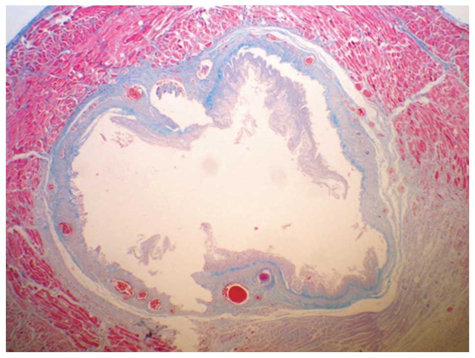
accumulation. The corrosive burn group was examined and prominent
damage in the mucosal, submucosal and muscular layers was observed.
In addition, a widespread accumulation of collagen in the
submucosal and muscular layers was identified (Fig. 1). A mild thickening of the
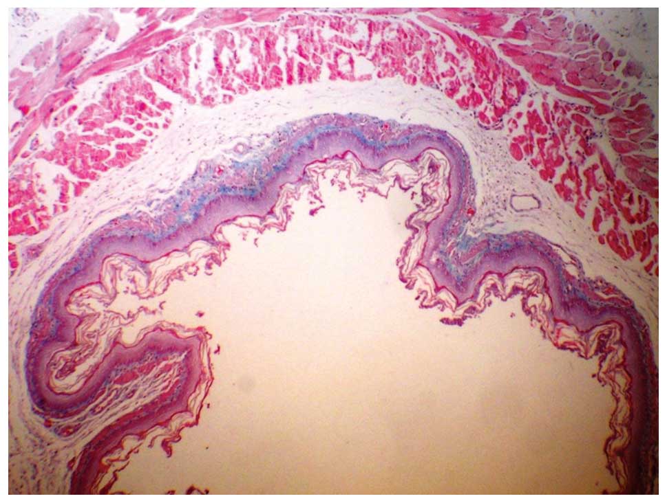
muscularis mucosa was observed during the histopathological
examination of the steroid-treated group. Furthermore, a mild
increase in collagen deposition was observed in the muscularis
mucosa, submucosa and the tunica muscularis of the S group when
compared with the B group (Fig.
2).
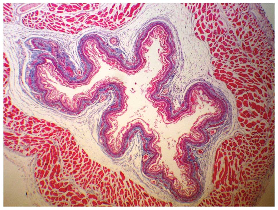
In the S/P-treated group, regular, preserved layers
of mucosal, submucosal and muscular structures were observed. In
the muscularis mucosa, a slight increase in collagen accumulation
was observed following staining with Masson’s Trichrome (Fig. 3).
Statistically significant differences with regard to
submucosal collagen accumulation, muscularis mucosa and tunica
muscularis injury were observed between the B group and the C group
(P<0.05). Similarly, statistically significant differences in
the three parameters were observed when comparing the S and
S/P-treated groups with the B group (P<0.05). In addition,
submucosal collagen accumulation increased (P=0.007), and injury to
the tunica muscularis was present (P=0.041) in the S-treated group
when compared with the S/P-treated group (Table III).
| Table IIIAnalysis of the stenosis index. |
Table III
Analysis of the stenosis index.
| Group | n | Stenosis index |
|---|
| Control | 8 | 0.32±0.05 |
| Burn | 6 | 0.95±0.07a |
| Steroid | 8 | 0.57±0.06a,b |
| Steroid +
palifermin | 8 | 0.41±0.03a,b,c |
Stenosis index
Parameters used to calculate the stenosis index are
shown in Table III.
Statistically significant differences were observed among all the
groups (P<0.05). The stenosis indexes in the C and S groups were
significantly lower when compared with the B group (P<0.05). In
addition, the stenosis index in the S/P group was significantly
lower compared with the steroid-treated group and the control group
(P<0.05).
Discussion
Ingestion of caustic materials is a serious problem
in children. Ingested alkaline materials penetrate rapidly into the
esophageal wall and cause liquefaction necrosis with destruction of
the mucosal, submucosal and, in severe cases, muscular layers of
the esophagus. Following ingestion of the caustic material,
hemorrhage, thrombosis of the submucosal vessels and inflammation
with tissue edema develop in the first 24 h. Thrombosis may further
increase the damage of the caustic burn and result in local
necrosis and gangrene, with inflammation extending through the
muscle layers and causing perforation. Following the sub-acute
phase, scar formation begins with fibroblastic proliferation in the
third week. Stricture formation occurs via the synthesis,
deposition and remodeling of collagen. Different treatment
strategies target different phases of the injury. During the early
period, the main aim is to prevent stricture formation (1).
The optimal management protocols for the treatment
of severe damage following the ingestion of caustic substances
remain controversial. The main aim of medical treatment is the
inhibition of any inflammatory reaction or stricture formation
caused by esophageal burning. It is hypothesized that stricture
formation may be overcome by inhibiting fibroplasia and scarring.
In numerous experimental and clinical studies, it has been
demonstrated that a combination of steroids and antibiotics are
effective for mild or moderate corrosive burns. However, the drugs
were unable to satisfactorily prevent stricture formation (13). Previous experimental studies have
indicated that the administration of miscellaneous agents,
including heparin, penicillamine, β-aminopropionitrile,
indomethacin, pentoxifylline, estradiol, antioxidants, epidermal
growth factor and vitamin E, may exert protective effects during
the early phase of burn injury (13–17).
In addition to these agents, esophageal stenting during the early
stages has been used without steroids in clinical and experimental
studies (18,19).
KGF is a growth factor that induces the
proliferation, differentiation and migration of epithelial cells. A
number of studies have demonstrated that KGF also plays key roles
in inflammation and repair processes, as well as exhibiting a cell
protective effect. Thus, KGF was approved by the FDA for use in
clinical settings for the treatment of oral mucositis and ulcers
(8,20). Using a rat model of an esophageal
ulcer, Baatar et al (21)
demonstrated that local administration of palifermin significantly
increased epithelial cell proliferation on the ulcer border and
promoted ulcer healing (21).
However, to the best of our knowledge, no previous studies have
reported the use of palifermin for the treatment of corrosive
esophageal burns.
Exogenous KGF treatment following various types of
injury significantly increases re-epithelization and causes major
stimulation during the repair process. This evidence indicates that
KGF treatment may reduce or prevent the deep penetration of damage
in the acute phase of corrosive damage by increasing the
epithelization in the esophageal lumen. Considering that steroid
treatment inhibits endogenous KGF expression in tissue injuries, it
may be hypothesized that the steroidal effect on delayed healing
partially occurs via this mechanism (22). However, steroid treatment does not
significantly affect the expression of KGF receptors. Therefore, it
was hypothesized that the addition of palifermin to the existing
treatment regime involving steroids may prevent the negative
effects of steroids. In addition, palifermin may aid steroids in
the anti-inflammatory response by downregulating the expression of
proinflammatory cytokines, including interferon-γ and tumor
necrosis factor-α, and may contribute to a reduction in the damage
that occurs during the acute phase of inflammation by upregulating
the expression of detoxifying enzymes that exert a protective
effect against free oxygen radicals (23–25).
The results of the present study indicate that the
administration of palifermin decreases the inflammatory response
and reduces esophageal damage, which are important considerations
for the sequelae of alkaline burns in esophageal injury.
The early inflammatory response and mucosal necrosis
in burn tissue are known to be major causes of stricture in
corrosive esophageal burns. During the early phase, the
inflammatory response leads to necrosis and the further destruction
of the esophageal wall. Bacterial translocation occurring on the
acutely injured esophageal wall causes a more severe clinical
situation. Following initiation of the burn, early and fast
re-epithelization of the esophageal lumen, as a result of
palifermin treatment, may have a protective effect against late
phase stricture development by preventing additional inflammatory
processes, including potential reflux esophagitis, bacterial and
fungal colonization and exposure to saliva and mucosal
secretions.
In conclusion, the results of the present study
demonstrate the efficacy of palifermin treatment as an adjunctive
therapy to the conventional treatment involving steroids plus
antibiotics in a corrosive esophageal burn model. Palifermin
treatment was shown to significantly decrease the degree of
fibrosis and ameliorate histopathological damage in an experimental
caustic burn model. However, since the present study was the first
to use palifermin in the treatment of corrosive esophageal burns,
further detailed animal and/or clinical studies, with a larger
sample size, are required to support the current results.
References
|
1
|
Millar AJW and Cywes S: Caustic strictures
of esophagus. Pediatric Surgery. O’Neill JJ, Rowe MI, Grosfeld JL,
et al: Mosby; St Louis, MO: pp. 969–979. 1998
|
|
2
|
Tiryaki T, Livanelioğlu Z and Atayurt H:
Early bougienage for relief of stricture formation following
caustic esophageal burns. Pediatr Surg Int. 21:78–80. 2005.
View Article : Google Scholar : PubMed/NCBI
|
|
3
|
Han Y, Cheng QS, Li XF and Wang XP:
Surgical management of esophageal strictures after caustic burns: a
30 years of experience. World J Gastroenterol. 10:2846–2849.
2004.PubMed/NCBI
|
|
4
|
Anderson KD, Rouse TM and Randolph JG: A
controlled trial of corticosteroids in children with corrosive
injury of the esophagus. N Engl J Med. 323:637–640. 1990.
View Article : Google Scholar : PubMed/NCBI
|
|
5
|
Cakmak M, Nayci A, Renda N, Erekul S,
Gökçora H and Yücesan S: The effect of corticosteroids and
pentoxifiline in caustic esophageal burns. Int Surg. 82:371–375.
1997.PubMed/NCBI
|
|
6
|
Ekingen G, Ozden M, Sözübir S, Maral H,
Müezzinoğlu B, Kahraman H and Güvenç BH: Effect of the prostacyclin
derivate iloprost in experimental caustic esophageal burn. Pediatr
Surg Int. 21:441–444. 2005. View Article : Google Scholar : PubMed/NCBI
|
|
7
|
Beawen AW and Shea TC: The effect of
palifermin on chemotherapy and radiation therapy induced mucositis:
A review of the current literature. Support Cancer Ther. 4:188–197.
2007. View Article : Google Scholar
|
|
8
|
Werner S: Keratinocyte growth factor: a
unique player in epithelial repair processes. Cytokine Growth
Factor Rev. 9:153–165. 1998. View Article : Google Scholar : PubMed/NCBI
|
|
9
|
Radtke ML and Kolesar JM: Palifermin
(Kepivance) for the treatment of oral mucositis in patients with
hematologic malignancies requiring hematopoietic stem cell support.
J Oncol Pharm Pract. 11:121–125. 2005. View Article : Google Scholar : PubMed/NCBI
|
|
10
|
Gehanno G and Guedon C: Inhibition of
experimental esophageal lye strictures by penicillamine. Arch
Otolaryngol. 107:145–147. 1981. View Article : Google Scholar : PubMed/NCBI
|
|
11
|
Liu AJ and Richardson MA: Effects of
N-acetylcysteine of experimentally induced esophageal lye injury.
Ann Otol Rhinol Laryngol. 94:477–482. 1985.PubMed/NCBI
|
|
12
|
Berthet B, Di Costanzo J, Arnaud C, et al:
Influence of epidermal growth factor and interferon gamma on
healing of oesophageal corrosive burns in the rat. Br J Surg.
81:395–398. 1994. View Article : Google Scholar : PubMed/NCBI
|
|
13
|
Temir ZG, Karkınel A, Karaca I, Ortaç R
and Ozdamar A: The effectiveness of sucralfate against stricture
formation in experimental corrosive esophageal burns. Surg Today.
35:617–622. 2005. View Article : Google Scholar : PubMed/NCBI
|
|
14
|
Kiyan G, Aktas S, Ozel K, Isbilen E,
Kotiloglu E and Dagli TE: Effects of hyperbaric oxygen therapy on
caustic esophageal injury in rats. J Pediatr Surg. 39:1188–1193.
2004. View Article : Google Scholar : PubMed/NCBI
|
|
15
|
Günel E, Cağlayan F, Cağlayan O, Canbilen
A and Tosun M: Effect of antioxidant therapy on collogen synthesis
in corrosive esophageal burns. Pediatr Surg Int. 18:24–27.
2002.PubMed/NCBI
|
|
16
|
Koltuksuz U, Mutuş M, Kutlu R, Ozyurt C,
et al: Effects of caffeic acid phenethyl ester and epidermal growth
factor on development of caustic esophageal stricture in rats. J
Pediatr Surg. 36:1504–1509. 2001. View Article : Google Scholar : PubMed/NCBI
|
|
17
|
Ozçelik MF, Pekmezci S, Saribeyoğlu K, et
al: The effect of halofuginone, a specific inhibitor of collogen
type 1 synthesis, in the prevention of esophageal strictures
related to caustic injury. Am J Surg. 187:257–260. 2004.PubMed/NCBI
|
|
18
|
Wijburg FA, Heymans HS and Urbanus NA:
Caustic esophageal lesions in childhood: prevention of stricture
formation. J Pediatr Surg. 24:171–173. 1989. View Article : Google Scholar : PubMed/NCBI
|
|
19
|
Fell SC, Denize A, Becker NH and Hurwitt
ES: The effect of intraluminal splinting in the prevention of
caustic stricture of the esophagus. J Thorac Cardiovasc Surg.
52:675–681. 1966.PubMed/NCBI
|
|
20
|
Rubin JS, Osada H, Finch PW, Taylor WG,
Rudikoff S and Aaronson SA: Purification and characterization of a
newly identified growth factor specific for epithelial cells. Proc
Natl Acad Sci USA. 86:802–806. 1989. View Article : Google Scholar : PubMed/NCBI
|
|
21
|
Baatar D, Kawanaka H, Szabo IL, et al:
Esophageal ulceration activates keratinocyte growth factor and its
receptor in rats: implications for ulcer healing. Gastroenterology.
122:458–468. 2002. View Article : Google Scholar : PubMed/NCBI
|
|
22
|
Brauchle M, Fässler R and Werner S:
Suppression of keratinocyte growth factor expression by
glucocorticoids in vitro and during wound healing. J Invest
Dermatol. 105:579–584. 1995. View Article : Google Scholar : PubMed/NCBI
|
|
23
|
Panoskaltsis-Mortari A, Taylor PA, Rubin
JS, et al: Keratinocyte growth factor facilitates alloengraftment
and ameliorates graft-versus-host disease in mice by a mechanism
independent of repair of conditioning-induced tissue injury. Blood.
96:4350–4356. 2000.
|
|
24
|
Ellison CA, Natuik SA, Fischer JM, et al:
Effect of recombinant human keratinocyte growth factor (rHuKGF) on
the immunopathogenesis of intestinal graft-vs. -host disease
induced without a preconditioning regimen. J Clin Immunol.
24:197–211. 2004. View Article : Google Scholar
|
|
25
|
Stout A, Gresser I and Thompson WD:
Inhibition of wound healing in mice by local interferon alpha/beta
injection. Int J Exp Pathol. 74:79–85. 1993.PubMed/NCBI
|