Risk of node metastasis of sentinel lymph nodes detected in level II/III of the axilla by single‑photon emission computed tomography/computed tomography

  • Authors:
    • Hiroaki Shima
    • Goro Kutomi
    • Fukino Satomi
    • Hideki Maeda
    • Tomoko Takamaru
    • Hidekazu Kameshima
    • Tosei Omura
    • Mitsuru Mori
    • Masamitsu Hatakenaka
    • Tadashi Hasegawa
    • Koichi Hirata
  • View Affiliations

  • Published online on: September 15, 2014     https://doi.org/10.3892/etm.2014.1968
  • Pages: 1447-1452
Metrics: Total Views: 0 (Spandidos Publications: | PMC Statistics: )
Total PDF Downloads: 0 (Spandidos Publications: | PMC Statistics: )


Abstract

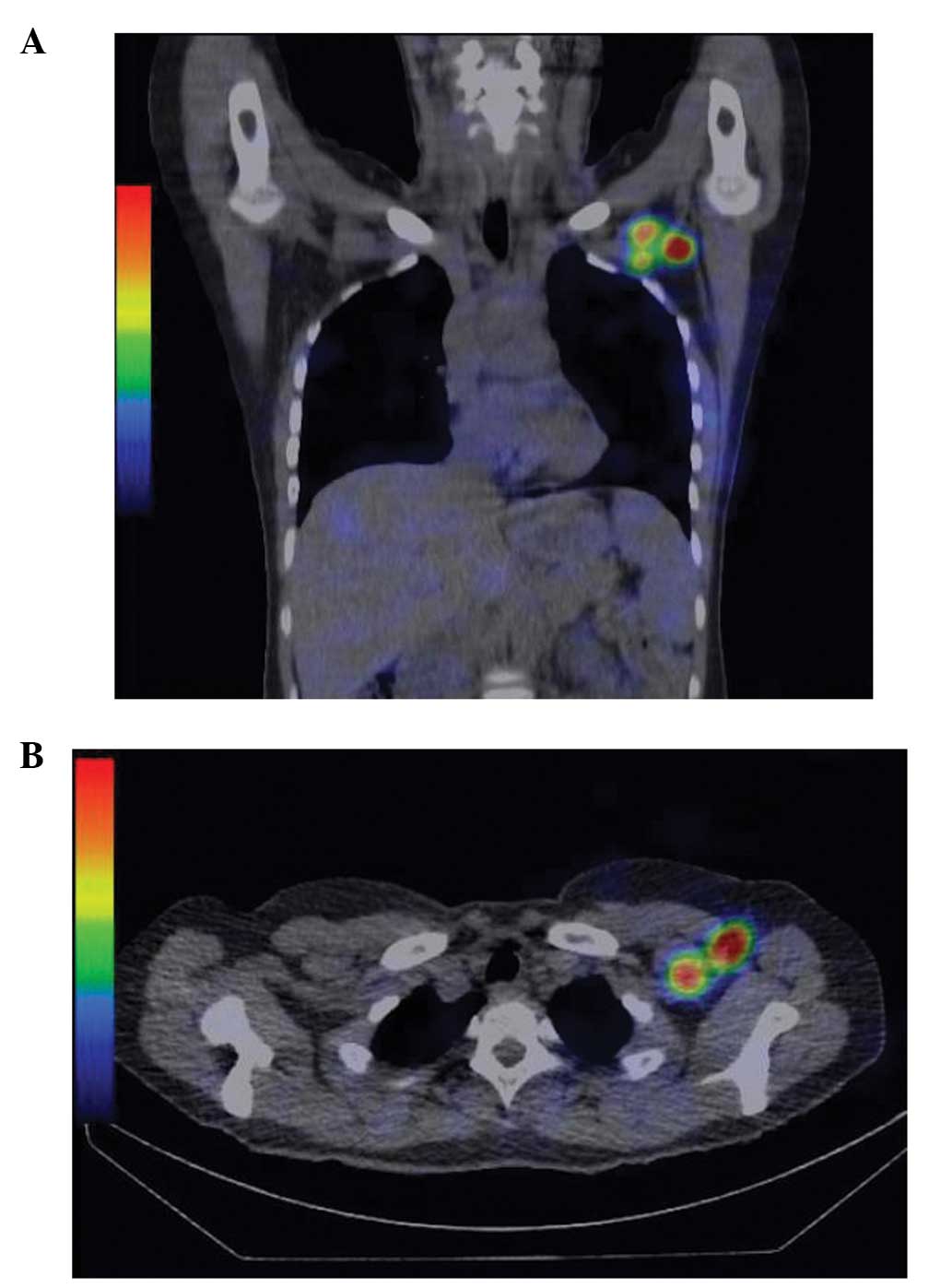
In breast cancer, single‑photon emission computed tomography/computed tomography (SPECT/CT) shows the exact anatomical location of sentinel nodes (SN). SPECT/CT mainly exposes axilla and partly exposes atypical sites of extra‑axillary lymphatic drainage. The mechanism of how the atypical hot nodes are involved in lymphatic metastasis was retrospectively investigated in the present study, particularly at the level II/III region. SPECT/CT was performed in 92 clinical stage 0‑IIA breast cancer patients. Sentinel lymph nodes are depicted as hot nodes in SPECT/CT. Patients were divided into two groups: With or without hot node in level II/III on SPECT/CT. The existence of metastasis in level II/III was investigated and the risk factors were identified. A total of 12 patients were sentinel lymph node biopsy metastasis positive and axillary lymph node dissection (ALND) was performed. These patients were divided into two groups: With and without SN in level II/III, and nodes in level II/III were pathologically proven. In 11 of the 92 patients, hot nodes were detected in level II/III. There was a significant difference in node metastasis depending on whether there were hot nodes in level II/III (P=0.0319). Multivariate analysis indicated that the hot nodes in level II/III and lymphatic invasion were independent factors associated with node metastasis. There were 12 SN‑positive patients followed by ALND. In four of the 12 patients, hot nodes were observed in level II/III. Two of the four patients with hot nodes depicted by SPECT/CT and metastatic nodes were pathologically evident in the same lesion. Therefore, the present study indicated that the hot node in level II/III as depicted by SPECT/CT may be a risk of SN metastasis, including deeper nodes.
View Figures
View References

Related Articles

Journal Cover

November-2014
Volume 8 Issue 5

Print ISSN: 1792-0981
Online ISSN:1792-1015

Sign up for eToc alerts

Recommend to Library

Copy and paste a formatted citation
x
Spandidos Publications style
Shima H, Kutomi G, Satomi F, Maeda H, Takamaru T, Kameshima H, Omura T, Mori M, Hatakenaka M, Hasegawa T, Hasegawa T, et al: Risk of node metastasis of sentinel lymph nodes detected in level II/III of the axilla by single‑photon emission computed tomography/computed tomography. Exp Ther Med 8: 1447-1452, 2014
APA
Shima, H., Kutomi, G., Satomi, F., Maeda, H., Takamaru, T., Kameshima, H. ... Hirata, K. (2014). Risk of node metastasis of sentinel lymph nodes detected in level II/III of the axilla by single‑photon emission computed tomography/computed tomography. Experimental and Therapeutic Medicine, 8, 1447-1452. https://doi.org/10.3892/etm.2014.1968
MLA
Shima, H., Kutomi, G., Satomi, F., Maeda, H., Takamaru, T., Kameshima, H., Omura, T., Mori, M., Hatakenaka, M., Hasegawa, T., Hirata, K."Risk of node metastasis of sentinel lymph nodes detected in level II/III of the axilla by single‑photon emission computed tomography/computed tomography". Experimental and Therapeutic Medicine 8.5 (2014): 1447-1452.
Chicago
Shima, H., Kutomi, G., Satomi, F., Maeda, H., Takamaru, T., Kameshima, H., Omura, T., Mori, M., Hatakenaka, M., Hasegawa, T., Hirata, K."Risk of node metastasis of sentinel lymph nodes detected in level II/III of the axilla by single‑photon emission computed tomography/computed tomography". Experimental and Therapeutic Medicine 8, no. 5 (2014): 1447-1452. https://doi.org/10.3892/etm.2014.1968