Introduction
Endothelial tissues are active and dynamic, lining
the entire vascular system and controlling important functions
(1). The endothelium serves as a
barrier between the blood and tissues, actively participates in the
regulation of vascular function and is critical to the biology of
normal tissues. Tissue health is often synonymous with endothelial
integrity. Vascular homeostasis and tone are controlled by the
endothelium via the synthesis and release of a number of
endothelium-derived relaxing and constricting substances (2), including nitric oxide (NO),
angiotensin II (Ang II) and endothelin-1 (ET-1) (3,4).
Quiescent endothelial cells suppress all phases of vascular
diseases, including the degree of injury, local thrombosis,
inflammation, proliferation and matrix remodeling, while an injured
or dysfunctional endothelium can promote such events (5). Impaired NO bioavailability represents
the central feature of endothelial dysfunction, which is a common
abnormality occurring in a number of vascular diseases (6).
Hyperuricemia is a complex metabolic disease that
can develop into gout (7). An
increased serum level of uric acid (UA) is hypothesized to be
independently associated with an increased risk of mortality from
cardiovascular diseases (8). The
serum levels of UA increase physiologically and gradually with
aging due to the excess dietary purine or ethanol intake (9,10).
Increasing evidence indicates that hyperuricemia is
associated with endothelial dysfunction (11,12).
The occurrence of abnormal endothelial function in individuals
suffering with hyperuricemia is a cause of health problems, since
endothelial dysregulation is a major determinant of atherosclerosis
in the early pathophysiological stages, and has been demonstrated
to occur in a time-dependent manner in patients with coronary
artery disease, hypertension and type 2 diabetes (13,14).
Thus, strategies for improving endothelial function are currently
under investigation.
Statins are 3-hydroxy-3-methylglutaryl coenzyme A
(HMG-CoA) reductase (HMGR) inhibitors, administered to inhibit
cholesterol synthesis, thereby lowering the serum levels of
cholesterol (15). In addition to
lowering the levels of low-density lipoproteins, statins exhibit
pleiotropic effects, such as improving endothelial function,
inhibiting the synthesis of endothelins, reducing coronary artery
thrombus formation and inhibiting the proliferation of smooth
muscle cells, as well as anti-inflammatory, antioxidant and
antiplatelet effects (16).
Endothelial function is critical for cardiovascular function, and
serves as a surrogate marker in monitoring the efficacy of statin
treatment, which indicates novel, potential applications for these
drugs in the primary and secondary prevention of acute
cardiovascular events (17).
Among the synthetic, type 2 or second-generation
statins, rosuvastatin is a sulphur-containing hydrophilic statin
with multiple binding sites that forms a strong interaction with
HMGR. Thus, rosuvastatin provides more potent enzyme inhibition
compared with other statins. Rosuvastatin, similar to other
statins, is a competitive antagonist of HMGR, competing directly
with the endogenous substrate for the active site cavity of the
enzyme. The drug has an affinity for the HMGR active site that is
>104-fold higher than the affinity for HMG-CoA
(18,19).
In the present study, yeast extract powder (YEP) and
oxonic acid (OA) potassium salt were used as revulsants to
establish a hyperuricemic rat model. Interventions with various
rosuvastatin calcium dosages were applied to analyze the effect of
rosuvastatin on the experimental animal model. In the present
study, a hyperuricemic rat model was established by intragastrical
feeding of yeast extract in combination with various doses of OA
potassium salt through intraperitoneal injection. The model was
treated with various rosuvastatin doses. By determining the drug
effect on various factors associated with the vascular endothelial
function of the rats, the present study may provide important
evidence on the application of rosuvastatin in the clinical
treatment of hyperuricemia.
Materials and methods
Animals
A total of 72 male Sprague-Dawley rats (age, 8
weeks; weight, 203.8±32.15 g) were provided by the Experimental
Animal Center of Xinjiang Medical University (Ürümqi, China). The
rats were fed a standard laboratory diet, available at the center,
prior to the initiation of the study. The body weight of the rats
was measured twice a week in order to adjust the drug dosage to the
rat weight. The study was conducted in strict accordance with the
recommendations in the Guide for the Care and Use of Laboratory
Animals of the National Institutes of Health (20). The animal use protocol was reviewed
and approved by the Ethical Committee and Institutional Animal Care
and Use Committee of the First Affiliated Hospital of Xinjiang
Medical University (approval no. 20100310003; Ürümqi, China).
Establishment of an animal model and drug
intervention
An experimental animal model of hyperuricemia was
established according to the reference data found in previous
studies (21). In the present
study, animal modeling and drug intervention were initiated
simultaneously after randomly dividing the 72 male rats into six
groups (12 rats per group); the drug intervention was continued for
six weeks. To establish the animal model, the rats were
intragastrically administered 21 g/kg/day YEP (production batch no.
20090705; Beijing Aoboxing Biological Technology Co., Ltd, Beijing,
China) mixed with standard feed at a proportion of 1:4, and
intraperitoneally injected with 200 mg/kg/day OA (production batch
no. 20120312; Sigma-Aldrich Co., Munich, Germany). A stock solution
of 5 mg/ml rosuvastatin (production batch no. EK188; AstraZeneca
Pharmaceuticals Co., Södertälje, Sweden) was prepared for the drug
intervention. The rats were administered 2.5, 5.0 and 10.0
mg/kg/day rosuvastatin by intravenous injection, while 53.57
mg/kg/day allopurinol (Beijing Double-Crane Pharmaceutical Co.,
Ltd, Beijing, China) was administered as the positive control.
Specimen collection
Anticoagulated blood samples (2 ml) were collected
by excising the eyeballs of the rats prior to the experiment (week
0) and at weeks 2, 4 and 6. Following separation through natural
blood clotting and centrifugation (1509.3 × g for 15 min; Allegra
64R, Beckmann Coulter, Miami, FL, USA), the plasma was collected
and stored at −80°C until required for analysis. Blood samples were
collected in the morning following a minimum fasting period of 10
h. After six weeks, the rats were sacrificed using 40 mg/kg sodium
pentobarbital. The entire thoracic aorta of each rat was removed,
snap frozen in liquid nitrogen, fixed in 1–4% neutral-buffered
formalin, embedded in paraffin and cut into 4–5-μm serial sections
for immunohistochemical staining.
Evaluated serum parameters
Lipids were extracted from 100-μl plasma samples and
analyzed to obtain the serum levels of UA, NO, ET-1 and Ang II
using commercially available ELISA detection kits (R&D Systems,
Inc., Minneapolis, MN, USA) and a DxC800 Synchron Biochemical
Analysis System (Beckman Coulter, Inc., Brea, CA, USA) according to
the manufacturer’s instructions and standard clinical
protocols.
Immunofluorescence staining
Tissue specimens were cut into 4-μm sections,
mounted on glass slides by heating at 56°C, dewaxed with standard
xylene and rehydrated using graded alcohol solutions and water. The
internal enzyme activity of the tissue sections was inhibited by
H2O2 treatment. Standard immunohistochemical
staining was performed using a rabbit antieNOS polyclonal primary
antibody (Santa Cruz Biotechnology, Inc., Santa Cruz, CA, USA)
followed by detection with a goat anti-rabbit polyclonal secondary
antibody conjugated with horseradish peroxidase (Beijing Zhongshan,
Beijing, China). Negative control sections of each specimen were
processed using the same method, with omission of the primary
antibodies.
The horseradish peroxidase antigen was positively
stained with a yellow, claybank or brown color in the nucleus. The
intensity and percentage of positive cells in the sections were
determined by selecting five scopes at a high magnification (×400)
under a light microscope (inverted fluorescence microscope; BA120,
Motic, HongKong, China). The percentage of positive cells was
scored as zero for 0–5%, one for 5–25%, two for 25–75% and three
for 75–100% (21). The staining
intensity was scored as zero, one, two or three, indicating the
absence of staining, weak yellow, claybank and brown staining,
respectively. The sum of the two scores was used to identify three
categories of expression: Total loss (<1), partial loss
(1–3) and normal (>4).
Statistical analysis
Statistical analysis was performed using SPSS 17.0
software (SPSS, Inc., Chicago, IL, USA). Values are presented as
the mean ± standard deviation. Statistical significance between two
groups or among multiple groups was evaluated using one-way
analysis of variance, followed by Fisher’s least significant
difference or Tamhane’s post hoc tests to correct for multiple
comparisons. P<0.05 was considered to indicate a statistically
significant difference.
Results
Effects of rosuvastatin treatment on the
serum UA level in hyperuricemic rats
In pilot experiments, the rats were treated with YEP
by intragastrical feeding, in combination with various doses of OA
by intraperitoneal injection, for six weeks. The optimal dose of OA
for the establishment of the experimental rat model of
hyperuricemia was found to be 200 mg/kg/day, which induced a
significant increase in the serum level of UA, accompanied with
evident morphological and pathological changes in the kidney, heart
and arteries of the rats. Following intervention with various
rosuvastatin doses, the drug was demonstrated to reduce or regulate
the serum levels of UA in a dose- and time-dependent manner
(Table I). The serum level of UA
increased markedly in the hyperuricemic rats when compared with the
normal animals after two weeks. However, no statistically
significant differences were observed between the
rosuvastatin-treated and untreated groups. After four weeks, the
serum level of UA continued to increase in the model hyperuricemic
rats when compared with the normal controls, indicating a
time-dependent effect of YEP and OA in the hyperuricemic rat model.
By contrast, a significant decrease was detected in the serum level
of UA in the hyperuricemic rats treated with rosuvastatin,
occurring in a dose- and time-dependent manner. The effect of
rosuvastatin was found to be statistically significant after six
weeks of treatment, since the serum UA level of
rosuvastatin-treated groups was recovered to the level of normal
animals or the allopurinol-treated group. The serum level of UA in
the hyperuricemic rats treated with rosuvastatin recovered to that
of the normal animals or the positive controls treated with
allopurinol, particularly at a high rosuvastatin dose (10
mg/kg/day). When compared with the rosuvastatin-treated groups, the
serum level of UA in the untreated hyperuricemic model rats
continued to increase after six weeks. Thus, rosuvastatin may serve
as a therapeutic agent against hyperuricemia, since the treatment
was shown to reduce the serum level of UA in the hyperuricemic
experimental rat model and produce an effect comparable with or
higher than the effect of allopurinol.
 | Table IChanges in the levels of uric acid in
the rats of the different groups (mean ± SD). |
Table I
Changes in the levels of uric acid in
the rats of the different groups (mean ± SD).
| Group | YEP + OA | Rosuvastatin
(mg/kg/day) | Allopurinol
(mg/kg/day) | Uric acid
(μmol/l) |
|---|
|
|---|
| Week 0 | Week 2 | Week 4 | Week 6 |
|---|
| Blank control | - | - | - | 43.99±0.59 | 49.50±2.64cegik | 45.31±1.66cdgi | 45.14±0.89cfi |
| Model | ✓ | - | - | 46.71±3.88 | 248.00±8.18begik | 352.25±5.35begik | 216.00±6.15begik |
| Rosuvastatin | ✓ | 2.50 | - | 43.29±0.90 | 124.75±16.79bc | 118.50±4.65begik | 89.75±2.04bcegk |
| ✓ | 5.00 | - | 44.63±1.09 | 129.00±5.05bc | 100.50±1.72begik | 62.25±6.20acdhj |
| ✓ | 10.00 | - | 42.28±0.61 |
112.75±13.89bc | 67.00±10.27ace | 41.75±4.76cfi |
| Allopurinol | ✓ | - | 53.75 | 40.69±5.51 |
103.75±10.03bc | 59.25±2.86ace | 44.75±4.32cfi |
Effects of rosuvastatin treatment on the
serum level of proteins associated with vascular endothelial
function in hyperuricemic rats
Changes in the serum levels of ET-1, Ang II and NO
reflect the function of vascular endothelial cells. An increase in
the serum levels of ET-1 and Ang II, and a decrease in the serum
level of NO, were detected following the establishment of the
hyperuricemic rat model (Table
II). Rosuvastatin treatment resulted in the recovery of the
serum levels of ET-1, Ang II and NO induced by hyperuricemia. In
addition, the effect was enhanced in a dose-dependent manner,
exhibiting a statistically significant decrease in the serum levels
of ET-1 and Ang II or an increase in the serum level of NO after
four weeks of treatment, when compared with the model group.
Recovery to normal levels occurred after six weeks of drug
treatment and was clearly enhanced compared with the persistently
increasing serum levels of the proteins in the model hyperuricemic
rats, particularly at a high rosuvastatin dose (10 mg/kg/day).
Treatment with allopurinol (positive control) also resulted in the
recovery of the serum levels; however, the recovery was to a lower
extent compared with that of rosuvastatin.
| Table IIChanges in the levels of
endothelin-1, angiotensin II and nitric oxide in the rats of the
different groups (mean ± SD). |
Table II
Changes in the levels of
endothelin-1, angiotensin II and nitric oxide in the rats of the
different groups (mean ± SD).
| Indicator | Blank control | Model (YEP+OA) | Rosuvastatin
(YEP+OA) | Allopurinol
(YEP+OA) 53.75 mg/kg/day |
|---|
|
|---|
| 2.5 mg/kg/day | 5 mg/kg/day | 10 mg/kg/day |
|---|
| Endothelin-1
(μmol/l) |
| Week 0 | 80.66±9.36 | 89.63±4.59 | 94.20±7.35 | 88.12±6.33 | 96.22±3.21 | 99.23±1.68 |
| Week 2 | 87.45±6.53bcefg |
127.37±10.36ae | 120.15±8.12ag | 124.93±4.09ag | 125.04±5.46ag | 138.55±3.30acef |
| Week 4 | 94.12±5.47bfg | 139.36±9.59ac | 137.07±6.45ac |
121.14±12.47a | 110.70±9.22bf |
126.29±13.21a |
| Week 6 | 85.37±3.85befg | 163.28±7.50aefg |
112.95±10.83abc | 99.03±14.49abc | 87.59±9.02befg | 96.62±8.12abc |
| Angiotensin II
(ng/l) |
| Week 0 | 221.96±39.22 | 219.49±45.61 | 223.20±29.39 | 247.23±26.91 | 200.50±4.46 | 239.13±16.85 |
| Week 2 |
231.43±41.99bcefg |
330.68±30.66a |
342.80±24.33a |
362.85±38.23a |
333.56±49.22a |
372.37±11.09a |
| Week 4 |
256.76±51.03bfg |
403.26±69.57acefg | 303.20±7.67ab |
305.00±30.34b |
274.22±48.68b |
323.26±25.96ab |
| Week 6 |
219.56±13.25bg |
419.69±50.99acefg |
282.78±52.35b |
250.07±64.71b |
201.46±54.84bg |
307.59±38.33abc |
| Nitric oxide
(mmol/l) |
| Week 0 | 21.36±2.67 | 25.32±2.19 | 22.39±3.56 | 20.98±3.94 | 22.22±3.31 | 23.96±1.38 |
| Week 2 | 25.21±2.90bcd | 23.06±2.59ce | 25.01±2.75cd | 32.37±2.74abfg | 33.87±5.29abfg | 25.23±1.38bce |
| Week 4 | 25.98±0.37d | 19.56±4.34cg | 25.60±1.31bc | 28.66±1.79 | 35.79±0.61abfg | 25.95±2.78c |
| Week 6 | 24.55±1.99b | 17.07±3.37ac | 21.95±1.88 | 26.35±3.53b | 28.32±8.71b | 20.63±1.45c |
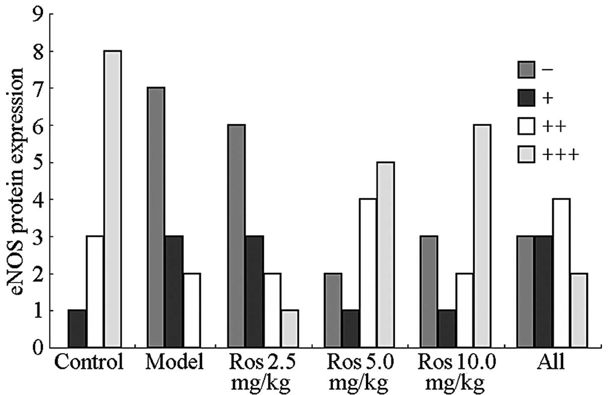
Effects of rosuvastatin on the expression
of endothelial NO synthase (eNOS) in the aortic tissues of
hyperuricemic rats
Effects of rosuvastatin on the expression level of
eNOS in the aortic tissues of the hyperuricemic rats are
demonstrated in Fig. 1.
Rosuvastatin was shown to affect the serum level of NO after two
weeks of treatment, indicating that the serum level of NO was a
fine-tuning factor in hyperuricemia and was more susceptible to
treatment compared with the other factors analyzed. To determine
the underlying mechanisms and confirm the aforementioned
conclusion, the expression level of eNOS, a rate-limiting enzyme in
NO metabolism, was determined in the aortic tissues of the rats by
an immunohistochemical assay.
Protein expression of eNOS was observed primarily in
the nucleus and partially in the cytoplasm of aortic tissue
endothelial cells. Positive expression was observed as granules
with colors ranging between yellow and brown, representing weak to
strong protein expression. Protein expression of eNOS was strongly
positive in the aortic specimens from the normal group, whereas the
expression was negative in the hyperuricemic model group (Fig. 1), correlating with the alteration
in the serum level of NO (Table
II). After six weeks of rosuvastatin treatment, the expression
level of eNOS in the aortic tissues of the hyperuricemic rats
increased gradually in a dose-dependent manner, and became fully
restored to normal levels when treated with a high dose. The effect
of rosuvastatin in improving the endothelial function of the aorta
in terms of eNOS expression was found to be significantly higher
compared with the allopurinol-treated group.
Discussion
In the present study, a hyperuricemic rat model was
generated using YEP and potassium oxonate to investigate the
pharmacological effect of rosuvastatin on the regulation of
endothelial function in hyperuricemic rats. Endothelial function
was assessed by analyzing the upregulation of UA, ET-1 and Ang II,
and the downregulation of NO, as well as the inhibition of eNOS
expression in aortic tissues.
The results of the present study support the
hypothesis that UA is a significant factor in the pathogenesis of
endothelial dysfunction, which is consistent with the results
obtained by previous studies (22,23).
A novel mechanism explaining this phenomenon was also proposed.
Xanthine dehydrogenases degrade purine metabolic products, whereas
xanthine oxidases are mainly released by vascular endothelial
cells. UA is a xanthine oxidase, and when the level of UA is
increased, xanthine oxidase-induced oxidative stress is enhanced,
generating and sequentially activating further oxygen free
radicals, including the endothelial superoxide anion, hydroxyl
radical and hydrogen peroxide. The oxygen free radicals
subsequently produce endoperoxide via the activation of epoxidase-1
and reactivate the tyrosine protein kinase receptor, leading to
acetylcholine endothelium-dependent vasoconstriction, increased
resistance, angiostatic disharmony and endothelial dysfunction
(24). In the present study, a
reduction in the levels of plasma nitrates and nitrites was
observed in the hyperuricemic rats, which was consistent with the
endothelial dysfunction and decreased NO production (25). UA can also increase renin
expression in vivo, stimulate the production of Ang II and
increase the expression of Ang II receptor type 1 in cultured
vascular smooth muscle cells. An increase in the expression levels
of UA can also increase the expression levels of ET-1 in cardiac
fibroblasts and vascular smooth muscle cells (26). An in vivo study demonstrated
that hyperuricemia may injure endothelial function via
resistin-dependent mechanisms (14).
The results of the present study indicate that
rosuvastatin may restore the expression levels of ET-1 and Ang II,
which are closely associated with endothelial function, in
hyperuricemic rats treated for two to six weeks in a dose- and
time-dependent manner. Previous studies hypothesized that an
interaction exists between ET-1 and Ang II, where ET-1 promotes the
conversion of Ang I into Ang II by reducing the activity of the
angiotensin-converting enzyme, whereas Ang II enhances the activity
of the endothelin-converting enzyme (27,28).
Statins may affect the endothelium through their antioxidant
effects, attenuating the production of Ang II-induced free radicals
in vascular smooth muscle cells by inhibiting the activity of
Rac1-mediated NAD(P)H oxidase and downregulating the expression of
the angiotensin receptor (29). In
addition, a decreased production of ET-1 may inhibit the
participation of the small G protein, RhoA (30). Takahashi et al demonstrated
that statins prevent Ang II-induced vascular remodeling and
oxidative stress, and suppress the Ang II-mediated activation of
the extracellular signal-regulated kinase 1/2 in rat mesenteric
arteries (31).
Experimental evidence has demonstrated that statins
exhibit lipid-independent effects on endothelial function (32). Animal models have also revealed
that statins can augment coronary blood flow, improve cardiac
contractile function and inhibit leukocyte-endothelial cell
interactions, primarily by enhancing the release of NO by
endothelial cells, which is mediated by eNOS. An experimental study
demonstrated the ability of statins to enhance the expression,
increase the activity or prevent the inactivation of eNOS (33).
Previous study has shown that statins can increase
the expression and activity of eNOS in vascular endothelial cells
(34). In the present study,
rosuvastatin was shown to increase the eNOS protein concentration
in hyperuricemic rat endothelial cells, and prevent the
hypoxia-dependent inhibition of eNOS synthesis. In addition, the
eNOS protein concentration increased after six weeks of
rosuvastatin treatment, particularly at a dosage of 10 mg/kg/day.
These results indicate a protective effect of rosuvastatin on the
synthesis of NO. Datar et al demonstrated that statins
inhibit endothelial-dependent vascular relaxation in the aorta and
increase the levels of reactive nitrogen and oxygen species
(35). The translocation of eNOS
to the intracellular structures may be responsible for the
increased peroxynitrite formation. The authors hypothesized that
lipophilic HMGR inhibitors may promote oxidative stress (36). Endothelium-derived NO is an
important mediator of endothelial function, and several mechanisms
have been reported for the upregulation of eNOS by statins
(35). A possible mechanism
involves the Rho/Rho-associated protein kinase signaling pathway,
through which statins increase the stability of eNOS mRNA, leading
to an increased expression of eNOS. A second important mechanism
through which statins activate eNOS is via the serine-threonine
protein kinase, Akt. Statins have been shown to rapidly promote the
activation of Akt in endothelial cells, resulting in eNOS
phosphorylation and increased angiogenesis, which is mediated by
the tyrosine phosphorylation of heat shock protein 90 that
activates eNOS (37). A third
mechanism reported hypothesizes that statins regulate the activity
of eNOS via their effects on caveolin-1. Caveolin-1 is an integral
membrane protein that binds to eNOS in caveolae, thereby directly
inhibiting the production of NO (38). The function of allopurinol, a
xanthine oxidase inhibitor, in the prevention and treatment of
hyperuricemia was limited due to the inhibitory effect of
allopurinol on the biosynthesis of UA, evident only when high
levels of UA accumulated in the blood circulation (39). In the present study, allopurinol
exhibited positive effects on endothelial function; however,
rosuvastatin was found to be more effective when compared with
allopurinol.
In conclusion, rosuvastatin was demonstrated to
reduce the serum level of UA and improve endothelial function in
hyperuricemic rats. Hyperuricemia was mainly associated with an
increase in the serum level of NO and reductions in the serum
levels of ET-1 and Ang II. The present study provides important
evidence on the clinical treatment of hyperuricemic patients with
rosuvastatin. However, further studies are required to determine
the mechanisms by which rosuvastatin reduces the serum level of UA
and improves endothelial function.
References
|
1
|
Ghisi GL, Durieux A, Pinho R and Benetti
M: Physical exercise and endothelial dysfunction. Arq Bras Cardiol.
95:e130–e137. 2010.(In English, Portuguese and Spanish).
|
|
2
|
Gao X, Martinez-Lemus LA and Zhang C:
Endothelium-derived hyperpolarizing factor and diabetes. World J
Cardiol. 3:25–31. 2011. View Article : Google Scholar : PubMed/NCBI
|
|
3
|
Oelze M, Schuhmacher S and Daiber A:
Organic nitrates and nitrate resistance in diabetes: the role of
vascular dysfunction and oxidative stress with emphasis on
antioxidant properties of pentaerithrityl tetranitrate. Exp
Diabetes Res. 2010:2131762010. View Article : Google Scholar : PubMed/NCBI
|
|
4
|
Sandoo A, van Zanten JJ, Metsios GS,
Carroll D and Kitas GD: The endothelium and its role in regulating
vascular tone. Open Cardiovasc Med J. 4:302–312. 2010.PubMed/NCBI
|
|
5
|
Franses JW, Baker AB, Chitalia VC and
Edelman ER: Stromal endothelial cells directly influence cancer
progression. Sci Transl Med. 3:66ra52011. View Article : Google Scholar : PubMed/NCBI
|
|
6
|
Barton M: Prevention and endothelial
therapy of coronary artery disease. Curr Opin Pharmacol.
13:226–241. 2013. View Article : Google Scholar : PubMed/NCBI
|
|
7
|
Mount DB: The kidney in hyperuricemia and
gout. Curr Opin Nephrol Hypertens. 22:216–223. 2013. View Article : Google Scholar : PubMed/NCBI
|
|
8
|
Bakris GL, Doghramji PP, Keenan RT and
Silber SH: CaseBook challenges: Managing gout, hyperuricemia and
comorbidities - dialogue with the experts. Am J Med. 127:S12014.
View Article : Google Scholar : PubMed/NCBI
|
|
9
|
Sautner J, Gruber J, Herold M, Zwerina J
and Leeb BF: Austrian 3e-recommendations for diagnosis and
management of gout 2013. Wien Klin Wochenschr. 126:79–89. 2014.(In
German).
|
|
10
|
Maruhashi T, Nakashima A, Soga J, et al:
Hyperuricemia is independently associated with endothelial
dysfunction in postmenopausal women but not in premenopausal women.
BMJ Open. 3:e0036592013. View Article : Google Scholar : PubMed/NCBI
|
|
11
|
Xu Y, Zhu J, Gao L, et al: Hyperuricemia
as an independent predictor of vascular complications and mortality
in type 2 diabetes patients: a meta-analysis. PLoS One.
8:e782062013. View Article : Google Scholar : PubMed/NCBI
|
|
12
|
Ndrepepa G, Cassese S, Braun S, et al: A
gender-specific analysis of association between hyperuricaemia and
cardiovascular events in patients with coronary artery disease.
Nutr Metab Cardiovasc Dis. 23:1195–1201. 2013. View Article : Google Scholar : PubMed/NCBI
|
|
13
|
Fan Y and Chen YE: Combined therapeutic
strategy to improve vascular endothelial function after
implantation of sirolimus-eluting stents. Circ J. 75:1051–1052.
2011. View Article : Google Scholar : PubMed/NCBI
|
|
14
|
Ghaffari N, Ball C, Kennedy JA, Stafford I
and Beltrame JF: Acute modulation of vasoconstrictor responses by
pravastatin in small vessels. Circ J. 75:1506–1514. 2011.
View Article : Google Scholar : PubMed/NCBI
|
|
15
|
Foody JM, Toth PP, Tomassini JE, et al:
Changes in LDL-C levels and goal attainment associated with
addition of ezetimibe to simvastatin, atorvastatin, or rosuvastatin
compared with titrating statin monotherapy. Vasc Health Risk Manag.
9:719–727. 2013. View Article : Google Scholar : PubMed/NCBI
|
|
16
|
Maji D, Shaikh S, Solanki D and Gaurav K:
Safety of statins. Indian J Endocrinol Metab. 17:636–646. 2013.
View Article : Google Scholar
|
|
17
|
Kones R: Rosuvastatin, inflammation,
C-reactive protein, JUPITER, and primary prevention of
cardiovascular disease - a perspective. Drug Des Devel Ther.
4:383–413. 2010. View Article : Google Scholar : PubMed/NCBI
|
|
18
|
Savoia C, Sisalli MJ, Di Renzo G,
Annunziato L and Scorziello A: Rosuvastatin-induced neuroprotection
in cortical neurons exposed to OGD/reoxygenation is due to nitric
oxide inhibition and ERK1/2 pathway activation. Int J Physiol
Pathophysiol Pharmacol. 3:57–64. 2011.PubMed/NCBI
|
|
19
|
Chen GL, Zhang QL, Ma XQ and Xu SY:
Hyperuricemia model induced by yeast in mice. Zhongguo Yao Li Xue
Tong Bao. 19:467–469. 2003.(In Chinese).
|
|
20
|
Guide for the care and use of laboratory
animals. 8th Edition. The National Academies Press; Washington,
D.C, USA: pp. 11–35. 2011
|
|
21
|
Xilifu D, Zhao P, Song LJ, Rehemu N and
Zhang XY: Development of secondary cardiovascular disease in
hyperuricemia model rats induced by yeast extract combined with
oteracil potassium. Zhongguo Zuzhi Gongcheng Yanjiu. 16:1994–1998.
2012.
|
|
22
|
Tomiyama H, Higashi Y, Takase B, et al:
Relationships among hyperuricemia, metabolic syndrome, and
endothelial function. Am J Hypertens. 24:770–774. 2011. View Article : Google Scholar : PubMed/NCBI
|
|
23
|
Hwa KS, Chung DM, Chung YC and Chun HK:
Hypouricemic effects of anthocyanin extracts of purple sweet potato
on potassium oxonate-induced hyperuricemia in mice. Phytother Res.
25:1415–1417. 2011.PubMed/NCBI
|
|
24
|
Wang Y and Bao X: Effects of uric acid on
endothelial dysfunction in early chronic kidney disease and its
mechanisms. Eur J Med Res. 18:262013. View Article : Google Scholar : PubMed/NCBI
|
|
25
|
Schwartz IF, Grupper A, Chernichovski T,
et al: Hyperuricemia attenuates aortic nitric oxide generation,
through inhibition of arginine transport, in rats. J Vasc Res.
48:252–260. 2011. View Article : Google Scholar : PubMed/NCBI
|
|
26
|
Zharikov SI, Swenson ER, Lanaspa M, et al:
Could uric acid be a modifiable risk factor in subjects with
pulmonary hypertension? Med Hypotheses. 74:1069–1074. 2010.
View Article : Google Scholar : PubMed/NCBI
|
|
27
|
Tsai IJ, Croft KD, Puddey IB, Beilin LJ
and Barden A: 20-Hydroxyeicosatetraenoic acid synthesis is
increased in human neutrophils and platelets by angiotensin II and
endothelin-1. Am J Physiol Heart Circ Physiol. 300:H1194–H1200.
2011. View Article : Google Scholar : PubMed/NCBI
|
|
28
|
Lagerqvist EL, Finnin BA, Pouton CW and
Haynes JM: Endothelin-1 and angiotensin II modulate rate and
contraction amplitude in a subpopulation of mouse embryonic stem
cell-derived cardiomyocyte-containing bodies. Stem Cell Res.
6:23–33. 2011. View Article : Google Scholar : PubMed/NCBI
|
|
29
|
Papageorgiou N, Tousoulis D, Antoniades C,
Giolis A, Briasoulis A and Stefanadis C: The impact of statin
administration in acute coronary syndromes. Hellenic J Cardiol.
51:250–261. 2010.PubMed/NCBI
|
|
30
|
Doi T, Sakoda T, Akagami T, et al:
Aldosterone induces interleukin-18 through endothelin-1,
angiotensin II, Rho/Rho-kinase, and PPARs in cardiomyocytes. Am J
Physiol Heart Circ Physiol. 295:H1279–H1287. 2008. View Article : Google Scholar : PubMed/NCBI
|
|
31
|
Takahashi Y, Satoh M, Tabuchi T and
Nakamura M: Prospective, randomized, single-blind comparison of
effects of 6 months’ treatment with atorvastatin versus pravastatin
on leptin and angiogenic factors in patients with coronary artery
disease. Heart Vessels. 27:337–343. 2012.
|
|
32
|
Ali F, Zakkar M, Karu K, et al: Induction
of the cytoprotective enzyme heme oxygenase-1 by statins is
enhanced in vascular endothelium exposed to laminar shear stress
and impaired by disturbed flow. J Biol Chem. 284:18882–18892. 2009.
View Article : Google Scholar : PubMed/NCBI
|
|
33
|
Lardizabal JA and Deedwania PC: The
anti-ischemic and anti-anginal properties of statins. Curr
Atheroscler Rep. 13:43–50. 2011. View Article : Google Scholar : PubMed/NCBI
|
|
34
|
Pasceri V, Willerson JT and Yeh ET: Direct
proinflammatory effect of C-reactive protein on human endothelial
cells. Circulation. 102:2165–2168. 2000. View Article : Google Scholar : PubMed/NCBI
|
|
35
|
Datar R1, Kaesemeyer WH, Chandra S, et al:
Acute activation of eNOS by statins involves scavenger receptor-B1,
G protein subunit Gi, phospholipase C and calcium influx. Br J
Pharmacol. 160:1765–1772. 2010. View Article : Google Scholar : PubMed/NCBI
|
|
36
|
Miyata R, Hiraiwa K, Cheng JC, et al:
Statins attenuate the development of atherosclerosis and
endothelial dysfunction induced by exposure to urban particulate
matter (PM10). Toxicol Appl Pharmacol. 272:1–11. 2013. View Article : Google Scholar : PubMed/NCBI
|
|
37
|
Meda C, Plank C, Mykhaylyk O, Schmidt K
and Mayer B: Effects of statins on nitric oxide/cGMP signaling in
human umbilical vein endothelial cells. Pharmacol Rep. 62:100–112.
2010. View Article : Google Scholar : PubMed/NCBI
|
|
38
|
Zhou Q and Liao JK: Pleiotropic effects of
statins. Basic research and clinical perspectives. Circ J.
74:818–826. 2010. View Article : Google Scholar : PubMed/NCBI
|
|
39
|
Yu ZF, Yang C, Qiu X, Chen Y, Kong LD and
Wang MS: The influence of daphnin on rats suffering hyperuricemia.
Zhongguo Yi Ke Da Xue Xue Bao. 33:142–145. 2002.(In Chinese).
|