Introduction
Diabetes mellitus (DM) is accompanied by a number of
complications due to the abnormal control of glycometabolism and
lipid metabolism. Diabetic cardiomyopathy (DCM), a condition
observed in diabetic individuals, is characterized by changes to
the myocardial structure and function, independent of coronary
artery disease and systemic hypertension (1,2). An
increase in the levels of blood lipoproteins and free fatty acids
facilitates the development of cardiovascular diseases, including
hyperlipidemia and coronary artery disease, which can lead to
further complications, such as retinopathy, nephropathy, neurosis,
nephrotoxicity and hyperglycemia-induced coma (3). However, the development of DCM
remains poorly understood and the underlying mechanisms have not
yet been clearly elucidated. Diabetic complications are generally
considered to be the result of oxidative stress (4), the excessive production of reactive
oxygen species (ROS) and the aberration of the antioxidant system
(5). In addition, diabetic
complications are interrelated with the inflammatory response, and
have been shown to be accelerated under a hyperglycemic state for
the production of acute response factors in fat cells (6–8).
Rutin is a phenolic compound and flavonoid glycoside
that is found in flowers and fruits as a major source. Rutin can be
broadly extracted from nature sources, including buckwheat,
oranges, grapes, lemons, limes, peaches and berries (9,10).
The compound has been reported to possess dynamic pharmacological
functions, including antioxidant, antibactericidal, antiviral
(11,12), antitumor (13), anti-inflammatory (14), myocardial protection (15) and hepatoprotective (16) effects. In addition, previous
studies have demonstrated the efficiency of the pharmacological
functions of rutin as an antioxidant (11,17,18).
In the present study, considering the potential
therapeutic properties of rutin, the aim was to investigate the
protective effects of rutin on DCM and its involvement in the
alterations of cardiac function and associated mechanisms in a rat
model of DM.
Materials and methods
Experimental animals
Two-month-old male Wistar rats were procured from
the Chinese People’s Liberation Army Military Academy of Medical
Sciences Animal Experiment Center (Beijing, China). In total, 24
male Wistar rats (weight, 70–90 g) were used for the experiment.
The animals were maintained with good ventilation and a 12-h
light/dark cycle. Prior to the experiments, the animals were
provided with food and water ad libitum. The animals were
treated in accordance with the Guide for the Care and Use of
Laboratory Animals published by the National Institutes of Health
(NIH Publication no. 85-23, revised 1996). All experiments were
approved by Institutional Animal Care and Use Committee of the
Affiliated Hospital of Qingdao University (Qingdao, China).
DM induction and rutin
administration
To induce DM, the rats were fasted for 12 h, after
which 65 mg/kg streptozotocin (STZ) dissolved in 0.1 M citrate
buffer (pH 4.5) was intraperitoneally administered. The rats were
fasted again for 12 h. At day 6 following STZ administration, the
level of blood glucose was measured by collecting whole blood from
the tail vein. Subsequently, the rats that had a blood glucose
level of >350 mg/dl were screened for further experiments. The
blood glucose level was measured using a glucometer (Accu-Chek Go
model GS; Roche Diagnostics GmbH, Mannheim, Germany).
For the experiments, the rats were divided into
three groups, which included the normal group (normal, n=8),
STZ-induced DM group (DM, n=8) and rutin-treated DM group (DM +
rutin, n=8). For the DM + rutin group, 8 mg/kg rutin dissolved in
soybean oil was orally administered at the same time every day for
one week following the induction of DM.
Hematological analysis
At 72 h following the STZ injection, blood glucose
levels were measured using a glucometer (Changsha Sinocare Inc.,
Changsha, China), following tail vein puncture blood sampling.
Serum triglyceride (TG) and total cholesterol (TC) levels were
determined using an auto-biochemical analysis system (AU2700;
Olympus, Tokyo, Japan). The body weight was recorded every day for
one week. After 12 days of rutin treatment, the experimental
animals were euthanized by CO2 inhalation.
Measurement of serum myocardial
enzymes
Blood samples were collected from the abdominal
artery and the serum was separated by centrifugation at 1,600 × g
for 10min at 4°C. The levels of creatine kinase-MB (CK-MB), lactate
dehydrogenase (LDH) and aspartate aminotransferase (AST) were
determined using an auto-biochemical analysis system (AU2700;
Olympus).
Estimation of the superoxide dismutase
(SOD) activity and malondialdehyde (MDA) level
Heart tissue samples were weighed and homogenized
(1:10, w/v) in 50 mmol/l phosphate buffer (pH 7.4). The SOD
activity and MDA level were measured using the appropriate
detection kits A001-4 for SOD and A003-1 for MDA purchased from
Nanjing Jiancheng Bioengineering Institute (Nanjing, China).
Immunohistochemical staining
Paraffin-embedded sections underwent
immunohistochemistry using a microwave-based antigen retrieval
method. The sections were incubated with primary rabbit polyclonal
anti-tumor necrosis factor-α (TNF-α; Abcam, Cambridge, MA, USA;
#ab9635; dilution: 1 μg/ml) and anti-interleukin-6 (IL-6; Abcam;
#ab6672; 1:500) antibodies overnight, and subsequently with a
corresponding biotinylated anti-rabbit (#7074) and anti-mouse IgG
(#7076) secondary antibody (Cell Signaling Technology, Inc.,
Danvers, MA, USA) for 30 min at 37°C. Negative controls were
performed with the omission of the primary antibody. The results
were viewed under a confocal FV1000 SPD laser-scanning microscope
(Olympus).
Western blot analysis
Frozen left ventricular tissue samples were
homogenized in ice-cold lysis buffer [20 mM Tris (pH 7.5), 150 mM
NaCl, 1 mM EDTA, 1 mM EGTA, 1% Triton X-100, 2.5 mM sodium
pyrophosphate, 1 mM β-glycerolphosphate, 1 mM
Na3VO4, 1 mg/ml aprotinin leupeptin and
pepstatin and 1 mM phenylmethylsulfonyl fluoride] and centrifuged
at 1,600 × g for 15 min at 4°C. A bicinchoninic acid (BCA) protein
assay (Beyotime Institute of Biotechnology, Haimen, China) was
utilized to measure the protein concentration in the supernatant.
Equal amounts of protein were used for western blot analysis, which
was performed with the following antibodies: Caspase-3 (#9661;
1:1000), Bcl-2 (#2870; 1:1,000) and BAX (#2772; 1:1000; all from
Cell Signaling Technology, Inc.), and β-actin (#sc-47778; 1:1000;
Santa Cruz Biotechnology, Inc., Dallas, TX, USA). The membrane was
incubated with a horseradish peroxidase-conjugated secondary
antibody for 1 h at 37°C. Blots were developed using an enhanced
chemiluminescence kit (Pierce Biotechnology, Inc. Rockford, IL,
USA).
Statistical analysis
Data are presented as the mean ± standard error of
the mean. SPSS software version 22 (SPSS, Inc., Chicago, IL, USA)
was used for the statistical analysis to perform one-way analysis
of variance, where P<0.05 was considered to indicate a
statistically significant difference.
Results
Rutin prevents metabolic
abnormalities
Hematological analysis revealed the metabolic
characteristics of the experimental animals (Table I). In the DM group, STZ-induced
diabetic rats exhibited a markedly lower body weight and higher
blood glucose levels when compared with the control group
(P<0.05). In addition, the heart-to-body weight ratio (HW/BW) in
the DM group was significantly (P<0.05) higher compared with the
control and rutin-treated groups, respectively (Table 1). Furthermore, rutin was shown to
significantly decrease the blood glucose levels (to similar values
to the control group) in the diabetic rats (P<0.05). In the
basal fasting state, the DM group exhibited significantly
(P<0.05) higher levels of TG and TC when compared with the
control group. However, the level of TG was decreased in the DM +
rutin group compared to the DM group. No statistically significant
difference in the TC level was observed between the DM and DM +
rutin groups.
 | Table IRutin prevents metabolic
abnormalities. |
Table I
Rutin prevents metabolic
abnormalities.
| Group | Body weight (g) | HW/BW (mg/g) | Blood glucose
(mmol/l) | TG (mmol/l) | TC (mmol/l) |
|---|
| Control | 415±16 | 2.77±0.16 | 5.3±0.3 | 0.79±0.07 | 1.26±0.07 |
| DM | 257±17a | 4.25±0.18a | 21.5±1.2a | 1.24±0.11a | 1.51±0.09a |
| DM + rutin | 345±21 | 3.32±0.19 | 9.2±1.9b | 0.93±0.04 | 1.35±0.12 |
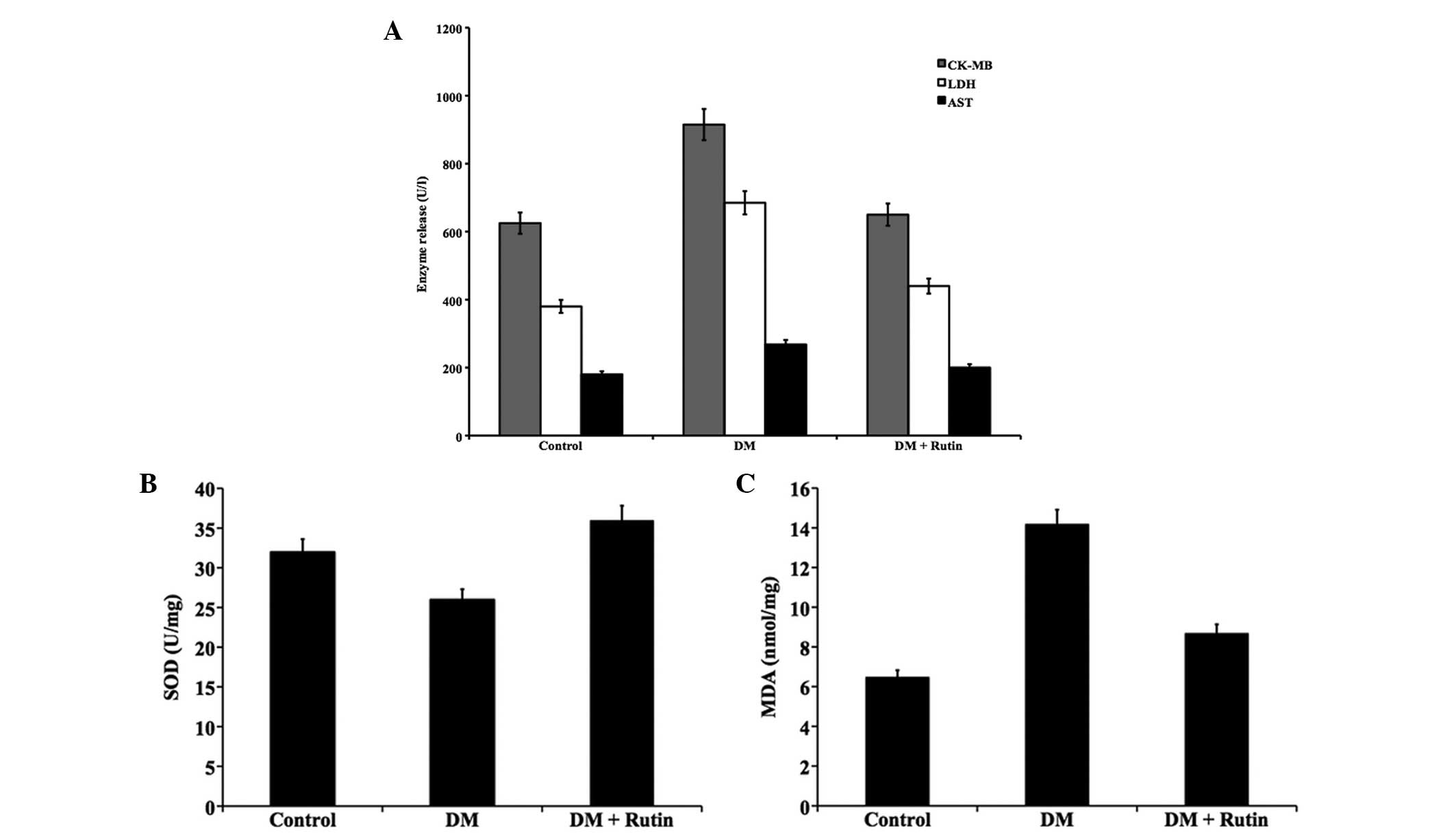
Rutin inhibits myocardial injury and
oxidative stress
The myocardial enzymes, CK-MB, LDH and AST, can be
used as biochemical indicators of myocardial injury (Fig. 1A). When compared with the control
group, the levels of the three enzymes were significantly increased
in the DM group (P<0.05). In the rutin-treated DM group,
decreased levels of myocardial enzymes were observed (P<0.05,
vs. DM group); thus, rutin was shown to protect the diabetic rats
against cardiac injury.
In the heart tissue samples of the DM group rats, a
decrease in the activity of SOD (Fig.
1B) and an increase in the accumulation of lipid peroxides with
a concordant increase in MDA content (Fig. 1C) were observed (P<0.05, vs.
control group). Following treatment with rutin in the diabetic
rats, the activity of SOD was found to be upregulated, while the
MDA content was markedly decreased (P<0.05, vs. DM group).
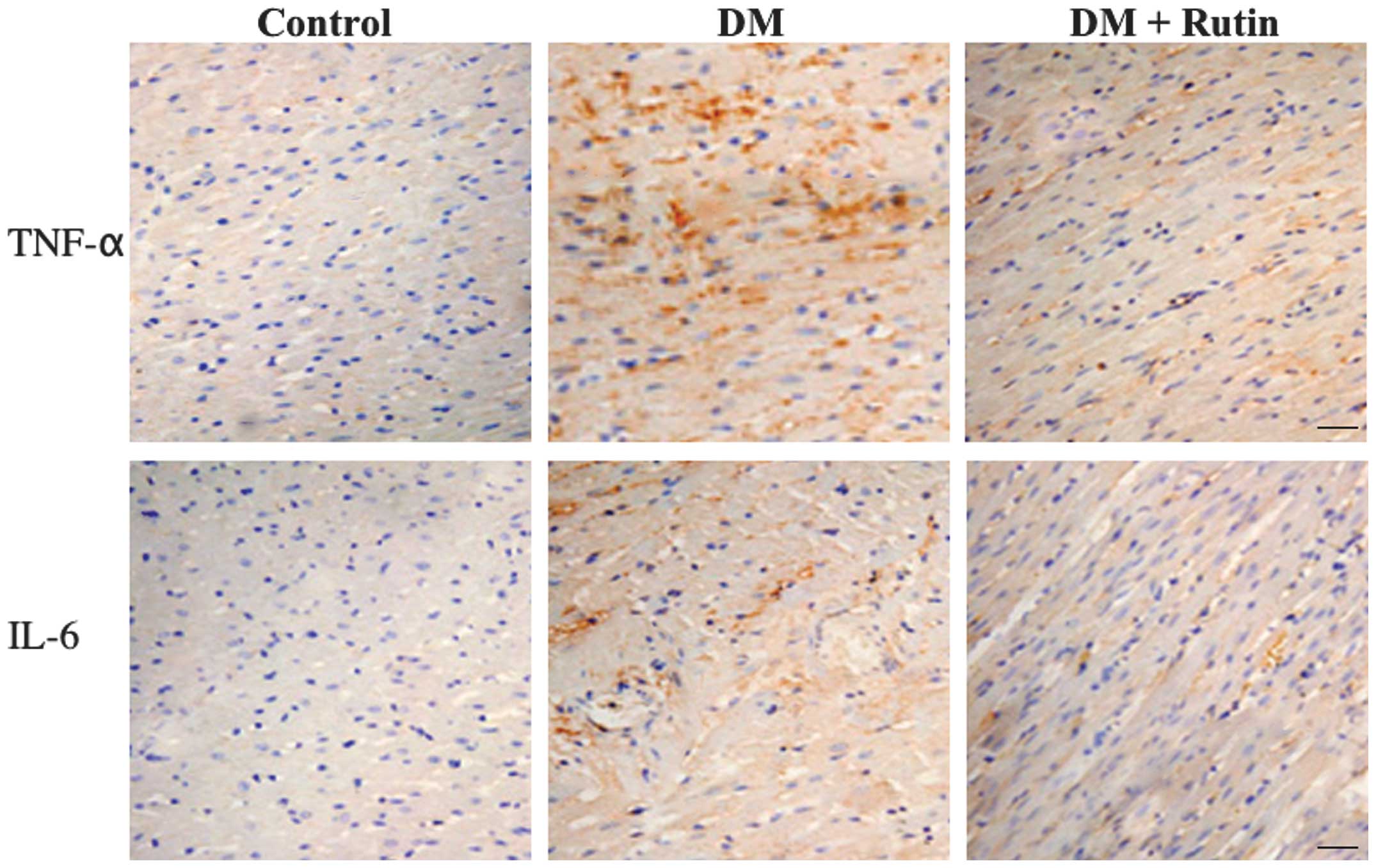
Rutin prevents the production of
inflammatory factors
Immunohistochemical analysis revealed increased
staining for the inflammatory factors, TNF-α and IL-6, in the DM
group when compared with the control group. However, decreased
levels of staining (TNF-α and IL-6) were observed in the
rutin-treated group when compared with the DM group (Fig. 2). These observations indicate the
protective effect of rutin against inflammation.
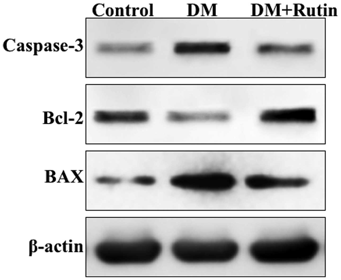
Rutin inhibits DM-induced apoptosis of
cardiomyocytes
The expression levels of the antiapoptotic protein,
Bcl-2, and proapoptotic proteins, BAX and caspase-3, were assessed
by immunoblotting. The blots revealed enhanced expression levels of
caspase-3 and BAX, but a reduced expression of Bcl-2 in the DM
group when compared with the control group. Notably, the diabetic
rats treated with rutin exhibited significantly increased protein
expression levels of Bcl-2, and downregulated protein expression
levels of caspase-3 and BAX (Fig.
3).
Discussion
The metabolic abnormalities observed in the DM
group, including the markedly higher concentrations of plasma and
serum glucose (P<0.05), were the result of insulin secretion
inhibition caused by the ROS produced by STZ. The ROS subsequently
repressed the function of the antioxidant system, while causing
oxidative damage to the pancreatic β-cells (19). Although the concentration levels of
plasma and serum glucose in the DM + rutin group were not reduced
to the same extent as that observed in the control group, the
levels were significantly decreased compared with the level in the
DM group (P<0.05). As shown in the study by Kamalakkannan and
Prince (20), the concentration
levels of plasma glucose and serum glucose decreased as rutin
removed free radicals and repressed lipid peroxidation, while
protecting the β-cells by impeding the oxidative stress caused by
STZ and increasing the level of insulin secretion. The different
forms of DM include type 1 (insulin-dependent), type 2
(non-insulin-dependent) and gestational diabetes. Using a low dose
of STZ combined with a high-energy intake is considered to be a
general strategy to obtain an animal model of type 2 DM, since
these factors simulate the real course of the human disease
(21,22). In the present study, STZ injections
were shown to be successful in inducing DM by markedly elevating
the levels of serum glucose, TG and TC. Through using this method,
the serum symptoms exhibited an increased similarity to those of
type 2 DM compared with those of type 1 DM.
DCM is classified as ventricular dysfunction with an
increased risk of cardiac failure, in the absence of hypertension,
coronary artery and valvular heart diseases (23). The condition is frequently observed
in humans and animals. Consistent with a previous study (24), the untreated DM rats in the present
study were characterized by a decreased or attenuated antioxidant
defense, as shown by the decreased SOD activity, accompanied with
increased myocardial lipid peroxidation and inactivation of
prosurvival pathways of Bcl-2, eventually culminating in cell
apoptosis and increased levels of inflammation. By contrast, the
administration of rutin was demonstrated to prevent the development
of these characteristic alterations of DCM. The beneficial effects
of rutin may be explained in part as follows. Firstly, rutin
treatment was shown to decrease the elevated levels of blood
glucose and TG. A previous study demonstrated that the onset of
cardiovascular complications may be delayed by controlling
metabolic abnormalities (25).
Secondly, rutin was found to attenuate oxidative
stress. The compound has previously been demonstrated to intercept
and neutralize ROS using its potential antioxidant function
(17). Oxidative stress is defined
as an imbalance between the production and elimination of free
radicals, which play a critical role in the development of heart
failure and left ventricular remodeling in DCM (26). Hyperglycemia has been shown to
exacerbate glucose oxidation and the generation of ROS in the
mitochondria (27), which
subsequently results in DNA damage and an accelerated rate of
apoptosis. NADPH oxidase is a critical determinant of myocardial
ROS generation (28). In the
present study, rutin was demonstrated to decrease the level of SOD
activity and reduce lipid peroxidation in a rat model of DM.
Thirdly, rutin was shown to suppress cardiac
inflammation, which is characterized by increased levels of
proinflammatory cytokines. Proinflammatory cytokines, including
IL-6 and TNF-α, are critical in the manifestation of DCM (29). Rutin has a number of properties,
including antioxidant activities, anti-inflammatory effects
(14), myocardial protection
(15) and hepatoprotective
activities (16), that enable the
suppression of cardiac inflammation. Rutin is hypothesized to exert
protective effects for various organs in DCM rats through dynamic
medical functions. In addition, according to a previous study,
antioxidants impede inflammation (30), and it is known that the
antioxidative activity and anti-inflammatory effects of rutin are
not an independent function.
In conclusion, the results demonstrated that rutin
may have great therapeutic potential in the treatment of DCM, and
possibly other cardiovascular disorders, by ameliorating metabolic
abnormalities, oxidative stress, inflammation and cellular
apoptosis pathways.
References
|
1
|
Acar E, Ural D, Bildirici U, Sahin T and
Yılmaz I: Diabetic cardiomyopathy. Anadolu Kardiyol Derg.
11:732–737. 2011.PubMed/NCBI
|
|
2
|
Battiprolu PK, Gillette TG, Wang ZV,
Lavandero S and Hill JA: Diabetic cardiomyopathy: Mechanisms and
therapeutic targets. Drug Discov Today Dis Mech. 7:e135–e143. 2010.
View Article : Google Scholar
|
|
3
|
West KM, Ahuja MM, Bennett PH, et al: The
role of circulating glucose and triglyceride concentrations and
their interactions with other ‘risk factors’ as determinants of
arterial disease in nine diabetic population samples from the WHO
multinational study. Diabetes Care. 6:361–369. 1983. View Article : Google Scholar : PubMed/NCBI
|
|
4
|
Baynes JW: Role of oxidative stress in
development of complications in diabetes. Diabetes. 40:405–412.
1991. View Article : Google Scholar : PubMed/NCBI
|
|
5
|
Coppey LJ, Gellett JS, Davidson EP, et al:
Effect of antioxidant treatment of streptozotocin-induced diabetic
rats on endoneurial blood flow, motor nerve conduction velocity,
and vascular reactivity of epineurial arterioles of the sciatic
nerve. Diabetes. 50:1927–1937. 2001. View Article : Google Scholar : PubMed/NCBI
|
|
6
|
Guha M, Bai W, Nadler JL and Natarajan R:
Molecular mechanisms of tumor necrosis factor alpha gene expression
in monocytic cells via hyperglycemia-induced oxidant
stress-dependent and -independent pathways. J Biol Chem.
275:17728–17739. 2000. View Article : Google Scholar : PubMed/NCBI
|
|
7
|
Lin Y, Rajala MW, Berger JP, et al:
Hyperglycemia-induced production of acute phase reactants in
adipose tissue. J Biol Chem. 276:42077–42083. 2001. View Article : Google Scholar : PubMed/NCBI
|
|
8
|
Aronson D, Bartha P, Zinder O, et al:
Obesity is the major determinant of elevated C-reactive protein in
subjects with the metabolic syndrome. Int J Obes Relat Metab
Disord. 28:674–679. 2004. View Article : Google Scholar : PubMed/NCBI
|
|
9
|
Kreft S, Knapp M and Kreft I: Extraction
of rutin from buckwheat (Fagopyrum esculentum Moench) seeds and
determination by capillary electrophoresis. J Agric Food Chem.
47:4649–4652. 1999. View Article : Google Scholar : PubMed/NCBI
|
|
10
|
Huang WY, Zhang HC, Liu WX and Li CY:
Survey of antioxidant capacity and phenolic composition of
blueberry, blackberry, and strawberry in Nanjing. J Zhejiang Univ
Sci B. 13:94–102. 2012. View Article : Google Scholar : PubMed/NCBI
|
|
11
|
Potter JD: Cancer prevention: epidemiology
and experiment. Cancer Lett. 114:7–9. 1997. View Article : Google Scholar : PubMed/NCBI
|
|
12
|
Rice-Evans C and Packer L: Flavonoids in
Health and Diseases. 13. Marcel Decker; New York, NY: pp. 483–522.
1998
|
|
13
|
Deschner EE, Ruperto J, Wong G and Newmark
HL: Quercetin and rutin as inhibitors of azoxymethanol-induced
colonic neoplasia. Carcinogenesis. 12:1193–1196. 1991. View Article : Google Scholar : PubMed/NCBI
|
|
14
|
Aleksandrov PN, Speranskaia TV, Bobkov
IuG, Zagorevskiĭ VA and Zykov DA: Effect of rutin and esculamine on
models of aseptic inflammation. Farmakol Toksikol. 49:84–86.
1986.(In Russian). PubMed/NCBI
|
|
15
|
Pozin VM, Skuratovskaia SG and Pocheptsova
GA: Changes in the vascular wall and ischemic damages to the
myocardium in reversible episodes of heart muscle ischemia. Fiziol
Zh. 42:10–16. 1996.(In Russian).
|
|
16
|
Janbaz KH, Saeed SA and Gilani AH:
Protective effect of rutin on paracetamol- and
CCl4-induced hepatotoxicity in rodents. Fitoterapia.
73:557–563. 2002. View Article : Google Scholar : PubMed/NCBI
|
|
17
|
Nagasawa T, Tabata N, Ito Y, et al:
Dietary G-rutin suppresses glycation in tissue proteins of
streptozotocin-induced diabetic rats. Mol Cell Biochem.
252:141–147. 2003. View Article : Google Scholar : PubMed/NCBI
|
|
18
|
Nagasawa T, Tabata N, Ito Y, et al:
Inhibition of glycation reaction in tissue protein incubations by
water soluble rutin derivative. Mol Cell Biochem. 249:3–10. 2003.
View Article : Google Scholar : PubMed/NCBI
|
|
19
|
Szkudelski T: The mechanism of alloxan and
streptozotocin action in B cells of the rat pancreas. Physiol Res.
50:537–546. 2001.
|
|
20
|
Kamalakkannan N and Prince PS:
Antihyperglycaemic and antioxidant effect of rutin, a polyphenolic
flavonoid, in streptozotocininduced diabetic wistar rats. Basic
Clin Pharmacol Toxicol. 98:97–103. 2006. View Article : Google Scholar : PubMed/NCBI
|
|
21
|
Wang HJ, Jin YX, Shen W, et al: Low dose
streptozotocin (STZ) combined with high energy intake can
effectively induce type 2 diabetes through altering the related
gene expression. Asia Pac J Clin Nutr. 16(Suppl 1): 412–417.
2007.PubMed/NCBI
|
|
22
|
Ti Y, Xie GL, Wang ZH, et al: TRB3 gene
silencing alleviates diabetic cardiomyopathy in a type 2 diabetic
rat model. Diabetes. 60:2963–2974. 2011. View Article : Google Scholar : PubMed/NCBI
|
|
23
|
Tarquini R, Lazzeri C, Pala L, Rotella CM
and Gensini GF: The diabetic cardiomyopathy. Acta Diabetol.
48:173–181. 2011. View Article : Google Scholar
|
|
24
|
Yu W, Wu J, Cai F, et al: Curcumin
alleviates diabetic cardiomyopathy in experimental diabetic rats.
PLosOne. 7:e520132012. View Article : Google Scholar
|
|
25
|
Boudina S and Abel ED: Diabetic
cardiomyopathy revisited. Circulation. 115:3213–3223. 2007.
View Article : Google Scholar : PubMed/NCBI
|
|
26
|
Rajesh M, Mukhopadhyay P, Bátkai S, et al:
Cannabidiol attenuates cardiac dysfunction, oxidative stress,
fibrosis, and inflammatory and cell death signaling pathways in
diabetic cardiomyopathy. J Am Coll Cardiol. 56:2115–2125. 2010.
View Article : Google Scholar : PubMed/NCBI
|
|
27
|
Wang J, Wang H, Hao P, et al: Inhibition
of aldehyde dehydrogenase 2 by oxidative stress is associated with
cardiac dysfunction in diabetic rats. Mol Med. 17:172–179.
2011.
|
|
28
|
Frantz S, Brandes RP, Hu K, et al: Left
ventricular remodeling after myocardial infarction in mice with
targeted deletion of the NADPH oxidase subunit gp91PHOX. Basic Res
Cardiol. 101:127–132. 2006. View Article : Google Scholar
|
|
29
|
Mano Y, Anzai T, Kaneko H, et al:
Overexpression of human C-reactive protein exacerbates left
ventricular remodeling in diabetic cardiomyopathy. Circ J.
75:1717–1727. 2011. View Article : Google Scholar : PubMed/NCBI
|
|
30
|
Cuzzocrea S, Riley DP, Caputi AP and
Salvemini D: Antioxidant therapy: a new pharmacological approach in
shock, inflammation, and ischemia/reperfusion injury. Pharmacol
Rev. 53:135–159. 2011.
|