Introduction
Goblet cells synthesize, store and secrete a complex
of high-molecular-weight glycoproteins. Mucins, included in the
glycoproteins, are essential for the maintenance of a normal
precorneal tear film and healthy ocular surface (1). It has been demonstrated that the
topical administration of rebamipide (Mucosta®), an
antiulcer agent, increases the mucin level of tear film, and
exclusively contributes to improvement of the ocular surface in
patients with dry eye (2). A study
has shown that rebamipide increases the number of goblet cells in
the bulbar conjunctiva of the rabbit in vivo (3). Rios et al demonstrated that
rebamipide led to the proliferation of cultured rat conjunctival
goblet cells (4), subsequently
stimulating secretion from the cells (5). Topical rebamipide has been shown to
significantly ameliorate corneal epithelial damage, by enhancing
the expression of mucin MUC5AC mRNA on the murine ocular
surface (6). Clinically, the
topical administration of rebamipide increases the mucin level of
tear film, and improves the ocular surface in dry eye (2). We recently revealed markedly
increased goblet cell numbers following the administration of
topical rebamipide in a patient with conjunctival dysplasia
(7). However, the mechanism by
which rebamipide exerts a strong action on goblet cell behaviors
and mucin secretion in human ocular surface remains largely unknown
how. To the best of our knowledge, the present study describes the
first case of nevus in the lacrimal caruncle showing a marked
increase in goblet cell number and secretion of mucin-like
substances following the administration of topical rebamipide.
Case report
A 62-year-old male complained of enlargement of a
pigmented lesion in the left eye. The patient was referred to Teine
Keijinkai Hospital (Sapporo, Japan) due to the observation of a
lacrimal caruncle tumor at a local clinic. The visual acuity of the
patient was 1.0 for the left eye with normal intraocular pressure.
Slit-lamp examination revealed a pigmented tumor located in the
caruncle. The right eye was normal. Since nevus or malignant
melanoma was initially suspected, an excisional biopsy was
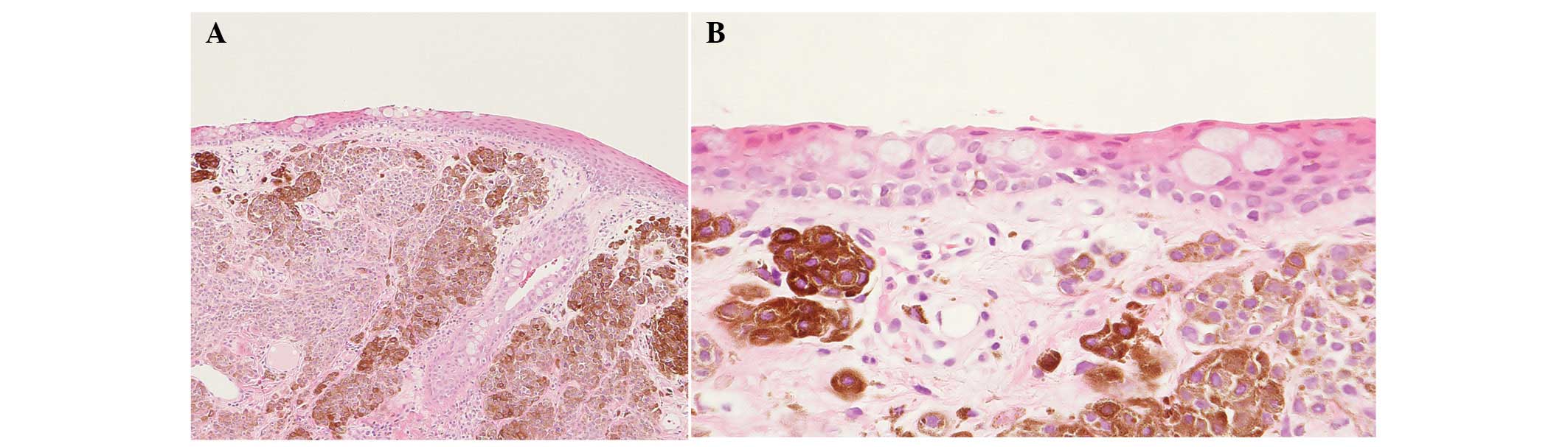
conducted on July 11, 2013. Histologically, since there were
pigmented nevus cells beneath the epithelium without cellular
atypia (Fig. 1A), the tumor was
diagnosed as pigmented nevus. A few goblet cells were noted within
the epithelium [4 cells/high power field (HPF); Fig 1B, arrows]. Mucin-like substances
were not observed on the epithelium (Fig. 1B). Nevus cells were present at the
surgical margin. The patient underwent treatment with topical
rebamipide eye drops four times a day for three months without any
other topical agents to support wound healing of the ocular
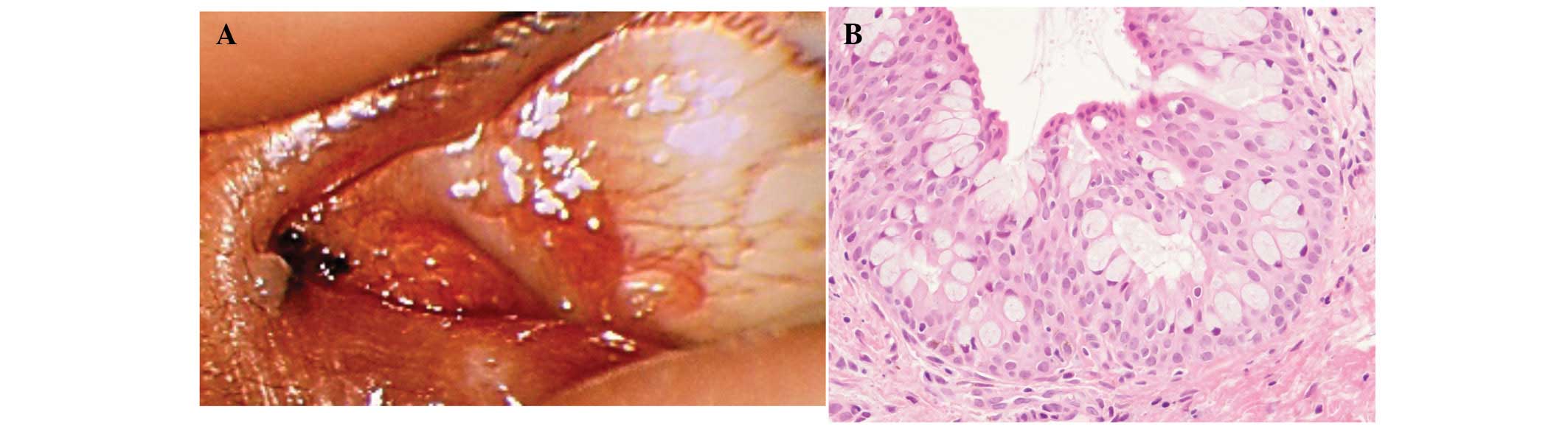
surface. Slit-lamp examination demonstrated a tiny pigmented lesion
of the left eye (Fig. 2A) three
months after the start of rebamipide treatment. After informed
consent was obtained from the patient, the pigmented lesion was
subsequently resected. Histologically, there was a small collection
of nevus cells without cellular atypia. The noncancerous epithelium
contained a large number of goblet cells (28 cells/HPF), where
marked secretion of mucin-like substances from the goblet cells was
noted (Fig. 2B). The surgical
margin was free of nevus cells. As of January 2014, the patient was
well without recurrence of the tumor. Informed consent was obtained
from the patient prior to publication of the present study and the
study was approved by the ethics committee of Hokkaido
University.
Discussion
Rebamipide has been used for patients with various
tissue injuries in not only the ocular surface but also systemic
mucosae such as the stomach. As one of its mechanisms of action,
rebamipide is considered to induce goblet cells in the tissues in
rabbit and mouse models (3,6). We
recently demonstrated that rebamipide clearly influenced mucosal
goblet cells in human conjunctival tissue (7), which verified in vitro
evidence that rebamipide led to an elevation of the number of
goblet cells in rat conjunctiva (4,5). The
histological findings in the present case, furthermore, revealed
that a 3-month administration of rebamipide resulted in not only in
an increased number of goblet cells but also large quantities of
mucin-like substances appearing on the ocular surface in a human
patient. Thus, this phenomenon is clearly due to the effect of
rebamipide and is not merely an epiphenomenon since the observed
increase in goblet cell number is in agreement with findings
obtained from animal experiments (3,6).
Clinical investigations have shown that topical
rebamipide is useful for the treatment of patients with various
corneal diseases, including dry eye (2,8),
persistent corneal erosion (9),
and lid wiper epitheliopathy (10). The present histological findings,
therefore, may support the clinical usefulness of topical
rebamipide in the ocular disorders described above. This study also
indicates that rebamipide is useful for promoting favorable tissue
repair in patients who have undergone surgery and/or biopsy in the
ocular surface.
References
|
1
|
Le QH, Wang WT, Hong JX, Sun XH, Zheng TY,
Zhu WQ and Xu JJ: An in vivo confocal microscopy and impression
cytology analysis of goblet cells in patients with chemical burns.
Invest Ophthalmol Vis Sci. 51:1397–1400. 2010. View Article : Google Scholar : PubMed/NCBI
|
|
2
|
Kinoshita S, Awamura S, Oshiden K,
Nakamichi N, Suzuki H and Yokoi N: Rebamipide (OPC-12759) in the
treatment of dry eye: a randomized, double-masked, multicenter,
placebo-controlled phase II study. Ophthalmology. 119:2471–2478.
2012. View Article : Google Scholar : PubMed/NCBI
|
|
3
|
Urashima H, Takeji Y, Okamoto T, Fujisawa
S and Shinohara H: Rebamipide increases mucin-like substance
contents and periodic acid Schiff reagent-positive cells density in
normal rabbits. J Ocul Pharmacol Ther. 28:264–270. 2012. View Article : Google Scholar : PubMed/NCBI
|
|
4
|
Rios JD, Shatos M, Urashima H, Tran H and
Dartt DA: OPC-12759 increases proliferation of cultured rat
conjunctival goblet cells. Cornea. 25:573–581. 2006. View Article : Google Scholar : PubMed/NCBI
|
|
5
|
Ríos JD, Shatos MA, Urashima H and Dartt
DA: Effect of OPC-12759 on EGF receptor activation, p44/p42 MAPK
activity, and secretion in conjunctival goblet cells. Exp Eye Res.
86:629–636. 2008. View Article : Google Scholar : PubMed/NCBI
|
|
6
|
Ohguchi T, Kojima T, Ibrahim OM, Nagata T,
Shimizu T, Shirasawa T, Kawakita T, Satake Y, Tsubota K, Shimazaki
J and Ishida S: The effects of 2% rebamipide ophthalmic solution on
the tear functions and ocular surface of the superoxide dismutase-1
(sod1) knockout mice. Invest Ophthalmol Vis Sci. 54:7793–7802.
2013. View Article : Google Scholar : PubMed/NCBI
|
|
7
|
Kase S, Shinohara T and Kase M: Effect of
topical rebamipide on human conjunctival goblet cells. JAMA
Ophthalmol. 132:1021–1022. 2014. View Article : Google Scholar : PubMed/NCBI
|
|
8
|
Koh S, Inoue Y, Sugmimoto T, Maeda N and
Nishida K: Effect of rebamipide ophthalmic suspension on optical
quality in the short break-up time type of dry eye. Cornea.
32:1219–1223. 2013. View Article : Google Scholar : PubMed/NCBI
|
|
9
|
Kashima T, Akiyama H, Miura F and Kishi S:
Resolution of persistent corneal erosion after administration of
topical rebamipide. Clin Ophthalmol. 6:1403–1406. 2012. View Article : Google Scholar : PubMed/NCBI
|
|
10
|
Itakura H, Kashima T, Itakura M, Akiyama H
and Kishi S: Topical rebamipide improves lid wiper epitheliopathy.
Clin Ophthalmol. 7:2137–2141. 2013. View Article : Google Scholar : PubMed/NCBI
|