Introduction
High blood cholesterol levels are considered to be
one of the most significant risk factors contributing to the
severity and prevalence of coronary heart disease (1,2).
Generally, a diagnosis of hyperlipidemia is confirmed in
individuals with blood cholesterol levels of >200 mg/dl or blood
triglyceride levels of >180 mg/dl. Furthermore, hyperlipemia can
be induced by the secondary effects of diabetes (3), and liver damage is often induced
under conditions of hyperlipemia, as shown by a marked increase in
the serum levels of aspartate aminotransferase (AST) and alanine
aminotransferase (ALT) (4).
In humans, atherosclerosis is a focal disease that
has been shown to evolve in a distinct pattern, resulting in
atheroma formation and vessel obstruction (5,6).
Considering the complexity of lesion development, the sequence of
events and underlying mechanisms that occur are difficult to
analyze in humans. One of the main challenges is the development of
a suitable animal model that closely imitates the human disease.
Although there is no perfect animal model, animal models can be
useful to sequentially investigate the pathological alterations,
from the initiation of the disease to the final stages of
atherosclerotic plaque development. Irrespective of the species,
the induction of vascular lesions is dependent upon
hypercholesterolemia. The elevation of plasma cholesterol levels
can be induced by a variety of methods, including dietary
supplementation, hepatic overproduction of lipoproteins or the
genetic mutation of receptors and/or receptor ligands that are
responsible for lipoprotein clearance. Previously, the golden
Syrian hamster has been successfully used to investigate vascular
changes that occur during atherogenesis (7). When compared with other animal
models, the hamster has a number of advantages. Firstly, similarly
to humans, the main plasma cholesterol carrier is low-density
lipoprotein (LDL), and lipoprotein metabolism exhibits similarities
to that of humans (8).
Furthermore, the hamster LDL receptor gene has been isolated and
characterized (9), and has been
demonstrated to have a number of similarities to the human gene. In
addition, atherosclerotic plaques develop with predilection in the
aortic arch, the aortic aspect of the sigmoid valves and the
coronary arteries, all lesion-prone areas, which allows reliable
assessment of the atherosclerotic process.
HMG-CoA reductase inhibitors have been used as
treatment for hyperlipemia (10),
and simvastatin (SIMVA) is one of the most prevalently used HMG-CoA
reductase inhibitors (11).
Therefore, SIMVA was used in the present study as a reference drug.
β-glucan is a fiber-type complex sugar (polysaccharide) derived
from the cell wall of baker’s yeast, oat and barley fiber, as well
as numerous medicinal mushrooms, including maitake. The two primary
uses of β-glucan are the enhancement of the immune system (12,13)
and to decrease the levels of blood cholesterol (14,15).
Previous clinical and animal studies have used concentrated
β-glucan preparations from oats and barley and have demonstrated
strong hypolipemic and associated anti-atherosclerosis effects on
hypercholesterolemic hamsters (16,17).
Although certain studies have demonstrated evidence of the direct
effects of β-glucan on hepatopathies (18,19),
the direct effects of β-glucan on hyperlipemic liver damage are
seldom. In addition, the effects of the β-glucan originating from
Aureobasidium on hypolipemia and associated
anti-atherosclerosis have not yet been reported. The β-glucan used
in the present study was extracted from Aureobasidium
pullulans SM-2001 (primarily β-1,3/1,6-glucans), which is a
UV-induced mutant of A. pullulans. Thus, the β-glucan is
known to demonstrate somewhat different characteristics from
β-glucan derived from other origins (20).
In the present study, the hypolipemic and associated
anti-atherosclerosis effects of polycan (β-glucan; Glucan
Corporation, Busan, Korea), originating from Aureobasidium,
were observed on a high-fat diet (HFD)-induced hamster model of
hyperlipemia, with possible effects on liver damage also assessed.
The effects were evaluated based on the serum levels of AST, ALT,
LDL, high-density lipoprotein (HDL), total cholesterol (T-CHOL) and
triglyceride, with changes in the histology and histomorphometry of
the liver and aorta (thoracic and abdominal) also analyzed
(5,21,22).
Materials and methods
Animals
In total, 30 male hamsters (age, 7 weeks; Samtako
Bio Korea Co., Ltd., Osan, Korea) were used in the study following
acclimatization for 19 days. The 30 hamsters were grouped into 6
groups. This was carried out by arranging the hamsters in order of
weight, the heaviest 6 hamsters were randomly assigned to each of
the 6 groups. The subsequent 6 heaviest hamsters were then randomly
assigned to each of the 6 groups; this division process was
continued until the 30 hamsters had been randomly assigned to the 6
groups. The animals were allocated five per polycarbonate cage in a
temperature (20–25°C) and humidity (40–45%) controlled room. A 12-h
light/dark cycle was applied, and food and water were supplied
ad libitum. The present study was approved by the ethics
committee of Daegu Haany University (Gyeongsan, Korea)
Preparations and administration of
drugs
Polycan (β-glucan extract from A. pullulans)
(20) and SIMVA (Sigma-Aldrich,
St. Louis, MO, USA) were used as test articles in the study.
Polycan was stored in a refrigerator at 4°C for protection against
light and degeneration. Polycan was diluted in distilled water and
dosed by oral gavage using a sonde attached to a 1-ml syringe,
which contained the test article at a dose of 31.25, 62.5 or 125
mg/kg in distilled water, once a day for 56 days. In addition,
SIMVA was orally administered at 10 mg/kg using distilled water as
a vehicle.
Hyperlipemia induction
To induce hyperlipemia, the animals were supplied
with free access to a HFD (Dyets, Inc., Bethlehem, PA, USA),
containing 1% cholesterol and 0.25% sodium cholate for 8 weeks,
after a 19-day acclimatization period (23–26).
The constituents of the HFD are listed in Table I. In the sham group, a normal
pellet diet (Samyang Foods Co., Ltd., Wonju, Korea) was supplied
ad libitum for the same time period.
 | Table IComposition of the high-fat diet used
in the study. |
Table I
Composition of the high-fat diet used
in the study.
| Ingredient | kcal/gm | g/kg | kcal/kg |
|---|
| Casen | 3.72 | 200 | 744 |
| DL-methionine | 4 | 3 | 12 |
| Cornstarch | 3.6 | 150 | 540 |
| Sucrose | 4 | 487.5 | 1950 |
| Cellulose | 0 | 50 | 0 |
| Corn oil | 9 | 50 | 450 |
| Mineral mix | 0.47 | 35 | 16.45 |
| Vitamin mix | 3.92 | 10 | 39.2 |
| Choline
bitartrate | 0 | 2 | 0 |
| Sodium cholate | 0 | 2.5 | 0 |
| Cholesterol | 0 | 10 | 0 |
Body weight change
Changes in the body weight of the hamsters were
calculated one day prior to drug administration (day −1), on the
day of drug administration (day 0) and at days 1, 7, 14, 21, 28,
35, 42, 49, 55 and 56 after administration of the test article and
HFD supply. On the first day of test article administration and at
sacrifice, all the experimental animals had fasted overnight (water
was not restricted) to reduce the erratum arousal from feeding. In
addition, the gain in body weight (body weight on day 56 - body
weight on day 0) was calculated.
Measurement of food consumption
Food consumption was calculated weekly during the
experimental period. The amount of food was measured prior to
supplying each cage, and the subsequent remnants were measured the
next day to calculate the difference, which was regarded as the
daily food consumption per group [mean food consumption (g/day per
animal) = daily food consumption per group/number of animals in
each group].
Liver weight changes
At sacrifice, the weight of the liver was
calculated. In order to reduce the erratum originating from
individual body weight differences, the relative weight (%) was
calculated by dividing the absolute weight by the body weight at
sacrifice and multiplying by 100.
Serum biochemistry
At sacrifice, a 2-ml sample of venous blood was
collected from the vena cava under anesthesia. All blood samples
were centrifuged at 600 × g for 10 min at room temperature using a
clotting activated serum tube. Serum AST and ALT levels were
detected with an automated blood analyzer (Toshiba 200FR; Toshiba
Medical Systems Corporation, Otawara-shi, Japan) and measured in
IU/l, using kinetic UV methods. Briefly, when the AST or ALT
enzymes reacted with the substrate, the NADH was oxidized to NAD.
By measuring the reduction of the UV absorbance of NADH, the levels
of AST or ALT were determined using the automatic blood analyzer.
Serum LDL, HDL, triglyceride and T-CHOL levels were detected with
an automated blood analyzer (AU400; Olympus Corporation, Tokyo,
Japan), in mg/dl units, using an enzyme assay.
Histopathological procedures
After measuring the liver weight, the liver and
thoracic and abdominal aorta were sampled. The sampled organs were
fixed in 10% neutral-buffered formalin. Following paraffin
embedding, 3–4-μm sections were prepared. Representative sections
were stained with hematoxylin and eosin for light microscopy
examination, following which the histological profiles of the
individual livers and aortas were observed (Eclipse 80i; Nikon
Corporation, Tokyo, Japan).
Histomorphometry
The percentage of degenerative regions (fatty
changes) in the hepatic parenchyma was calculated as the percentage
change between one randomly selected field of the liver (%/200
μm2 hepatic parenchyma) using an automated image
analysis system (analySIS Image Processing; SiS Sensoren
Instrumente Systeme GmbH, Schwentinental, Germany). The percentage
of atherosclerotic plaque regions on the aorta surface was
calculated as the percentage in a 1-mm section of the aorta surface
(%/1 mm aorta surface), using an automated image analysis system
(DMI-300; DMI, Seoul, South Korea).
Statistical analysis
All data are expressed as the mean ± standard
deviation. Statistical analyses were conducted with a
Mann-Whitney-Wilcoxon test (MW test), using SPSS software for
Windows (Release 14K; SPSS, Inc., Chicago, IL, USA). The inhibition
rate compared with the vehicle control group was calculated to aid
understanding of the efficacy of test materials on the differences
between the sham and vehicle control [percentage change vs. sham
(%) = (data of vehicle control - data of sham)/data of sham ×100]
and vehicle control and test groups [percentage change vs. vehicle
control (%) = (data of test group - data of vehicle control)/data
of vehicle control ×100]. P<0.05 was considered to indicate a
statistically significant difference.
Results
Changes to the body weight
A statistically significant (P<0.05) increase in
body weight was detected between the hamsters that were supplied
with the HFD for 49 days compared with those in the sham group that
were fed a normal diet. In addition, the body weight gain
throughout the whole experimental period significantly (P<0.05)
increased. In the SIMVA group, a non-significant decrease in the
body weight was detected from day 42 after administration, with a
statistically significant (P<0.05) decrease observed in the body
weight gain when compared with the vehicle control. However, no
statistically significant differences were detected in any of the
polycan groups (Table II) when
compared with the sham or vehicle control groups.
| Table IIChanges to the body weight (g) in the
HFD-induced hyperlipemic hamster model. |
Table II
Changes to the body weight (g) in the
HFD-induced hyperlipemic hamster model.
| | | | Polycan groups
(mg/kg) |
|---|
| | | |
|
|---|
| Time point | Sham | Control | SIMVA | 31.25 | 62.5 | 125 |
|---|
| Day -1 | 97.62±12.08 | 95.44±4.43 | 96.40±8.75 | 95.82±2.06 | 95.32±8.73 | 96.66±12.72 |
| Day 0a | 93.48±12.33 | 92.20±4.19 | 91.36±8.24 | 92.30±2.92 | 90.24±8.33 | 87.68±17.28 |
| Day 1 | 96.00±13.34 | 90.54±4.34 | 90.46±8.20 | 90.34±2.30 | 86.98±7.25 | 89.60±14.36 |
| Day 7 | 100.86±14.69 | 90.24±12.58 | 95.66±8.21 | 92.00±5.19 | 84.22±10.08 | 94.86±15.28 |
| Day 14 | 103.76±7.64 | 94.90±12.09 | 95.24±5.45 | 96.24±7.90 | 91.62±11.15 | 100.32±15.93 |
| Day 21 | 107.10±8.00 | 100.26±13.92 | 101.86±6.87 | 101.60±11.55 | 96.64±18.65 | 105.02±16.48 |
| Day 28 | 118.30±9.63 | 110.92±14.70 |
103.08±11.45d | 110.24±16.12 | 106.78±15.91 | 112.44±15.78 |
| Day 35 | 123.34±9.10 | 116.28±13.41 |
106.02±15.58d | 119.46±19.53 | 112.88±17.21 | 124.18±9.85 |
| Day 42 | 131.58±7.41 | 133.64±14.08 | 117.96±16.38 | 130.76±13.34 | 126.84±9.26 | 129.98±8.82 |
| Day 49 | 133.00±6.94 | 145.06±9.66d | 129.64±20.84 | 139.74±20.31 | 144.90±13.18 | 138.48±7.75 |
| Day 55 | 134.30±7.56 |
158.42±12.57d | 126.38±25.79 | 148.10±21.14 | 152.16±19.10 | 149.56±16.77 |
| Day 56b | 126.70±6.87 |
147.58±10.93d | 116.80±24.84 | 136.70±16.41 | 140.38±17.49 | 138.14±15.70 |
| Gainc | 33.22±10.97 | 55.38±8.95d | 25.44±18.38e | 44.40±14.60 | 50.14±15.42 | 50.46±27.13 |
In the vehicle control group, the body weight gain
throughout the experimental period was shown to increase by 66.71%
when compared with the sham group. In the SIMVA, polycan 31.25,
62.5 and 125 mg/kg groups, the changes in the body weight gain over
the experimental period were found to be −54.06, −19.83, −9.46, and
−8.88% when compared with vehicle control group, respectively.
Food consumption
Statistically significant (P<0.01 or P<0.05)
decreases were detected in food consumption when comparing all the
HFD supplied groups, including the vehicle control, with the sham
group who were fed a normal diet. However, no significant changes
were identified in food consumption when comparing the treatment
groups with the vehicle control (Table III).
| Table IIIChanges in the food consumption (g)
in the high-fat diet-induced hyperlipemic hamster model. |
Table III
Changes in the food consumption (g)
in the high-fat diet-induced hyperlipemic hamster model.
| Time points | Sham | Control | SIMVA | Polycan groups
(mg/kg) |
|---|
|
|---|
| 31.25 | 62.5 | 125 |
|---|
| Day 1 | 6.74 | 5.12 | 4.98 | 5.10 | 5.36 | 5.58 |
| Day 7 | 6.10 | 4.04 | 5.94 | 5.34 | 5.76 | 5.08 |
| Day 14 | 5.94 | 4.64 | 5.28 | 5.34 | 4.54 | 5.70 |
| Day 21 | 6.02 | 5.66 | 5.34 | 6.12 | 4.30 | 4.30 |
| Day 28 | 8.14 | 4.26 | 4.54 | 5.72 | 4.44 | 4.92 |
| Day 35 | 8.44 | 6.30 | 4.25 | 5.20 | 5.20 | 5.78 |
| Day 42 | 8.93 | 5.66 | 5.71 | 4.80 | 5.90 | 6.43 |
| Day 49 | 7.22 | 5.80 | 6.22 | 4.22 | 5.80 | 5.75 |
| Day 55 | 7.12 | 6.23 | 5.89 | 4.33 | 5.72 | 5.16 |
| Meana | 7.18±1.11 | 5.30±0.83b | 5.35±0.66b | 5.13±0.61b | 5.22±0.64b | 5.41±0.62b |
The mean daily food consumption per animal was
detected as 7.18±1.11, 5.30±0.83, 5.35±6.16, 5.13±0.61, 5.22±0.64
and 5.41±0.62 g/day per animal in the sham, vehicle control, SIMVA,
polycan 31.25, 62.5 and 125 mg/kg groups, respectively.
Changes in the liver weight
Statistically significant (P<0.01) increases were
identified in the absolute and relative liver weights when
comparing the vehicle control and the normal diet supplied sham
group. In the SIMVA group, a statistically significant (P<0.05)
increase in absolute liver weight was observed when compared with
the vehicle control. However, statistically significant (P<0.05)
decreases were observed in the absolute liver weight when comparing
the polycan 62.5 and 125 mg/kg groups with the vehicle control,
with a non-significant decrease in the 31.25 mg/kg group. In
addition, the differences between the relative liver weights in all
the polycan groups and the vehicle control group were
non-significant, but were shown to dose-dependently decrease
(Table IV).
| Table IVChanges in the absolute and relative
liver weights in the high-fat diet-induced hyperlipemic hamster
model. |
Table IV
Changes in the absolute and relative
liver weights in the high-fat diet-induced hyperlipemic hamster
model.
| Group | Absolute weight
(g) | Relative weight
(%) |
|---|
| Sham | 3.682±0.405 | 2.907±0.277 |
| Control | 6.413±1.063a | 4.372±0.876a |
| SIMVA | 6.485±1.137a | 5.605±0.562a,c |
| Polycan 31.25 | 5.356±0.797a | 3.955±0.745b |
| Polycan 62.5 | 4.939±0.571a,c | 3.560±0.582a |
| Polycan 125 | 4.828±0.427b,c | 3.530±0.479b |
In the vehicle control group, the absolute liver
weight was shown to change by 74.15% when compared with the sham
group. In the SIMVA, polycan 31.25, 62.5 and 125 mg/kg groups, the
absolute liver weight changed by 1.12, −16.49, −22.98 and −24.71%
when compared with vehicle control group, respectively. In the
vehicle control group, the relative liver weight was found to
increase by 50.37% when compared with the sham group. In addition,
the relative liver weights in the SIMVA, polycan 31.25, 62.5 and
125 mg/kg groups were found to change by 28.20, −9.53, −18.57 and
−19.25% when compared with vehicle control group, respectively.
Changes in the serum levels of AST and
ALT
Statistically significant (P<0.01) increases in
the serum levels of AST and ALT were detected when comparing the
vehicle control group with the sham group, who were supplied a
normal diet. In the SIMVA group, non-significant increases in the
serum levels of AST and ALT were detected when compared with the
vehicle control. However, statistically significant (P<0.01 or
P<0.05) and dose-dependent decreases in the serum AST and ALT
levels were demonstrated in all the polycan treatment groups when
compared with the vehicle control group (Table V).
| Table VChanges in the serum biochemistry of
the high-fat diet-induced hyperlipemic hamster model. |
Table V
Changes in the serum biochemistry of
the high-fat diet-induced hyperlipemic hamster model.
| Group | AST (IU/l) | ALT (IU/l) | Triglyceride
(mg/dl) | T-CHOL (mg/dl) | LDL (mg/dl) | HDL (mg/dl) |
|---|
| Sham | 51.80±5.01 | 61.20±7.40 | 79.20±20.89 | 99.40±17.05 | 18.60±0.89 | 65.76±15.47 |
| Control |
165.00±15.28a |
165.80±38.92a |
218.40±38.00a |
269.80±40.06a | 83.40±5.18a |
112.94±19.84b |
| SIMVA |
189.60±26.00a |
180.20±24.02a | 91.20±20.02b,c |
138.80±38.15c | 43.80±10.33a,c |
123.30±38.77b |
| Polycan 31.25 | 91.80±34.59a,c |
109.40±37.00a,d |
133.20±30.81b,c |
200.60±43.19a,d | 67.00±12.92a,d |
114.58±14.38a |
| Polycan 62.5 | 73.20±7.89a,c | 99.60±9.37a,c |
107.00±35.55c |
182.60±32.85a,d | 64.20±5.63a,c |
117.40±18.85b |
| Polycan 125 | 71.20±24.35a,c | 96.20±8.58a,c | 96.80±26.45c |
168.80±53.44b,c | 63.00±9.57a,c |
122.56±18.97a |
In the vehicle control group, the serum AST level
was shown to increase by 218.53% when comparing with the sham
group. In addition, the AST levels in the SIMVA, polycan 31.25,
62.5 and 125 mg/kg groups were demonstrated to change by 14.91,
−44.36, −55.64 and −56.85% when compared with vehicle control
group, respectively. Furthermore, in the vehicle control group, the
serum ALT level was shown to increase by 170.92% of that observed
in the sham group. In the SIMVA, polycan 31.25, 62.5 and 125 mg/kg
groups, the changes in the ALT levels were 8.69, −34.02, −39.93 and
−41.98% when compared with the vehicle control group,
respectively.
Changes in the serum levels of
triglyceride and T-CHOL
Statistically significant (P<0.01) increases in
the levels of serum triglyceride and T-CHOL were detected when
comparing the vehicle control group with the normal diet supplied
sham group. However, the serum triglyceride and T-CHOL levels in
all the test article administration groups were significantly
(P<0.01 or P<0.05) decreased when compared with the vehicle
control. In the polycan groups, evident dose-dependent decreases
were observed (Table V).
In the vehicle control group, the serum triglyceride
level was found to increase by 175.76% when compared with the sham
group. The triglyceride levels in the SIMVA, polycan 31.25, 62.5
and 125 mg/kg groups were demonstrated to change by −58.24, −39.01,
−51.01 and −55.68% when compared with the vehicle control group,
respectively. Furthermore, in the vehicle control group, the serum
T-CHOL levels were shown to increase by 171.43% when comparing with
the sham group. In the SIMVA, polycan 31.25, 62.5 and 125 mg/kg
groups, the changes in the T-CHOL levels were −48.55, −25.65,
−32.32 and −37.44% when compared with the vehicle control group,
respectively.
Changes in the serum levels of LDL and
HDL
Statistically significant (P<0.01 or P<0.05)
increases in the serum levels of LDL and HDL were detected in the
vehicle control group when compared with the sham group, who were
supplied with a normal diet. However, the serum LDL levels in all
the test article administration groups were found to significantly
(P<0.01) decrease when compared with the vehicle control group.
No statistically significant differences were identified with
regard to the serum HDL levels in all the test article
administration groups when compared with the vehicle control group.
In the polycan groups, an evident dose-dependent decrease in the
serum LDL levels was observed (Table
V).
In the vehicle control group, the serum LDL levels
were found to be 348.39% of those in the sham group. Furthermore,
the LDL levels in the SIMVA, polycan 31.25, 62.5 and 125 mg/kg
groups were shown to change by −47.48, −19.66, −23.02 and −24.46%
when compared with the vehicle control group, respectively. In the
vehicle control group, the serum HDL levels increased by 71.75%
when compared with the sham group. In the SIMVA, polycan 31.25,
62.5 and 125 mg/kg groups, the HDL levels were shown to change by
9.17, 1.45, 3.95 and 8.52% when compared with the vehicle control
group, respectively.
Changes in the histopathology and
histomorphometry of the liver
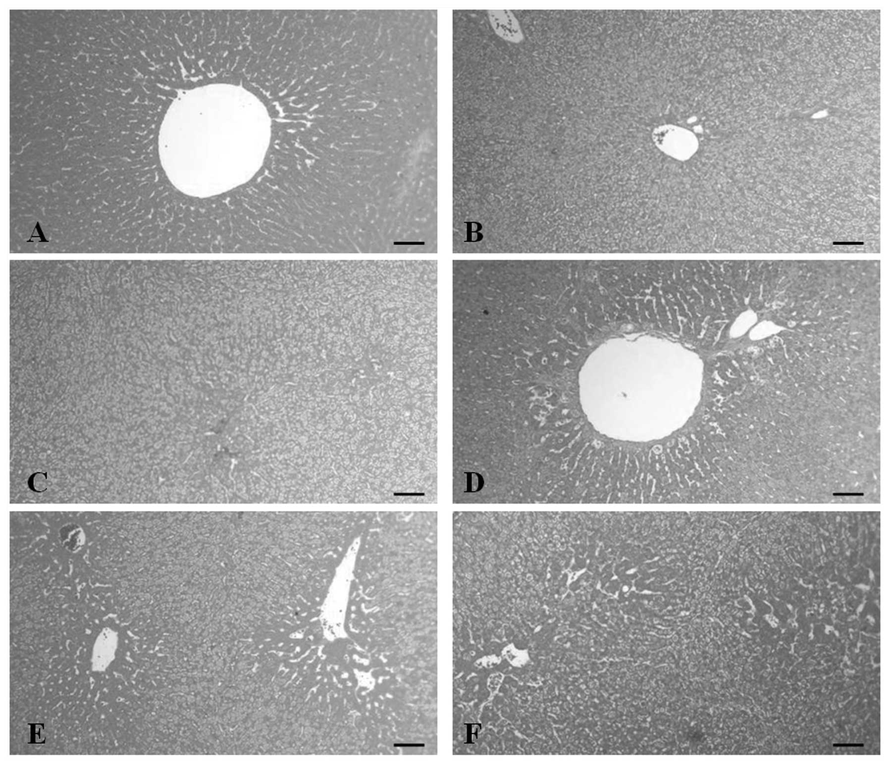
Fatty changes were detected throughout all the
hepatic lobules in all groups (Fig.
1) and similar fatty changes were detected in all the treatment
groups. In addition, the percentage of fatty changes in the hepatic
regions significantly (P<0.01) increased in the vehicle control
group when compared with the sham group. The degenerative regions
in the SIMVA group were quite similar to those in the vehicle
control group. However, statistically significant (P<0.01) and
dose-dependent decreases were observed in the percentage of regions
exhibiting fatty changes in the liver parenchyma when comparing all
the polycan groups with the vehicle control group (Table VI). In addition, the severity of
liver steatosis markedly and dose-dependently decreased in all the
polycan groups when compared with the vehicle control group
(Fig. 1).
| Table VIChanges to the histomorphometry of
the liver and aorta in the high-fat diet-induced hyperlipemic
hamster model. |
Table VI
Changes to the histomorphometry of
the liver and aorta in the high-fat diet-induced hyperlipemic
hamster model.
| Group | Fatty change
regions (%/200 μm2 liver parenchyma) | Atherosclerotic
plaques (%/1 mm aorta surface) |
|---|
|
|---|
| Abdominal
aorta | Thoracic aorta |
|---|
| Sham | 0.86±0.39 | 0.64±0.56 | 0.43±0.17 |
| Control | 93.94±2.88a | 8.12±1.89a | 8.32±2.33a |
| SIMVA | 94.61±2.73a | 1.55±0.48b,c | 1.02±0.19a,c |
| Polycan 31.25 | 84.76±4.93a,c | 4.54±1.20a,c | 2.83±0.87a,c |
| Polycan 62.5 | 77.60±8.62a,c | 2.70±1.13a,c | 2.11±0.69a,c |
| Polycan 125 | 76.61±8.74a,c | 2.40±1.36b,c | 1.60±0.93a,c |
In the vehicle control group, the percentage of
fatty change hepatic regions was shown to be 10,874.07% of those
observed in the sham group. In the SIMVA, polycan 31.25, 62.5 and
125 mg/kg groups, the percentage of degenerative regions was shown
to change by 0.71, −9.77 −17.39 and −18.45% when compared with the
vehicle control group, respectively.
Changes in the histopathology and
histomorphometry of the aorta
Atherosclerotic plaques consisting of foam cells
were detected throughout the whole aortic surface of the vehicle
control. However, the incidence of these atherosclerotic plaques in
all the treatment groups was shown to markedly decrease compared
with the vehicle control group, regardless of whether the aorta was
thoracic or abdominal (Figs. 2 and
3). In addition, the percentage of
atherosclerotic plaques on the aorta surface was significantly
(P<0.01) increased in the vehicle control group when compared
with the sham group. However, the percentage of atherosclerotic
plaques significantly (P<0.01) decreased in all the treatment
groups when compared with the vehicle control group. In the polycan
groups, an evident dose-dependent decrease was observed in the
percentage of atherosclerotic plaques (Table VI).
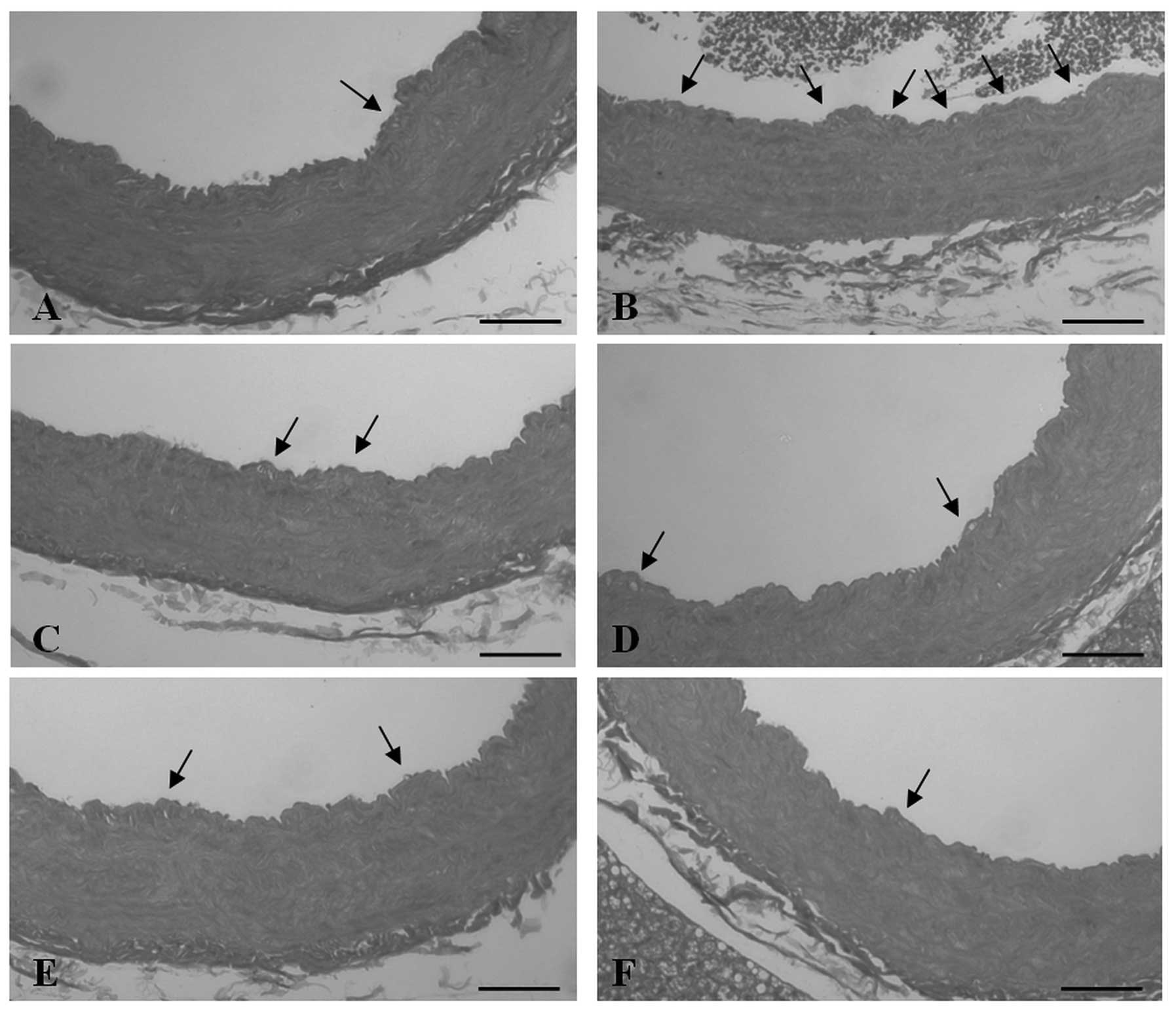
 | Figure 2Changes to the histological profiles
of the abdominal aorta in the (A) sham, (B) vehicle control, (C)
simvastatin (SIMVA), (D) polycan 31.25 mg/kg, (E) polycan 62.5
mg/kg and (F) polycan 125 mg/kg groups at sacrifice. Small
atherosclerotic plaques, consisting of foam cells, were detected in
the sham group at a low frequency; however, numerous and relatively
broad atherosclerotic plaques were detected in the tunica intima of
the vehicle control group. A marked and dose-dependent decrease was
observed in the polycan groups, and in the SIMVA group, when
compared with the vehicle control. Arrows indicate the
atherosclerotic plaques. All the images were stained with
hematoxylin and eosin (scale bars, 100 μm). |
 | Figure 3Changes to the histological profiles
of the thoracic aorta in the (A) sham, (B) vehicle control, (C)
simvastatin (SIMVA), (D) polycan 31.25 mg/kg, (E) polycan 62.5
mg/kg and (F) polycan 125 mg/kg groups at sacrifice. Small
atherosclerotic plaques, consisting of foam cells, were detected in
the sham group at a low frequency; however, numerous and relatively
broad atherosclerotic plaques were detected in the tunica intima of
the vehicle control group. A marked and dose-dependent decrease in
the number of plaques was observed in the polycan groups, and in
the SIMVA group, when compared with the vehicle control. Arrows
indicate the atherosclerotic plaques. All the images were stained
with hematoxylin and eosin (scale bars, 100 μm). |
In the vehicle control group, the percentage of
atherosclerotic plaques in the thoracic aorta was shown to be
1,826.85% greater when compared with the sham group. In the SIMVA,
polycan 31.25, 62.5 and 125 mg/kg groups, the percentage of
atherosclerotic plaques in the thoracic aorta was shown to change
by −87.77, −66.00, −74.60 and −80.80% when compared with the
vehicle control group, respectively.
In the vehicle control group, the percentage of
atherosclerotic plaques in the abdominal aorta was shown to
increase by 1,164.80% when compared with the sham group.
Furthermore, in the SIMVA, polycan 31.25, 62.5 and 125 mg/kg
groups, the percentage of plaques were shown to change by −80.96,
−44.04, −66.70 and −70.42% when compared with the vehicle control
group, respectively.
Discussion
Hypolipemic and associated anti-atherosclerosis
effects of β-glucan have been previously investigated (16,17),
with evidence demonstrating the favorable effects of β-glucan on
the hepatopathies (18,19). However, the direct effects of
β-glucan on hyperlipemic liver damage, and the effects on
hypolipemia and associated anti-atherosclerosis using the β-glucan
originating from Aureobasidium, have not yet been
determined. In the present study, the β-glucan used was extracted
from A. pullulans SM-2001 (mainly β-1,3/1,6-glucans), a
UV-induced mutant of A. pullulans that demonstrates
different characteristics compared with other types of β-glucan,
derived from various origins (20). In the present study, the
hypolipemic and associated anti-atherosclerosis effects of the test
articles were observed on a HFD-induced hyperlipemia hamster model,
with regard to assessing the possible effects on liver damage. The
effects were evaluated based on the serum levels of AST, ALT, LDL,
HDL, T-CHOL and triglyceride, with changes to histology and
histomorphometry in the liver and aorta (thoracic and abdominal)
also observed. As a result of the HFD supply, statistically
significant (P<0.01 or P<0.05) decreases were observed in
food consumption, while hyperlipemia and associated liver steatosis
(damages) markedly increased. In addition, increases were observed
in the body weight, liver weight and serum levels of AST, ALT, LDL,
HDL, triglyceride and T-CHOL in all the HFD supplied groups when
compared with the sham group who were fed a normal pellet diet.
Furthermore, severe fatty changes and atherosclerotic plaques were
observed on the liver and aorta surface, respectively, with the
percentage of fatty change regions in the liver and the percentages
of atherosclerotic plaques increasing significantly. However, the
effects on hyperlipemia were shown to markedly and dose-dependently
decrease in all the test article administration groups, with the
exception of the HDL level in the SIMVA group. No statistically
significant differences were observed in the HDL levels. More
severe hepatic damage was detected in the SIMVA group when compared
with the vehicle control group, with the SIMVA group showing
significantly increased serum AST and ALT levels. However,
statistically significant and dose-dependent decreases were
observed with regard to the hyperlipemia-associated liver damage in
the polycan treatment groups, with changes in the histology and
serum biochemistry compared with the vehicle control group. Based
on the results of the present study, polycan was demonstrated to
exert favorable effects with regard to decreasing HFD-induced
hyperlipemia and associated atherosclerosis, with relatively good
protective effects on liver damage.
An increase in body weight following hyperlipemia is
generally observed (27), and this
can be used as a type of animal model in the development of
antiobesity agents (28). Similar
to the observations of previous studies (27,29),
a significant increase was detected in the body weight of the HFD
supplied groups when compared with the sham group in the present
study. However, no statistically significant differences were
observed when comparing the body weights of the hamsters in the
polycan groups with the vehicle control group. The change detected
in the SIMVA group was considered to be a secondary effect caused
by hepatotoxicity since more substantial hepatic damage was
detected in this group compared with the vehicle control.
A decrease in food consumption was detected in all
the HFD supplied groups, which was considered to be the result of
the HFD and the time schedule of the study. In the present study,
to evaluate the preventative effects of the study treatments, the
acclimatization period to HFD was excluded. However, food
consumption did not differ among the HFD supplied groups, and the
body weight was not found to decrease compared with that in the
normal diet supplied sham group, as previously discussed (17).
In general, liver damage is accompanied with
hyperlipemia (30), and changes to
the liver weight and the serum levels of AST and ALT function as
serum markers of liver damage, which are generally monitored in
HFD-induced hyperlipemia. Although a number of materials exhibiting
hypolipemic effects have been reported to show hepatoprotective
effects, particularly in herbal extracts (30–32),
SIMVA has been demonstrated to increase the serum levels of AST and
ALT (33). In the present study,
although similar histological profiles of the liver and
histomorphometrical changes to the fatty change regions were
observed, an increase in the serum levels of AST and ALT were
detected in the SIMVA group, with an increase in liver weight also.
Thus, SIMVA was considered to aggravate the hepatic damage induced
by the HFD supply. However, the hyperlipemia-associated hepatic
damages were significantly and dose-dependently decreased in all
the polycan treatment groups, with changes observed in the liver
weight, serum AST and ALT levels, and histology and
histomorphometry of the liver parenchyma. Therefore, polycan was
considered to exert a number of favorable effects in preventing
hyperlipemia-associated hepatic damage. Previous studies have
revealed that certain antioxidants may scavenge free radicals and
inhibit lipid peroxidation; thus, treatment with several
antioxidants has been shown to protect against free radical-induced
hepatic damage (34,35). In addition to chemically
synthesized antioxidants, dietary antioxidants have also been shown
to protect against CCl4-induced lipid peroxidation
(36). Therefore, the mechanisms
underlying the hepatoprotective effects detected in the present
study were considered to be associated with the antioxidative and
free radical scavenging activities of β-glucan (37,38).
Generally, the most critical problem in hyperlipemia
is the increase in the serum levels of LDL, triglyceride and
T-CHOL, with a decrease in the HDL level (39–41).
The efficacy of hypolipemic agents is generally evaluated based on
the decrease in serum LDL, triglyceride and T-CHOL levels, and the
increase in the HDL level (42–44).
In the present study, the serum HDL levels in the vehicle control
group were shown to increase compared with the sham group, which
differs to previous studies (3,39,43).
These differences are considered to be the result of using an
animal model, as shown previously with the use of animals (45). In the present study, marked and
dose-dependent decreases were observed in the serum levels of LDL,
triglyceride and T-CHOL, which demonstrates the evident hypolipemic
effects of polycan. No statistically significant differences were
detected in the serum HDL levels compared with the vehicle control,
similar to the observations of the SIMVA group.
In addition, atherosclerotic plaques, consisting of
foam cells, have been previously detected on the aortic surface of
hyperlipemic animals (5), and
these atherosclerotic plaques have been used as index for
determining anti-atherosclerosis effects (46,47).
In the present study, the incidence of these atherosclerotic
plaques in the polycan and SIMVA treatment groups were shown to
markedly and dose-dependently decrease when compared with the
vehicle control group, regardless of whether the plaques were
identified in the thoracic or abdominal aorta. In addition, the
percentage of atherosclerotic plaques on the aorta surfaces was
shown to significantly decrease in the treatment groups when
compared with the vehicle control group. Thus, the present study
provides direct evidence that polycan exerts a number of
anti-atherosclerosis effects following HFD induction.
A number of mechanisms are hypothesized to
contribute to the ability of soluble fibers and their specific
components to lower the serum cholesterol levels. Previous studies
have demonstrated that the consumption of β-glucan inhibits the
absorption of cholesterol from the gut, as demonstrated by a
significant increase in the excretion of fecal cholesterol and
neutral sterols (48–50). Thus, the cholesterol-lowering
properties of β-glucan, at least in part, are considered to be the
result of the inhibition of cholesterol absorption from the gut. In
addition, fibers containing β-glucan have been reported to increase
the excretion of bile acids, indicating a causative role in the
decrease of the plasma cholesterol concentration (49,51,52).
Therefore, similar concentrations of bile acid, relative to the
fecal weight, may represent an increase in the bulk excretion of
bile acids. However, the present study did not evaluate fecal
output.
In conclusion, the results of the present study
demonstrated that polycan exerts favorable effects in decreasing
the extent of HFD-induced hyperlipemia and associated
atherosclerosis. In addition, polycan was shown to exert relatively
good protective effects on liver damage.
Acknowledgements
This study was fully funded by Glucan Corporation
(Busan, Korea).
References
|
1
|
Grundy SM: Cholesterol and coronary heart
disease. A new era. JAMA. 256:2849–2858. 1986. View Article : Google Scholar : PubMed/NCBI
|
|
2
|
Neaton JD, Kuller LH, Wentworth D and
Borhani NO: Total and cardiovascular mortality in relation to
cigarette smoking, serum cholesterol concentration, and diastolic
blood pressure among black and white males followed up for five
years. Am Heart J. 108:759–769. 1984. View Article : Google Scholar : PubMed/NCBI
|
|
3
|
Tan BK, Tan CH and Pushparaj PN:
Anti-diabetic activity of the semi-purified fractions of Averrhoa
bilimbi in high fat diet fed-streptozotocin-induced diabetic rats.
Life Sci. 76:2827–2839. 2005. View Article : Google Scholar : PubMed/NCBI
|
|
4
|
el-Saadany SS, el-Massry RA, Labib SM and
Sitohy MZ: The biochemical role and hypocholesterolaemic potential
of the legume Cassia fistula in hypercholesterolaemic rats.
Nahrung. 35:807–815. 1991. View Article : Google Scholar : PubMed/NCBI
|
|
5
|
Sima A, Stancu C, Constantinescu E,
Ologeanu L and Simionescu M: The hyperlipemic hamster - a model for
testing the anti-atherogenic effect of amlodipine. J Cell Mol Med.
5:153–162. 2001. View Article : Google Scholar
|
|
6
|
Stary HC: The sequence of cell and matrix
changes in atherosclerotic lesions of coronary arteries in the
first forty years of life. Eur Heart J. 11(Supple E): 3–19. 1990.
View Article : Google Scholar : PubMed/NCBI
|
|
7
|
Sima A, Bulla A and Simionescu N:
Experimental obstructive coronary atherosclerosis in the
hyperlipidemic hamster. J Submicrosc Cytol Pathol. 22:1–16.
1990.PubMed/NCBI
|
|
8
|
Sullivan MP, Cerda JJ, Robbins FL, Burgin
CW and Beatty RJ: The gerbil, hamster, and guinea pig as rodent
models for hyperlipidemia. Lab Anim Sci. 43:575–578.
1993.PubMed/NCBI
|
|
9
|
Bishop RW: Structure of the hamster low
density lipoprotein receptor gene. J Lipid Res. 33:549–557.
1992.PubMed/NCBI
|
|
10
|
Vickers S, Duncan CA, Vyas KP, Kari PH,
Arison B, Prakash SR, Ramjit HG, Pitzenberger SM, Stokker G and
Duggan DE: In vitro and in vivo biotransformation of simvastatin,
an inhibitor of HMG CoA reductase. Drug Metab Dispos. 18:476–483.
1990.PubMed/NCBI
|
|
11
|
Steinmetz EF, Buckley C, Shames ML, Ennis
TL, Vanvickle-Chavez SJ, Mao D, Goeddel LA, Hawkins CJ and Thompson
RW: Treatment with simvastatin suppresses the development of
experimental abdominal aortic aneurysms in normal and
hypercholesterolemic mice. Ann Surg. 241:92–101. 2005.
|
|
12
|
Czop JK: The role of beta-glucan receptors
on blood and tissue leukocytes in phagocytosis and metabolic
activation. Pathol Immunopathol Res. 5:286–296. 1986. View Article : Google Scholar : PubMed/NCBI
|
|
13
|
Estrada A, Yun CH, Van Kessel A, Li B,
Hauta S and Laarveld B: Immunomodulatory activities of oat
beta-glucan in vitro and in vivo. Microbiol Immunol. 41:991–998.
1997. View Article : Google Scholar : PubMed/NCBI
|
|
14
|
Bell S, Goldman VM, Bistrian BR, Arnold
AH, Ostroff G and Forse RA: Effect of beta-glucan from oats and
yeast on serum lipids. Crit Rev Food Sci Nutr. 39:189–202. 1999.
View Article : Google Scholar : PubMed/NCBI
|
|
15
|
Lia A, Hallmans G, Sandberg AS, Sundberg
B, Aman P and Andersson H: Oat beta-glucan increases bile acid
excretion and a fiber-rich barley fraction increases cholesterol
excretion in ileostomy subjects. Am J Clin Nutr. 62:1245–1251.
1995.PubMed/NCBI
|
|
16
|
Delaney B, Nicolosi RJ, Wilson TA, Carlson
T, Frazer S, Zheng GH, Hess R, Ostergren K, Haworth J and Knutson
N: Beta-glucan fractions from barley and oats are similarly
antiatherogenic in hypercholesterolemic Syrian golden hamsters. J
Nutr. 133:468–475. 2003.PubMed/NCBI
|
|
17
|
Wilson TA, Nicolosi RJ, Delaney B,
Chadwell K, Moolchandani V, Kotyla T, Ponduru S, Zheng GH, Hess R,
Knutson N, Curry L, Kolberg L, Goulson M and Ostergren K: Reduced
and high molecular weight barley beta-glucans decrease plasma total
and non-HDL-cholesterol in hypercholesterolemic Syrian golden
hamsters. J Nutr. 134:2617–2622. 2004.PubMed/NCBI
|
|
18
|
Pereira FE, Motta L and Cardoso AA:
Kupffer cell activation with BCG. Corynebacterium parvum or zymosan
protects against acute liver injury induced by carbon tetrachloride
in rats. Arq Gastroenterol. 34:157–162. 1997.PubMed/NCBI
|
|
19
|
Kutina SN and Zubakhin AA: Liver
resistance to CCl (4)-induced injury after stimulation of
macrophages with various preparations. Bull Exp Biol Med.
129:524–526. 2000. View Article : Google Scholar : PubMed/NCBI
|
|
20
|
Seo HP, Kim JM, Shin HD, Kim TK, Chang HJ,
Park BR and Lee JW: Production of β-1,3/1,6-glucan by Aureobasidium
pullulans SM-2001. Kor J Bitechnol Bioeng. 17:376–380. 2002.
|
|
21
|
Auger C, Caporiccio B, Landrault N,
Teissedre PL, Laurent C, Cros G, Besançon P and Rouanet JM: Red
wine phenolic compounds reduce plasma lipids and apolipoprotein B
and prevent early aortic atherosclerosis in hypercholesterolemic
golden Syrian hamsters (Mesocricetus auratus). J Nutr.
132:1207–1213. 2002.PubMed/NCBI
|
|
22
|
Kim HG, Une M, Kuramoto T, Noshiro M and
Fujimura K: Hypocholesterolemic effect of bile acid sulfonate
analogs in hamsters. Biol Pharm Bull. 24:218–220. 2001. View Article : Google Scholar : PubMed/NCBI
|
|
23
|
Jiang CY, Yang KM, Yang L, Miao ZX, Wang
YH and Zhu HB: A 1H NMR-based metabonomic investigation of
time-related metabolic trajectories of the plasma, urine and liver
extracts of hyperlipidemic hamsters. PLoS One. 8:e667862013.
View Article : Google Scholar :
|
|
24
|
Zhang X, Wu C, Wu H, Sheng L, et al:
Anti-hyperlipidemic effects and potential mechanisms of action of
the caffeoylquinic acid-rich Pandanus tectorius fruit extract in
hamsters fed a high fat-diet. PLoS One. 8:e619222013. View Article : Google Scholar : PubMed/NCBI
|
|
25
|
Gao H, Long Y, Jiang X, Liu Z, Wang D,
Zhao Y, Li D and Sun BL: Beneficial effects of Yerba Mate tea (Ilex
paraguariensis) on hyperlipidemia in high-fat-fed hamsters. Exp
Gerontol. 48:572–578. 2013. View Article : Google Scholar : PubMed/NCBI
|
|
26
|
Naples M, Baker C, Lino M, Iqbal J,
Hussain MM and Adeli K: Ezetimibe ameliorates intestinal
chylomicron overproduction and improves glucose tolerance in a
diet-induced hamster model of insulin resistance. Am J Physiol
Gastrointest Liver Physiol. 302:G1043–G1052. 2012. View Article : Google Scholar : PubMed/NCBI
|
|
27
|
Han LK, Zheng YN, Yoshikawa M, Okuda H and
Kimura Y: Anti-obesity effects of chikusetsusaponins isolated from
Panax japonicus rhizomes. BMC Complement Altern Med. 5:92005.
View Article : Google Scholar : PubMed/NCBI
|
|
28
|
Olsson B, Bohlooly-Y M, Fitzgerald SM,
Frick F, Ljungberg A, Ahrén B, Törnell J, Bergström G and Oscarsson
J: Bovine growth hormone transgenic mice are resistant to
diet-induced obesity but develop hyperphagia, dyslipidemia, and
diabetes on a high-fat diet. Endocrinology. 146:920–930. 2005.
View Article : Google Scholar
|
|
29
|
Yamashita J and Hayashi S: The effect of
dietary protein source on plasma cholesterol level and fecal
steroid excretion in obese mice. J Nutr Sci Vitaminol (Tokyo).
36:545–558. 1990. View Article : Google Scholar
|
|
30
|
Mukai M, Ozasa K, Hayashi K and Kawai K:
Various S-GOT/S-GPT ratios in nonviral liver disorders and related
physical conditions and life-style. Dig Dis Sci. 47:549–555. 2002.
View Article : Google Scholar : PubMed/NCBI
|
|
31
|
Hoyos M, Guerrero JM, Perez-Cano R, Olivan
J, Fabiani F, Garcia-Pergañeda A and Osuna C: Serum cholesterol and
lipid peroxidation are decreased by melatonin in diet-induced
hypercholesterolemic rats. J Pineal Res. 28:150–155. 2000.
View Article : Google Scholar : PubMed/NCBI
|
|
32
|
Lal JJ, Sreeranjit Kumar CV, Suresh MV,
Indira M and Vijayammal PL: Effect of in utero exposure of Toddy
(coconut palm wine) on liver function and lipid metabolism in rat
fetuses. Plant Foods Hum Nutr. 52:209–219. 1998. View Article : Google Scholar
|
|
33
|
Blé-Castillo JL, Rodríguez-Hernández A,
Miranda-Zamora R, Juárez-Oropeza MA and Díaz-Zagoya JC: Arthrospira
maxima prevents the acute fatty liver induced by the administration
of simvastatin, ethanol and a hypercholesterolemic diet to mice.
Life Sci. 70:2665–2673. 2002. View Article : Google Scholar : PubMed/NCBI
|
|
34
|
Campo GM, Squadrito F, Ceccarelli S, et
al: Reduction of carbon tetrachloride-induced rat liver injury by
IRFI 042, a novel dual vitamin E-like antioxidant. Free Radic Res.
34:379–393. 2001. View Article : Google Scholar : PubMed/NCBI
|
|
35
|
Mansuy D, Sassi A, Dansette PM and Plat M:
A new potent inhibitor of lipid peroxidation in vitro and in vivo,
the hepatoprotective drug anisyldithiolthione. Biochem Biophys Res
Commun. 135:1015–1021. 1986. View Article : Google Scholar : PubMed/NCBI
|
|
36
|
Taylor SL and Tappel AL: Effect of dietary
antioxidants and phenobarbital pretreatment on microsomal lipid
peroxidation and activation by carbon tetrachloride. Life Sci.
19:1151–1160. 1976. View Article : Google Scholar : PubMed/NCBI
|
|
37
|
Krizková L, Duracková Z, Sandula J,
Slamenová D, Sasinková V, Sivonová M and Krajcovic J: Fungal
beta-(1–3)-D-glucan derivatives exhibit high antioxidative and
antimutagenic activity in vitro. Anticancer Res. 23:2751–2756.
2003.
|
|
38
|
Sener G, Toklu H, Ercan F and Erkanli G:
Protective effect of beta-glucan against oxidative organ injury in
a rat model of sepsis. Int Immunopharmacol. 5:1387–1396. 2005.
View Article : Google Scholar : PubMed/NCBI
|
|
39
|
Forrester JS, Makkar R and Shah PK:
Increasing high-density lipoprotein cholesterol in dyslipidemia by
cholesteryl ester transfer protein inhibition: an update for
clinicians. Circulation. 111:1847–1854. 2005. View Article : Google Scholar : PubMed/NCBI
|
|
40
|
Kamada T, Hata J, Kusunoki H, Ito M,
Tanaka S, Kawamura Y, Chayama K and Haruma K: Eradication of
Helicobacter pylori increases the incidence of hyperlipidaemia and
obesity in peptic ulcer patients. Dig Liver Dis. 37:39–43. 2005.
View Article : Google Scholar : PubMed/NCBI
|
|
41
|
Milionis HJ, Kakafika AI, Tsouli SG,
Athyros VG, Bairaktari ET, Seferiadis KI and Elisaf MS: Effects of
statin treatment on uric acid homeostasis in patients with primary
hyperlipidemia. Am Heart J. 148:635–640. 2004. View Article : Google Scholar : PubMed/NCBI
|
|
42
|
Cheng JW: Rosuvastatin in the management
of hyperlipidemia. Clin Ther. 26:1368–1387. 2004. View Article : Google Scholar : PubMed/NCBI
|
|
43
|
Pirat B, Korkmaz ME, Eroǧlu S, Tayfun E,
Yildirir A, Uluçam M, Ozin B and Müderrisoǧlu H: The effects of
simvastatin combined with different antioxidant vitamin regimens on
serum lipid profile in patients with low HDL cholesterol levels.
Anadolu Kardiyol Derg. 4:318–322. 2004.(In Turkish). PubMed/NCBI
|
|
44
|
Zdrenghea D, Gligor E, Ossian V and Pop D:
The effect of simvastatin associated with ranitidine and alcohol
upon serum lipids. Rom J Intern Med. 42:143–148. 2004.PubMed/NCBI
|
|
45
|
Morishita S, Saito T, Mishima Y, Mizutani
A, Hirai Y, Koyama S and Kawakami M: Strains and species
differences in experimental hyperlipidemia. Nihon Yakurigaku
Zasshi. 87:259–264. 1986.(In Japanese). View Article : Google Scholar : PubMed/NCBI
|
|
46
|
Ausman LM, Rong N and Nicolosi RJ:
Hypocholesterolemic effect of physically refined rice bran oil:
studies of cholesterol metabolism and early atherosclerosis in
hypercholesterolemic hamsters. J Nutr Biochem. 16:521–529. 2005.
View Article : Google Scholar : PubMed/NCBI
|
|
47
|
Deepa PR and Varalakshmi P:
Atheroprotective effect of exogenous heparin-derivative treatment
on the aortic disturbances and lipoprotein oxidation in
hypercholesterolemic diet fed rats. Clin Chim Acta. 355:119–130.
2005. View Article : Google Scholar : PubMed/NCBI
|
|
48
|
Illman RJ and Topping DL: Effects of
dietary oat bran on faecal steroid excretion, plasma volatile fatty
acids and lipid synthesis in the rat. Nutr Res. 5:839–846. 1985.
View Article : Google Scholar
|
|
49
|
Judd PA and Truswell AS: The effects of
rolled oats on blood lipids and fecal steroid excretion in man. Am
J Clin Nutr. 34:2061–2067. 1981.PubMed/NCBI
|
|
50
|
Rieckhoff D, Trautwein EA, Mälkki Y and
Ebersdobler HF: Effects of different cereal fibers on cholesterol
and bile acid metabolism in the Syrian golden hamster. Cereal Chem.
76:788–795. 1999. View Article : Google Scholar
|
|
51
|
Anderson JW, Story L, Sieling B, Chen WJ,
Petro MS and Story J: Hypocholesterolemic effects of oat-bran or
bean intake for hypercholesterolemic men. Am J Clin Nutr.
40:1146–1155. 1984.PubMed/NCBI
|
|
52
|
Kirby RW, Anderson JW, Sieling B, Rees ED,
Chen WJ, Miller RE and Kay RM: Oat-bran intake selectively lowers
serum low-density lipoprotein cholesterol concentrations in
hypercholesterolemic men. Am J Clin Nutr. 34:824–829.
1981.PubMed/NCBI
|