Introduction
Rapid midpalatal expansion (RME) is a common
treatment for patients with a narrow maxillary dental arch. It has
been effectively used to correct transverse maxillary discrepancies
in children and adolescents up to the pubertal stage (1). The maxillary dental arch increases
rapidly with active tissue remodeling in the palate (2). Surgical assistance, such as measured
tipping of the anchor teeth, is often used to eliminate
side-effects (3). Favorable
orthopedic responses can be achieved for patients prior to and/or
during pubertal growth (4,5). Certain clinicians also use expansion
treatments to treat adult patients who have a narrow maxillary arch
with increasing skeletal resistance (6,7).
Although the effectiveness of this treatment has been reported, the
long-term dental and skeletal stability remains uncertain (8).
Retention appliances are typically used to maintain
the results of tooth movement until the architectural environment
achieves equilibrium. However, even following the retention period,
the expanded maxillary dental arch has a strong tendency to rebound
to its previous form (9). A
greater relapse tendency has been reported for midpalatal expansion
in adults due to the decreasing rate of bone regeneration.
Orthodontists and biologists have studied the mechanism of
stretch-mediated osteogenesis in the expanded suture for decades;
however, relapse continues to be unavoidable (10).
Accelerating bone formation during midpalatal
expansion would be beneficial in preventing relapse and shortening
the retention period. Oztürk et al (11) found that zoledronic acid had
positive effects on bone formation in response to expansion; it was
able to decrease the relapse rate following expansion in rats.
Transforming growth factor-β1 also been reported to stimulate bone
formation in the expanding suture (12). Results of recent animal studies
have demonstrated the potential utility of stem cell
transplantation for skeletal regeneration (13–15).
Stem cell transplantation may also shed light on midpalatal
expansion in adult patients as the impaired bone formation activity
can be attributed to the partial (16) or reduced osteo-formation
capabilities of osteoprogenitor cells in aged individuals (17).
In the present study, a rat midpalatal expansion
model was used to elucidate the mechanism underlying bone
remodeling and determine whether bone marrow cell transplantation
was able to accelerate it.
Materials and methods
Animals and grouping
A total of 48 male Sprague-Dawley (SD) rats (Vital
River Laboratory, Beijing, China) were used in this study. Their
mean weight was 208.36±7.32 g. The animal protocol was approved by
the Institutional Animal Care and Use Committee of Capital Medical
University (Beijing, China).
Three rats that did not receive any intervention
were observed as a blank control (BC) for histological morphology.
The remaining rats were divided into five groups (n=9 per group):
Non-expansion control group (NC), in which the midpalatal suture of
the rats was cut; expansion group (Exp), in which the rats
underwent midpalatal expansion for 2 weeks; expansion and
transplantation group (EaT), in which rats underwent midpalatal
expansion for 2 weeks with midpalatal incision and bone marrow
mononuclear cell (BMMC) transplantation: expansion and relapse
group (ExR); and expansion, transplantation and relapse group
(EtR). In the two relapse groups, the rats underwent midpalatal
expansion for 2 weeks with midpalatal incision without or with BMMC
transplantation, respectively, and two weeks after the removal of
the expansion appliance, the rats were sacrificed for the
observation of palatal changes. Samples from six rats from each
group were used for reverse transcription-quantitative polymerase
chain reaction (RT-qPCR) analysis and the other three were used for
histological and immunohistochemical assessment.
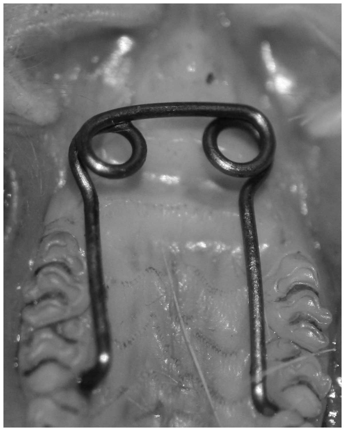
Expansion of the midpalatal suture
The rats were subjected to midpalatal expansion as
described in a previous study (18). The distal ends of the midpalatal
expansion appliance (0.45 mm stainless steel A.J. Wilcock
Australian Wire; A.J. Wilcock PTY., Ltd., Melbourne, Australia)
were placed into the interproximate space between the second and
third molars, and were activated through the ends of the
compression helices to exert an initial expansion force of 150 g
(Fig. 1). A 1.5-cm midsagittal
incision was made anteroposteriorly. Expansion appliances were
activated twice every other day to achieve maxillary expansion.
BMMC culture and transplantation
Approximately 0.5 ml bone marrow aspirate was
collected from the tibias of 2-month old BALB/C mice (Vital River
Laboratory). The marrow cells were transferred to a 15-ml conical
tube containing 1-ml Percoll (1.073 g/l; GE Healthcare, Piscataway,
NJ, USA). The tube was centrifuged at 1,500 × g for 25 min. The
mononuclear cells at the interface were collected, resuspended,
then plated at 1.0×10⁴ cells/cm2 in
Dulbecco’s modified Eagle’s medium (DMEM)/F12 medium supplemented
with 20% fetal bovine serum (Gibco Life Technologies, Carlsbad, CA,
USA) and antibiotics at 37°C in humid air with 5%
CO2.
The cells were labeled with bromodeoxyuridine (BrdU;
Sigma-Aldrich, St. Louis, MO, USA). Expanded and marked BMMCs
(1×10⁶ cells/ml, 0.5 ml) were suspended in sterile
medium, then were injected intra-orally into the masticatory muscle
area opposite the first molar 2 days after the second activation of
the expansion appliance; the expansion appliances were kept in
place for 2 weeks. A 9% sodium chloride solution (5 mg/kg) was
injected instead of labeled cells into the animals of the Exp and
ExR groups.
Posterior dentition arch width
To identify whether there was an expansion-induced
widening effect of the midpalatal suture, three inter-molar
distances of each rat were measured. This measurement was taken
three times at the end of the experimental period to calculate the
average increased posterior dentition width.
Observation of histological changes
Dissected samples, including the midpalatal suture
from the first to second molars of rats from all groups, were fixed
in 10% formalin solution for 48 h. Tissue blocks were demineralized
and embedded with paraffin routinely. Sections (5 μm) were cut and
mounted on poly-L-lysine-coated glass slides. Cut sections were
incubated at 60°C for 1 h, held in xylene and rehydrated through a
series of ethanol solutions, then stained with hematoxylin and
eosin for observation under a fluorescence microscope (Olympus
BX61; Olympus, Tokyo, Japan).
Osteogenic activities during midpalatal
suture expansion
Osteocalcin expression indicated the commencement of
active bone formation (18,19).
Immunofluorescent staining for osteocalcin was performed. The
paraffin sections were incubated with a polyclonal rabbit
anti-osteocalcin antibody (1:100 dilution; cat. no. BA1677-1,
Boster Biological Technology, Ltd., Wuhan, China) in a moist
chamber at 4°C for 18 h, followed by
tetramethyl-rhodamine-isothiocyanate-conjugated goat anti-rabbit
IgG (1:50 dilution; cat. no. BA1090, Boster Biological Technology
Co., Ltd.) at 37°C for 1 h. To visualize the cell nuclei in the
palatal tissue, 4′,6-diamidino-2-phenylindole (Molecular
Probes®, Life Technologies, Carlsbad, CA, USA) staining
was used. Fluorescent images were captured using an Olympus BX61
fluorescence microscope with an excitation wavelength of 550
nm.
Osteogenic capability of transplanted
BMMCs
The transplanted BMMCs labeled with BrdU were
identified and tracked by immunofluorescent staining with a mouse
monoclonal antibody targeting BrdU (1:100 dilution; cat. no. B8434,
Sigma-Aldrich). Double immunohistochemical staining for osteocalcin
and BrdU was carried out using DouMax Vision™ (Maixin
Biotechnology, Fuzhou, China), according to the manufacturer’s
instructions, in order to confirm the osteogenic capability of the
transplanted BMMCs in the palatal bone. The blue-black
BrdU-positive areas were visualized with
5-bromo-4-chloro-3-indolyl-phosphate/nitro-blue-tetrazolium,
whereas the salmon pink osteocalcin-positive areas were visualized
with 3-amino-9-ethylcarbazole. Sections were counterstained with
hematoxylin prior to mounting, and were visualized under an Olympus
BX61 microscope.
Receptor activator of nuclear factor κB
ligand (RANKL) and osteoprotegerin (OPG) expression in midpalatal
expansion
RANK, which is produced by osteoblasts, promotes
osteoclastogenesis when it binds to its ligand RANKL. OPG acts as a
decoy receptor, and can bind RANKL and prevent RANK signaling. The
OPG-RANK-RANKL signaling pathway has been attributed a decisive
role in the regulation of osteoblast and osteoclast activity to
ensure normal bone turnover (20).
In this experiment, RT-qPCR was performed to determine the
expression of RANKL and OPG. The tissue blocks from the first to
the second molars were harvested free of the overlying soft tissue.
Specimens were powdered under liquid nitrogen and homogenized in
TRIzol solution (Invitrogen Life Technologies, Carlsbad, CA, USA)
using a homogenizer. The yield and purity of RNA were estimated
spectrophotometrically using the ratio of absorbances at 260 and
280 nm (A260:A280). RNA was reverse transcribed to cDNA using the
SuperScript reverse transcriptase system (Invitrogen Life
Technologies) according to the manufacturer’s instructions. The
primers used are listed in Table
I. The RT-qPCR processes were run on the 7300 RT PCR detection
system (Applied Biosystems, Foster City, CA, USA), and the results
were analyzed using the software supplied with the system. The
running conditions were incubation at 50°C for 2 min and 95°C for
10 min, followed by 60 cycles of incubation at 94°C for 15 sec and
60°C for 1 min. β-actin was used as an internal control. The
expression levels of the target genes were correlated with β-actin
using the 2−ΔΔCt method. The RANKL/OPG ratio was
calculated to reflect the equilibrium between bone resorption and
formation.
 | Table IPrimer sequences used for RT-qPCR. |
Table I
Primer sequences used for RT-qPCR.
| Gene | Forward primer
sequence | Reverse primer
sequence | Accession number |
|---|
| RANKL |
5′-AGCGCTTCTCAGGAGTTCCA-3′ |
5′-GCCGGGCCACATCGA-3′ | NM_057149 |
| OPG |
5′-GCTGGCACACGAGTGATGAA-3′ |
5′-CGGTCTGCAGTTCCTTGCA-3′ | U94330.1 |
| β-actin |
5′-CTTCAACACCCCAGCCATGT-3′ |
5′-CAGAGGCATACAGGGACAACA C-3′ | NM_031144.3 |
Statistical analysis
Data are expressed as mean ± standard deviation. The
data were subjected to Fisher’s protected least significant
difference test and one-way analysis of variance. Statistical
analyses were performed using SPSS version 17.0 software (SPSS,
Inc., Chicago, IL, USA). P<0.05 was considered to indicate a
statistically significant difference.
Results
General condition of the rats
None of the rats in these groups experienced a
significant loss of body weight during the experiment. Their food
intake was disturbed during rapid expansion of the suture at the
beginning of the experiment; however, the body weights recovered
afterward.
Expanded posterior dentition arch width
in the EaT group
The posterior dentition arch width values for each
group were as follows: NC, 7.44±0.29 mm; Exp, 7.55±0.18 mm; EaT,
8.37±0.32 mm; ExR 7.33±0.21 mm; and EtR 7.69±0.19 mm. The width of
the maxillary dentition arch was expanded significantly in the EaT
group compared with that in the other four groups. The width of the
maxillary dentition arch in the EtR group was expanded
significantly more than that in the NC and ExR groups.
Midpalatal suture changes
A complete palatal shelf structure revealed by
hematoxylin and eosin staining in a normal SD rat is shown in
Fig. 2A. The chondrocytes on each
side of the suture gradually rose to mature laterally toward
bone-marrow-like cavities and the compact bone of the maxilla. Two
weeks after an incision was made in the midpalatal suture in the NC
group (Fig. 2B), the cartilage at
the midpalatal suture remained separated. Some of the mesenchymal
spindle cells had crawled along the midline of the disconnected
surface of the midpalatal suture area. In the Exp group (Fig. 2C), the chondrocytes were arranged
more laterally with mesenchymal cells migrating and bridging
one-third of the midpalatal suture 2 weeks later. In the ExR group
(Fig. 2E), the midpalatal suture
was almost bridged by a large quantity of mononuclear cells. The
chondrocytes remained evident and involved in the endochondral-type
bone formation. The palatine vessels and nerve bundles in these
groups were stretched, and the compact bone on both sides of the
midpalatal suture did not exhibit evident changes.
 | Figure 2Palatal shelf structure observed after
hematoxylin and eosin staining. BMMC transplantation accelerated
bone remodeling through transferring endochondral bone formation
into intramembraneous bone formation. Images from the (A) normal
(BC group) rats and the rats of the (B) NC, (C) Exp, (D) EaT, (E)
ExR and (F) EtR groups. BMMC, bone marrow mononuclear cell; BC,
blank control; NC, non-expansion control; Exp, expansion; EaT,
expansion and transplantation; ExR, expansion and relapse; EtR,
expansion, transplantation and relapse. |
The morphology of the midpalatal suture changed
significantly in the EaT group (Fig.
2D). All of the chondrocytes disappeared and were replaced by
fibrous-like tissues, which contained many mesenchymal cells
migrating from the submucosal tissue. The submucosal layer became
thicker and a large number of mesenchymal cells entered into the
surrounding compact palatal bone and transformed it into trabecular
bone. In the EtR group (Fig. 2F),
the suture and surrounding tissues had organized from an earlier
cancellous form (Fig. 2D) into a
mature compact structure with several small vessels.
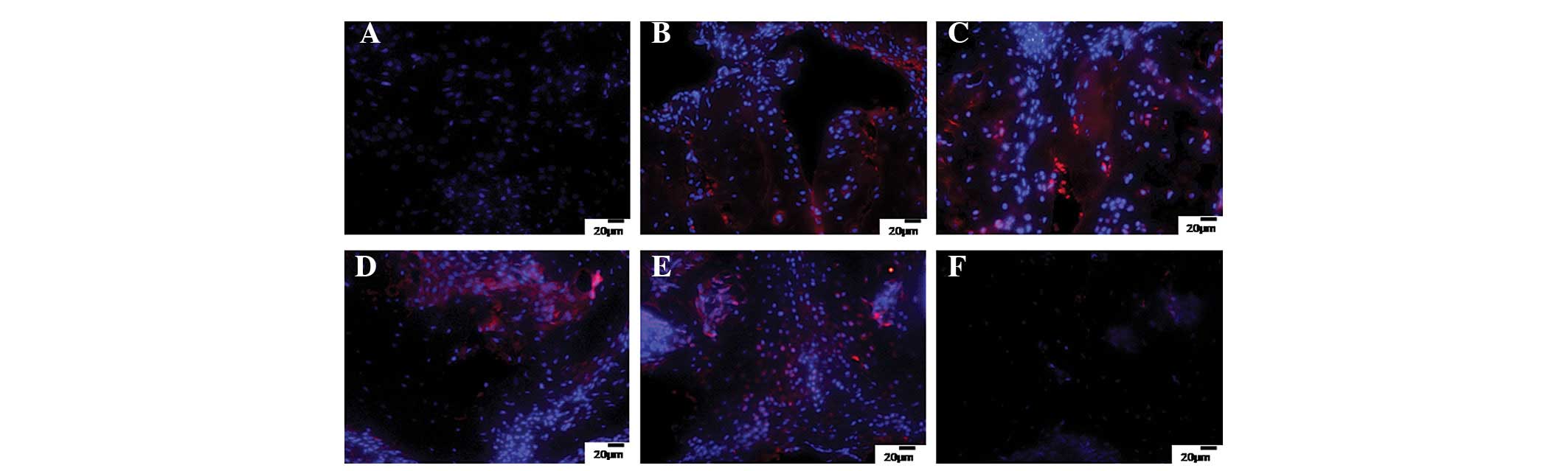
Elevated osteocalcin expression in the
EaT group
Fig. 3 exhibits the
immunofluorescent staining results of osteocalcin. In normal SD
rats (BC group), no osteocalcin expression was found in the
midpalatal suture and surrounding bone (Fig. 3A). In the NC (Fig. 3B), Exp (Fig. 3C), and ExR (Fig. 3E) groups, osteocalcin expression
was observed in the chondrocytes adjacent to the suture. The mature
chondrocytes toward the bone marrow cavity exhibited an increased
expression, indicating endochondral bone formation. The cells
migrating from the mucosa were not positive for osteocalcin after 2
weeks (Fig. 3C) but exhibited
moderate signals during the relapse period (Fig. 3E). Heterologous BMMC
transplantation in the EaT group (Fig.
3D) resulted in a large quantity of spindle cells with strong
osteocalcin expression, and when presented in the fibrous-like
tissue at the midline of the midpalatal suture area, they appeared
as intramembranous bone formation. However, no osteocalcin
expression was observed when the suture and palatal bone
transformed into a mature compact structure in the EtR group
(Fig. 3F).
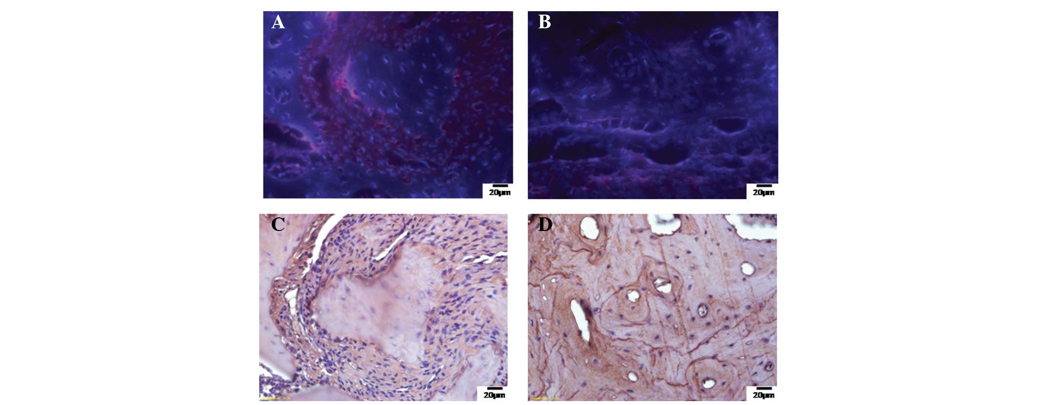
Osteogenic differentiation of BMMCs
transplanted into the midpalatal suture
The transplanted BMMCs labeled with BrdU were
tracked in the fibrous-like tissues at the suture of the EaT group
(Fig. 4A) and at the junction of
the bone and submucosal tissue in the EtR group (Fig. 4B). Double immunostaining showed
that in the EaT group (Fig. 4C),
certain proliferating cells were positive for both BrdU and
osteocalcin; the cells were adjacent to the palatal bone in the
midpalatal suture. In the EtR group (Fig. 4D), certain osteocytes in the newly
forming bone showed co-localization of BrdU and osteocalcin,
indicating that the transplanted BMMCs were involved in the new
bone formation induced by mechanical expansion.
RT-qPCR results of RANKL and OPG gene
expression
Table II shows the
ratio of RANKL/OPG expression. A significant reduction in the
RANKL/OPG ratio directly resulting from inhibition of RANKL
expression was observed in the EaT group and indicated bone
formation as the principal activity. The maximal RANKL/OPG ratio
appeared in the ExR group, indicating that bone resorption remained
evident after 4 weeks of midpalatal expansion.
| Table IIRT-qPCR analysis of RANKL/OPG
expression in midpalatal expansion (mean ± SD). |
Table II
RT-qPCR analysis of RANKL/OPG
expression in midpalatal expansion (mean ± SD).
| Group | RANKL | OPG | RANKL/OPG ratio |
|---|
| NC | 1.20±0.12a | 1.59±0.56 | 1.12±0.52 |
| Exp | 1.03±0.32a | 0.48±0.26 | 2.04±0.75 |
| EaT | 0.00±0.00 | 0.31±0.19 | 0.02±0.01 |
| ExR | 0.69±0.28a | 0.41±0.23 | 2.89±1.00 |
| EtR | 0.29±0.13 | 1.24±0.70 | 0.99±0.58 |
Discussion
Orthodontists often use midpalatal expansion to
treat malocclusion caused by a narrow maxillary dental arch. With
the aim of accelerating the speed of palatal remodeling, the
present study investigated the transplantation of BMMCs into the
intraoral muscles of rats subjected to midpalatal expansion. The
cartilage adjacent to the suture was replaced by a large quantity
of fibrous tissue after 2 weeks of expansion. This result may
explain why the distance between the molars increased to the
greatest extent in the EaT group, as a consequence of the reduction
in skeletal resistance. Heterologous BMMC transplantation
significantly reduced the extent of relapse 4 weeks after
expansion. Compared with the endochondral bone formation 2 weeks
after expansion without BMMC transplantation, intramembranous bone
formation was evident in the BMMC transplantation group. Although
intramembranous bone formation appeared in the expansion group 4
weeks later, BMMC transplantation promoted the remodeling activity
and completed midpalatal suture reconstruction with compact
bone.
A number of studies have shown the potential benefit
of bone marrow-derived mesenchymal stem cell transplantation in the
enhancement of bone repair (21–23).
Certain researchers have attributed osteopenia/osteoporosis to
allogeneic stem cell transplantation (24), whereas others have argued that
cultured bone marrow stem cells help reduce the severity of
osteoporosis and increase bone mineral density by decreasing
markers of resorption (25). In
the present study, BMMC transplantation in midpalatal sutures
accelerated the bone remodeling process over 4 weeks through
absorption into the suture cartilage. With invasion by a number of
mesenchymal cells derived from local stromal cell populations,
transplanted BMMCs healed the dissected and expanded midpalatal
suture with completely ossified bone.
Although emerging evidence suggests that bone marrow
mesenchymal stem cells may have utility for skeletal repair, the
related mechanisms remain unclear. The present study indicates that
these cells were involved directly in bone remodeling through
migration, proliferation and differentiation into osteoblasts with
osteocalcin expression. Another study has also reported that BMMC
transplantation is able to enhance bone formation indirectly by
secreting paracrine factors and recruiting perivascular stem cells
to healing wounds (12).
The maintenance of bone structure is usually
considered as a process in which a balance between osteoclastic
bone resorption and osteoblastic bone formation is achieved to
fulfill its function. The OPG-RANK-RANKL signaling pathway is a key
regulator of osteoblasts and osteoclasts to ensure normal bone
turnover (26). An increasing
RANKL/OPG ratio indicates an imbalance that favors resorption over
weight formation during bone remodeling (20). Kon et al (27) demonstrated that RANKL and OPG are
involved in fracture healing and the regulation of endochondral
resorption and bone remodeling. Bone loss is a common complication
following allogeneic stem cell transplantation, possibly through an
increased RANKL/OPG ratio in the bone microenvironment, which
results in the promotion of bone resorption activities (28). In the present study, the
temporal-spatial expression patterns of RANKL and OPG in the
midpalatal expansion groups (Exp and ExR) are in agreement with
those reported in the study by Zhu (29).
In the present study, BMMC transplantation
significantly reduced the RANKL/OPG ratio 2 weeks after midpalatal
expansion by inhibiting RANKL expression, indicating that intensive
osteogenic activities occurred prior to this time in the midpalatal
expansion group without BMMCs. The RANKL/OPG ratio recovered in the
EtR group, suggesting that osteoblast and osteoclast activities
regained equilibrium 4 weeks later, during relapse. However,
further studies are required to clarify the long-term effect of
BMMC transplantation on the whole body.
Acknowledgements
The authors would like to their colleagues for help
provided during this study. The present study was supported by the
National Natural Science Foundation of China (grant no. 81070804),
Hubei-MOST KLOS&KLOMBE (grant no. 201101) and Shandong Province
Science and Technique Foundation (grant no. 2014GSF118093).
References
|
1
|
de Gurgel JA, Malmström MF and
Pinzan-Vercelino CR: Ossification of the midpalatal suture after
surgically assisted rapid maxillary expansion. Eur J Orthod.
34:39–43. 2012. View Article : Google Scholar
|
|
2
|
Ahrari F and Eslami N: Nonsurgical
treatment of maxillary deficiency using tongue guard appliance: a
case report. J Dent Res Dent Clin Dent Prospects. 5:136–140.
2011.
|
|
3
|
Verstraaten J, Kuijpers-Jagtman AM,
Mommaerts MY, et al: A systematic review of the effects of
bone-borne surgical assisted rapid maxillary expansion. J
Craniomaxillofac Surg. 38:166–174. 2010. View Article : Google Scholar
|
|
4
|
Petrick S, Hothan T, Hietschold V, et al:
Bone density of the midpalatal suture 7 months after surgically
assisted rapid palatal expansion in adults. Am J Orthod Dentofacial
Orthop. 139(4 Suppl): S109–S116. 2011. View Article : Google Scholar : PubMed/NCBI
|
|
5
|
Wertz RA: Skeletal and dental changes
accompanying rapid midpalatal suture opening. Am J Orthod.
58:41–66. 1970. View Article : Google Scholar : PubMed/NCBI
|
|
6
|
Chung CH and Font B: Skeletal and dental
changes in the sagittal, vertical, and transverse dimensions after
rapid palatal expansion. Am J Orthod Dentofacial Orthop.
126:569–575. 2004. View Article : Google Scholar : PubMed/NCBI
|
|
7
|
Lines PA: Adult rapid maxillary expansion
with corticotomy. Am J Orthod. 67:44–56. 1975. View Article : Google Scholar : PubMed/NCBI
|
|
8
|
Lagravere MO, Major PW and Flores-Mir C:
Long-term dental arch changes after rapid maxillary expansion
treatment: a systematic review. Angle Orthod. 75:155–161.
2005.PubMed/NCBI
|
|
9
|
Vardimon AD, Brosh T, Spiegler A,
Lieberman M and Pitaru S: Rapid palatal expansion: Part 1.
Mineralization pattern of the midpalatal suture in cats. Am J
Orthod Dentofacial Orthop. 113:371–378. 1998.PubMed/NCBI
|
|
10
|
Bianchi A, Amadori S, Pironi M and
Marchetti C: Maxillary expansion and stability in the
orthodontic-surgical treatment of skeletal anterior open bites.
Prog Orthod. 10:26–37. 2009.PubMed/NCBI
|
|
11
|
Oztürk F, Babacan H, Inan S and Gümüş C:
Effects of bisphosphonates on sutural bone formation and relapse: A
histologic and immunohistochemical study. Am J Orthod Dentofacial
Orthop. 140:e31–e41. 2011. View Article : Google Scholar : PubMed/NCBI
|
|
12
|
Sawada M and Shimizu N: Stimulation of
bone formation in the expanding mid-palatal suture by transforming
growth factor-beta 1 in the rat. Eur J Orthod. 18:169–179.
1996.PubMed/NCBI
|
|
13
|
Khosla S, Westendorf JJ and Mödder UI:
Concise review: Insights from normal bone remodeling and stem
cell-based therapies for bone repair. Stem Cells. 28:2124–2128.
2010. View
Article : Google Scholar : PubMed/NCBI
|
|
14
|
Li Z, Liao W, Zhao Q, et al: Angiogenesis
and bone regeneration by allogeneic mesenchymal stem cell
intravenous transplantation in rabbit model of avascular necrotic
femoral head. J Surg Res. 183:193–203. 2013. View Article : Google Scholar : PubMed/NCBI
|
|
15
|
Li F, Wang X and Niyibizi C: Bone marrow
stromal cells contribute to bone formation following infusion into
femoral cavities of a mouse model of osteogenesis imperfecta. Bone.
47:546–555. 2010. View Article : Google Scholar : PubMed/NCBI
|
|
16
|
Quarto R, Thomas D and Liang CT: Bone
progenitor cell deficits and the age-associated decline in bone
repair capacity. Calcif Tissue Int. 56:123–129. 1995. View Article : Google Scholar : PubMed/NCBI
|
|
17
|
Liang CT, Barnes J, Seedor JG, et al:
Impaired bone activity in aged rats: alterations at the cellular
and molecular levels. Bone. 13:435–441. 1992. View Article : Google Scholar : PubMed/NCBI
|
|
18
|
Lee K, Sugiyama H, Imoto S and Tanne K:
Effects of bisphosphonate on the remodeling of rat sagittal suture
after rapid expansion. Angle Orthod. 71:265–273. 2001.PubMed/NCBI
|
|
19
|
Kobayashi ET, Hashimoto F, Kobayashi Y, et
al: Force-induced rapid changes in cell fate at midpalatal suture
cartilage of growing rats. J Dent Res. 78:1495–1504. 1999.
View Article : Google Scholar : PubMed/NCBI
|
|
20
|
Pérez-Sayáns M, Somoza-Martin JM,
Barros-Angueira F, Rey JM and García-García A: RANK/RANKL/OPG role
in distraction osteogenesis. Oral Surg Oral Med Oral Pathol Oral
Radiol Endod. 109:679–686. 2010. View Article : Google Scholar : PubMed/NCBI
|
|
21
|
Hernigou P, Poignard A, Beaujean F and
Rouard H: Percutaneous autologous bone-marrow grafting for
nonunions. Influence of the number and concentration of progenitor
cells. J Bone Joint Surg Am. 87:1430–1437. 2005. View Article : Google Scholar : PubMed/NCBI
|
|
22
|
Ma JT, Yu M, Zhang MC, et al: Clinical
observation on percutaneous autologous bone marrow grafting for
treatment of fracture nonunion. Zhongguo Gu Shang. 22:862–864.
2009.(In Chinese).
|
|
23
|
Hatzokos I, Stavridis SI, Iosifidou E,
Karataglis D and Christodoulou A: Autologous bone marrow grafting
combined with demineralized bone matrix improves consolidation of
docking site after distraction osteogenesis. J Bone Joint Surg Am.
93:671–678. 2011. View Article : Google Scholar : PubMed/NCBI
|
|
24
|
Yao S, McCarthy PL, Dunford LM, et al:
High prevalence of early-onset osteopenia/osteoporosis after
allogeneic stem cell transplantation and improvement after
bisphosphonate therapy. Bone Marrow Transplant. 41:393–398. 2008.
View Article : Google Scholar
|
|
25
|
No authors listed. The treatment of
osteoporosis through transplantation of bone marrow stem cells in
the experiments performed on rats. Georgian Med News. 204:88–92.
2012.PubMed/NCBI
|
|
26
|
Trouvin AP and Goëb V: Receptor activator
of nuclear factor-kappaB ligand and osteoprotegerin: maintaining
the balance to prevent bone loss. Clin Interv Aging. 5:345–354.
2010.
|
|
27
|
Kon T, Cho TJ, Aizawa T, et al: Expression
of osteoprotegerin, receptor activator of NF-kappaB ligand
(osteoprotegerin ligand) and related proinflammatory cytokines
during fracture healing. J Bone Miner Res. 16:1004–1014. 2001.
View Article : Google Scholar : PubMed/NCBI
|
|
28
|
Ricci P, Tauchmanova L, Risitano AM, et
al: Imbalance of the osteoprotegerin/RANKL ratio in bone marrow
microenvironment after allogeneic hemopoietic stem cell
transplantation. Transplantation. 82:1449–1456. 2006. View Article : Google Scholar : PubMed/NCBI
|
|
29
|
Zhu WQ, Wang X, Wang XX and Wang ZY:
Temporal and spatial expression of osteoprotegerin and receptor
activator of nuclear factor -kappaB ligand during mandibular
distraction in rats. J Craniomaxillofac Surg. 35:103–111. 2007.
View Article : Google Scholar : PubMed/NCBI
|