Introduction
Extract of Panax quinquefolius and
Corydalis tuber (EPC), which is primarily composed of P.
quinquefolius saponins and tetrahydropalmatine, has previously
shown efficacy in the treatment of ischemic cardiovascular diseases
in the clinic (1). In Traditional
Chinese Medicine, P. quinquefolius is known to invigorate Qi
and nourish Yin, clear fire and generate body fluid. C.
tuber, which has an acrid taste, activates blood circulation
and regulates Qi to alleviate pain (2,3).
These two ingredients accentuate each other. Our recent study
revealed that, following myocardial infarction (MI), EPC exerted
significant protective effects against oxidative stress injury in
the myocardium by increasing superoxide dismutase activity and
decreasing levels of 8-iso-prostaglandin F2α (1). Furthermore, moderate-to-high doses of
EPC significantly decreased the mRNA and protein expression of
78-kDa glucose-regulated protein and C/EBP-homologous protein when
compared with the control group, indicating that EPC could
alleviate injury to the myocardium following MI by suppressing
excessive endoplasmic reticulum stress (1). In another study, EPC treatment
significantly inhibited ERS and oxidatedive stress, balanced the
Bcl-2/bax ratio, suppressed the activation of caspase-3 and exerted
anti-apoptotic effects in pigs with larger anterior wall AMI
(4).
Abnormalities in the coagulation and fibrinolytic
system and platelet activation are the principal pathophysiological
features of coronary heart disease, particularly for MI (5). The aim of the present study was to
investigate the hypothesis that EPC acts to protect against MI by
inhibiting platelet activation and improving the hypercoagulable
state, in order to elucidate part of the pharmacological mechanism
of EPC.
Materials and methods
Animals
One hundred male Wistar rats, weighing 180±20 g,
were purchased from the Institute of Laboratory Animal Sciences,
Chinese Academy of Medical Sciences (certificate no. SCXK Beijing
2005–0013; Beijing, China). All rats were housed in
humidity-controlled rooms (55±5%) at 22±2°C with a 12-h light/dark
cycle and were fed standard rat chow. All animals were cared for in
accordance with the policies and guidelines released by the Animal
Care and Ethics Committee of the China Academy of Chinese Medical
Sciences (Beijing, China).
Preparation of EPC
EPC was provided by the Institute of Chinese Materia
Medica, China Academy of Chinese Medical Sciences. The main active
components of the extract are shown in Table I, as measured by the
high-performance liquid chromatography (HPLC) method (1,6). The
main active components of the extract were separated using a
Kromasil 100-5C18 column (4 μml 4.5×150 mm; EKA Nobel,
Bohus, Sweden) at a flow rate of 1.0 μl/l. Gradient elution was
performed, at a ratio of 81:19 (A:B, v/v, A:0.01 mol/l sodium
dihydrogen phosphate and disodium gydrogen phosphate, B:
acetonitrile).
 | Table IQuality evaluation of extract of
Panax quinquefolius and Corydalis tuber. |
Table I
Quality evaluation of extract of
Panax quinquefolius and Corydalis tuber.
| Major
constituent | Content (%) |
|---|
| Ginsenoside Rg1 | 0.11 |
| Ginsenoside Re | 1.88 |
| Ginsenoside Rb1 | 5.30 |
|
Tetrahydropalmatine | 0.07 |
Animal model establishment and
grouping
Ten rats were randomly selected as a sham group, and
the remaining rats were randomly divided into five groups: Control,
metoprolol, low-dose EPC, moderate-dose EPC and high-dose EPC
(n=18/group). Anesthesia was induced with an intraperitoneal
injection of urethane solution (20%) at a dose of 0.6 ml/kg. The
left anterior descending coronary artery (LAD) was ligated to
establish the MI model according to the methods of Olivetti, as
described previously (7–9). The rats in the sham group did not
undergo ligation.
Treatment methods
Following the MI surgery, group-specific treatments
were given to the surviving rats. The metoprolol group was
administered metoprolol (9 mg/kg; batch no. 1012055; AstraZeneca
Pharmaceutical Co., Ltd., London, UK) and the low-, moderate- and
high-dose EPC groups were administered EPC at doses of 0.54, 1.08
and 2.16 g/kg, respectively, by gastrogavage once every 24 h for
two weeks. An equal volume of normal saline was given to the sham
and control groups. The rats were sacrificed at the end of the 2
week period using intraperitoneal injection of 20% urethane
Pathomorphological analysis
Two left ventricular myocardia from each group were
selected randomly following blood collection, fixed in 10% neutral
formalin buffer and paraffin-embedded subsequent to dehydration to
carry out the hematoxylin and eosin (HE) staining. The other
ischemic hearts were rinsed in normal saline to remove the blood,
cut into four pieces parallel to the coronary sulcus, and then
incubated in 10% nitroblue tetrazolium (NBT; 50 mg/100 ml; Sigma,
St. Louis, MO, USA) at 37°C for 10 min. The infarct size was
quantified using Image Pro Plus software (version 4.0; Media
Cybernetics, Inc., Rockville, MD, USA), and was expressed as the
proportion of infarct in the left ventricular.
Enzyme-linked immunosorbent assay (ELISA)
for the detection of serum levels of von Willebrand factor (vWF),
D-dimer (DD), platelet membrane glycoproteins IIb-IIIa (GPIIb-IIIa)
and CD62P
The serum levels of vWF, DD, GPIIb-IIIa and CD62P
were detected using ELISA with kits provided by the Sino-American
Biotechnology Co., Ltd. (Wuhan, China), according to the
manufacturer’s instructions. A Multiskan™ MK3 microplate reader
(Thermo Fisher Scientific, Inc., Waltham, MA, USA) was used for the
detection.
Statistical analysis
Data from at least nine independent experiments are
presented as the mean ± standard deviation. One-way analysis of
variance was performed for the comparison of the means. All
statistical analyses were carried out with SPSS software (version
11.0; SPSS, Inc., Chicago, IL USA), and P<0.05 were considered
to indicate a statistically significant difference.
Results
General observations
Twenty-four hours after LAD ligation, the surviving
rats with ST segment elevation (monitored by lead II of the
electrocardiogram) included nine rats in the control group, 12 in
the metoprolol group, nine in the low-dose EPC group, 11 in the
moderate-dose EPC group and 10 in the high-dose EPC group, in
addition to the 10 rats in the sham group. The rats in the
different groups exhibited normal physical appearance and behavior
during the two-week gavage period.
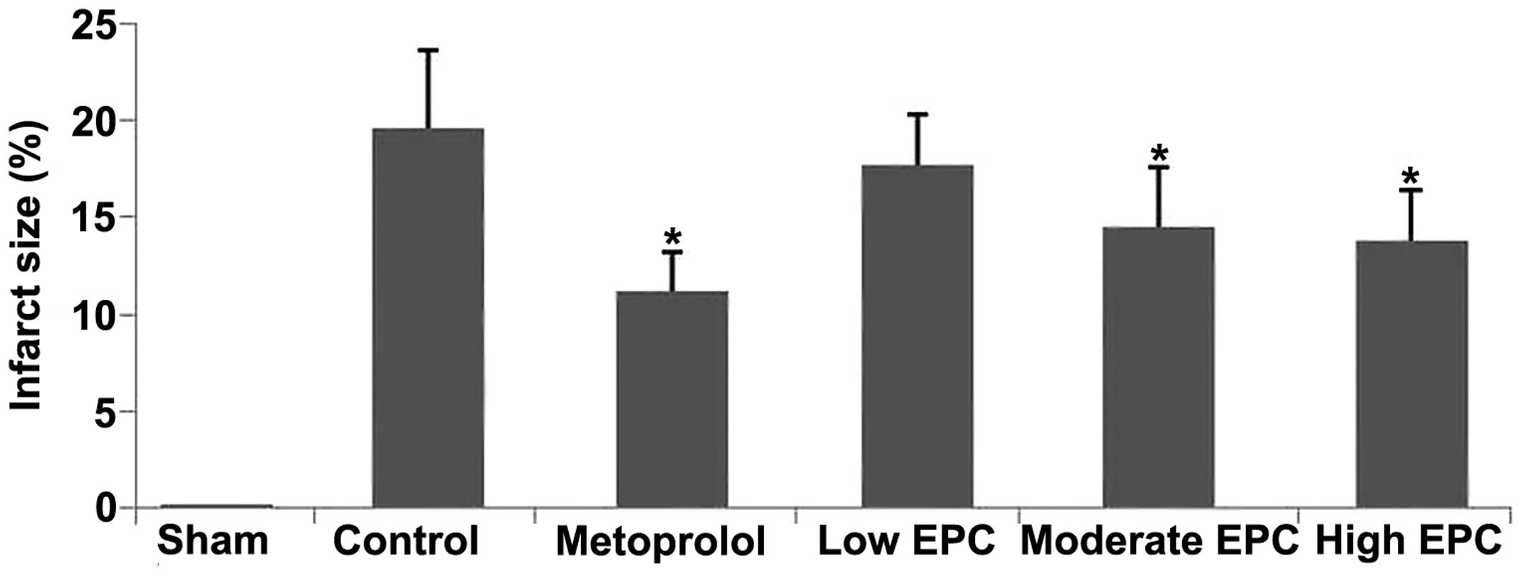
Morphological changes in the myocardium
among the groups
NBT stained the normal myocardium dark blue, while
the infarcted myocardium exhibited no staining. The myocardial
infarct size was larger in the control group when compared with
that in the sham group (Fig. 1).
Furthermore, the myocardial infarct size was decreased in the
metoprolol and low-, moderate- and high-dose EPC groups when
compared with that in the control group (Figs. 1).
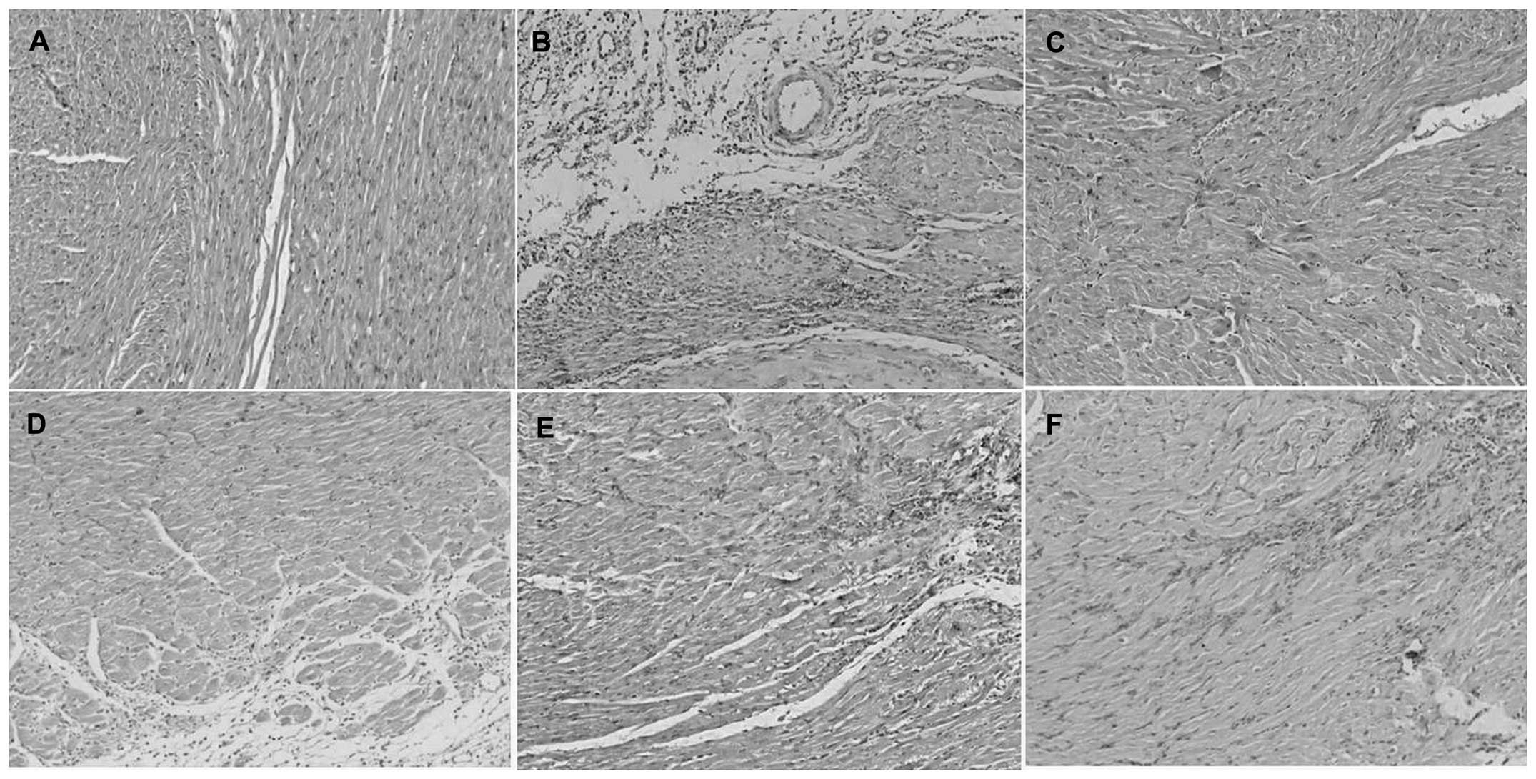
HE staining showed that the myocardial cells of the
sham group were arranged in an orderly manner, with normal
morphology and texture (Fig. 2A).
The myocardial cells in the control group exhibited a swollen
appearance and formed a wave shape with vacuolar degeneration and
fibrosis (Fig. 2B). These changes
in cardiac structure were significantly attenuated in the
metoprolol group (Fig. 2C) and
with all doses of EPC (Fig. 2D–F
for the low-, medium- and high-dose groups, respectively).
Expression of vWF and DD in the
serum
Compared with levels in the sham group, the serum
vWF and DD levels in the control group were significantly increased
(P<0.01, Table II). Metoprolol
and high-dose EPC decreased the serum concentration of vWF when
compared with the control group (P<0.01). Moderate- and
high-dose EPC decreased the DD level in serum when compared with
the control group (P<0.05 and P<0.01, respectively).
| Table IIComparison of the levels of serum vWF
and DD among the groups. |
Table II
Comparison of the levels of serum vWF
and DD among the groups.
| Group | n | vWF (ng/ml) | DD (ng/ml) |
|---|
| Sham | 10 |
4549.75±844.76b | 24.67±8.64b |
| Control | 9 | 6163.22±1045.94 | 55.62±15.42 |
| Metoprolol | 12 |
4621.94±1002.79b | 45.36±15.15 |
| Low-dose EPC | 9 | 5624.18±1034.12 | 43.21±13.06 |
| Moderate-dose
EPC | 11 | 5672.15±965.41 | 25.85±4.60b |
| High-dose EPC | 10 |
4093.56±977.52b | 32.30±10.28a |
Expression of GPIIb-IIIa and CD62P in the
serum
Compared with levels in the sham group, the serum
GPIIb-IIIa and CD62P levels in the control group were significantly
increased (P<0.05, Table
III). Moderate and high doses of EPC decreased the GPIIb-IIIa
level when compared with the control group (P<0.01). EPC reduced
the CD62P serum level gradually at all doses, with dose escalation
(P<0.05 and P<0.01).
| Table IIIComparison of the levels of serum
GPIIb-IIIa and CD62P among the groups. |
Table III
Comparison of the levels of serum
GPIIb-IIIa and CD62P among the groups.
| Group | n | GPIIb-IIIa
(ng/ml) | CD62P (ng/ml) |
|---|
| Sham | 10 | 5.31±1.06a | 31.28±8.92a |
| Control | 9 | 7.71±1.16 | 51.57±9.52 |
| Metoprolol | 12 | 5.56±1.05 | 34.24±11.35 |
| Low-dose EPC | 9 | 6.97±1.13 | 30.55±12.76a |
| Moderate-dose
EPC | 11 | 3.91±1.11b | 25.52±10.60b |
| High-dose EPC | 10 | 4.59±1.08b | 23.26±14.16b |
Discussion
Platelet activation is a key pathophysiological
feature of coronary heart disease, particularly for MI (10). The formation of a platelet plug at
sites of atherosclerotic lesion rupture is the most common
mechanism leading to MI (11). In
the present study the LAD was ligated to establish an MI model. The
results showed that the serum GPIIb-IIIa and CD62P levels in the MI
rats were significantly increased compared with those in the sham
group. Since GPIIb-IIIa plays an essential role in platelet
aggregation and CD62P is the marker of late-phase platelet
activation, and can only be expressed on the degranulated platelet
surface (12,13), these proteins are specific indices
presently known to be able to most directly reflect the degree of
platelet activation (14,15). The present results therefore
indicated that the platelets were activated in the rats following
MI.
Abnormalities in the coagulation and fibrinolytic
system have been associated with an increased risk of coronary
heart disease in observational studies and meta-analyses (5,10).
In the process of platelet activation, the regulation of binding
between vWF and the platelet receptor GPIbα is one of the key steps
for platelet adhesion (11,16,17).
The present results showed that the serum vWF level was increased
in the MI model when compared with that in the sham group, which
indicated an enhancement of platelet adhesion and thrombogenesis.
Fibrin DD is the primary degradation product of cross-linked fibrin
and is a marker of activated coagulation and fibrinolysis (18). The rise in DD at the early stage of
acute MI (AMI) may be useful to indicate the critical condition of
patients with AMI (19). The
present results showed that the serum vWF and DD levels in the
control group were significantly increased compared with those in
the sham group, indicating that, subsequent to MI, a
hypercoagulable state was induced in the rat model, followed by
platelet activation, thrombosis and subsequent fibrinolysis. This
may explain the severe pathological damages in the control group,
as shown by NBT and HE staining.
P. quinquefolius has the effects of
invigorating Qi and nourishing Yin, clearing fire and generating
body fluid. C. tuber, which has an acrid taste, activates
blood circulation and regulates Qi to alleviate pain, as stated in
the Chinese Pharmacopoeia (3).
EPC, an extract of P. quinquefolius and C. tuber, has
long been used for the treatment of ischemic cardiovascular
diseases in the clinic. P. quinquefolius saponins and
tetrahydropalmatine are the active components of EPC, as determined
by HPLC. Previous animal experiments and clinical trials have found
that P. quinquefolius saponins possess varied
pharmacological properties, and exert anti-anoxia, anti-ischemic
and antioxidation effects (4,20).
Furthermore, tetrahydropalmatine has been demonstrated to have
analgesic and sedative effects (21). The present results showed that
high-dose EPC decreased the serum concentration of vWF when
compared with the control group. Moderate and high doses of EPC
decreased the DD and GPIIb-IIIa levels, and the CD62P levels were
gradually decreased with EPC dose escalation. It can therefore be
concluded from the findings with this rat model of AMI that EPC has
a therapeutic role in inhibiting platelet activation and improving
the hypercoagulable state by suppressing the expression of
GPIIb-IIIa, CD62P, vWF and DD.
Metoprolol, which is a key drug in the therapy of
post-infarct hearts, has an important effect in decreasing
mortality in patients following AMI (22). Metoprolol was thus used as the
control drug in the present study. Infarct size predicts
post-infarction mortality (22).
In this study, NBT and HE staining showed that metoprolol and EPC
reduced myocardial infarct size, and metoprolol decreased the serum
concentration of vWF when compared with control group; however,
metoprolol showed no effects on platelet activation and oxidative
injury. We therefore speculated that the cardioprotective
mechanisms of metoprolol were achieved predominantly by blocking
cardiac β1-receptors and thereby slowing the heart rate and
reducing myocardial contractility and oxygen consumption, as
previously reported (23).
Therefore, EPC has a therapeutic effect in a rat model of AMI by
attenuating the pathological changes of the myocardium, inhibiting
platelet activation and improving the hypercoagulable state.
Acknowledgements
This study was supported by the National Science and
Technology Major Project (no. 2009ZX09103-441) and the National
Natural Science Foundation of China (nos. 81030063, 81102722 and
81273933).
References
|
1
|
Xue M, Liu ML, Zhu XY, et al: Effective
components of Panax quinquefolius and Corydalis tuber protect
myocardium through attenuating oxidative stress and endoplasmic
reticulum stress. Evid Based Complement Alternat Med.
2013:4823182013. View Article : Google Scholar : PubMed/NCBI
|
|
2
|
Zhang XR, Zhao YL, Wang JB, et al:
Differences between cold and hot natures of processed Radix ginseng
rubra and Panax quinquefolius L. based upon mice temperature
tropism. Zhonghua Yi Xue Za Zhi. 89:1994–1998. 2009.(In Chinese).
PubMed/NCBI
|
|
3
|
People’s Republic of China Pharmacopoeia
Commission, . Pharmacopoeia of the People’s Republic of China.
Chemical Industry Press; Beijing, China: 2000
|
|
4
|
Zhu XY, Zhang ZL, Li P, et al: Shenyuan,
an extract of American Ginseng and Corydalis Tuber formula,
attenuates cardiomyocyte apoptosis via inhibition of endoplasmic
reticulum stress and oxidative stress in a porcine model of acute
myocardial infarction. J Ethnopharmacol. 150:672–681. 2013.
View Article : Google Scholar : PubMed/NCBI
|
|
5
|
Abe S, Maruyama I, Arima S, et al:
Increased heparin-releasable platelet factor 4 and D dimer in
patients one month after the onset of acute myocardial infarction:
persistent activation of platelets and the coagulation/fibrinolytic
system. Int J Cardiol. 47:S7–12. 1994. View Article : Google Scholar : PubMed/NCBI
|
|
6
|
Liu YM, Zhang J, Shi F, et al: Studies on
extracting methods of shenyuan tablet (effective components of
American Ginseng and Corydalis tuber).). Zhong Guo Shi Yan Fang Ji
Xue Za Zhi. 17:18–21. 2012.(In Chinese).
|
|
7
|
Abbate A, Salloum FN, Vecile E, et al:
Anakinra, a recombinant human interleukin-1 receptor antagonist,
inhibits apoptosis in experimental acute myocardial infarction.
Circulation. 117:2670–2683. 2008. View Article : Google Scholar : PubMed/NCBI
|
|
8
|
Olivetti G, Capasso JM, Meggs LG, et al:
Cellular basis of chronic ventricular remodeling after myocardial
infarction in rats. Circ Res. 68:856–869. 1991. View Article : Google Scholar : PubMed/NCBI
|
|
9
|
Xue M, Yin HJ, Zhang L, et al: Dynamic
expression of the main related indicators to thrombosis,
inflammation reaction and tissue damage in a rat model of
myocardial infarction. Mol Med Rep. 4:693–696. 2011.PubMed/NCBI
|
|
10
|
Whincup PH, Danesh J, Walker M, et al: von
Willebrand factor and coronary heart disease: prospective study and
meta-analysis. Eur Heart J. 23:1764–1770. 2002. View Article : Google Scholar : PubMed/NCBI
|
|
11
|
Wang Y, Andrews M, Yang Y, et al:
Platelets in thrombosis and hemostasis: old topic with new
mechanisms. Cardiovasc Hematol Disord Drug Targets. 12:126–132.
2012. View Article : Google Scholar : PubMed/NCBI
|
|
12
|
Xue M, Yin HJ, Wu CF, Ma XJ, et al: Effect
of Chinese drugs for activating blood circulation and detoxifying
on indices of thrombosis, inflammatory reaction and tissue damage
in a rabbit model of toxin-heat and blood stasis syndrome. Chin J
Integr Med. 19:42–47. 2013. View Article : Google Scholar
|
|
13
|
Xue M, Zhang L, Yang L, et al: Effect of
Chinese herbal medicine for activating blood circulation and
detoxifying on expression of inflammatory reaction and tissue
damage related factors in experimental carotid artery thrombosis
rats. Chin J Integr Med. 16:247–251. 2010. View Article : Google Scholar : PubMed/NCBI
|
|
14
|
Chen KJ, Xue M and Yin HJ: The
relationship between platelet activation and coronary heart disease
and blood-stasis syndrome. Shou Du Yi Ke Da Xue Xue Bao. 29(29):
266–269. 2008.(In Chinese).
|
|
15
|
Bledzka K, Smyth SS and Plow EF: Integrin
αIIbβ3: from discovery to efficacious therapeutic target. Circ Res.
112:1189–1200. 2013. View Article : Google Scholar : PubMed/NCBI
|
|
16
|
Andrews RK and Berndt MC: Platelet
physiology and thrombosis. Thromb Res. 114:447–453. 2004.
View Article : Google Scholar : PubMed/NCBI
|
|
17
|
Szántó T, Joutsi-Korhonen L, Deckmyn H, et
al: New insights into von Willebrand disease and platelet function.
Semin Thromb Hemost. 38:55–63. 2012. View Article : Google Scholar : PubMed/NCBI
|
|
18
|
Danesh J, Whincup P, Walker M, et al:
Fibrin D-dimer and coronary heart disease: prospective study and
meta-analysis. Circulation. 103:2323–2327. 2001. View Article : Google Scholar : PubMed/NCBI
|
|
19
|
Yang J and Li CS: The predictive values of
D-dimer for the early prognosis of the acute myocardial infarction
(AMI): a review of 3134 AMI patients. Zhongguo Wei Zhong Bing Ji
Jiu Yi Xue. 24:725–729. 2012.(In Chinese). PubMed/NCBI
|
|
20
|
Yang L, Miao Y, YIN HJ, et al:
Identification of ginsenosides from Panax quinquefolius in Xinyue
Capsule by LC-ESI-MSn. Zhong Cao Yao. 41:1942–1947. 2010.(In
Chinese).
|
|
21
|
Sun S, Chen Z, Li L, et al: The two
enantiomers of tetrahydropalmatine are inhibitors of P-gp, but not
inhibitors of MRP1 or BCRP. Xenobiotica. 42:1197–1205. 2012.
View Article : Google Scholar : PubMed/NCBI
|
|
22
|
Ibanez B, Fuster V, Macaya C, et al: Study
design for the ‘effect of METOprolol in CARDioproteCtioN during an
acutemyocardial infarction’ (METOCARD-CNIC): a randomized,
controlled parallel-group, observer-blinded clinical trial of early
pre-reperfusion metoprolol administration in ST-segment elevation
myocardial infarction. Am Heart J. 164:473–480. 2012. View Article : Google Scholar
|
|
23
|
Wu A, Zhai J, Zhang D, et al: Effect of
wenxin granule on ventricular remodeling and myocardial apoptosis
in rats with myocardial infarction. Evid Based Complement Alternat
Med. 2013:9679862013. View Article : Google Scholar : PubMed/NCBI
|