Introduction
Thyroid cancer is one of the most common tumors
derived from endocrine cells and its incidence rate has been rising
(1). In order to improve the early
diagnosis and treatment of patients, an in-depth study of the
pathogenesis of thyroid cancer is urgently required.
Extracellular matrix metalloproteinase inducer
(EMMPRIN) belongs to the immunoglobulin super family, which can
promote tumor invasion and metastasis (2). Human epidermal growth factor receptor
(HER)-2 gene is a type of proto-oncogene. As in vivo studies
have shown, HER-2 oncogene is a neoplasm metastasis driving factor;
malignant tumors with overexpressed HER-2 have a higher risk for
tumor invasion and metastasis, with a poorer prognosis (3). Little has been reported on the
detection of EMMPRIN and HER-2 protein expression in papillary
thyroid carcinoma by the quantum dot (QD)-based immunofluorescence
technique in tissue chips. In the current study, a QD-based
immunofluorescence tissue analysis technique was adopted to detect
EMMPRIN and HER-2 protein expression in tissue chips of papillary
thyroid carcinoma from human sources. The correlation between the
expression of EMMPRIN and HER-2 proteins was investigated in order
to further understand the mechanism of the occurrence, development
and prognosis of papillary thyroid carcinoma.
Materials and methods
Chips
Four tissue chips of papillary thyroid carcinoma
were provided by Fanpu Biotech, Inc. (Guilin, China). These had a
dot matrix of 15×10, a dot diameter of 1.1 mm and thickness of 4
μm, and included 70 cases of papillary thyroid carcinoma tissues
and five peri-tumor tissues, in a dual chip matrix. Tissues were
surgically resected during clinical surgery and then fixed for 4 h
with neutral buffered formaldehyde. Among the patients with
papillary thyroid carcinoma, there were 22 male and 48 female cases
(age range, 25–80 years; mean, 57.2 years). There were 42 cases
with lymph node metastasis (LNM) and 28 cases of papillary thyroid
carcinoma tissues without LNM. The diagnosis of the patients was
according to the World Health Organization Classification of
papillary thyroid carcinoma (4).
Antibodies
The primary antibodies were rabbit anti-human
EMMPRIN polyclonal antibody (#sc-13976) and mouse anti-human HER-2
polyclonal antibody (#sc-33684; Santa Cruz Biotechnology Inc.,
Dallas, TX, USA; concentration, 1:100). The secondary antibodies
were goat anti-rabbit/mouse biotinylated secondary antibodies
(#AS-28175-05; AnaSpec, Inc., Fremont, CA, USA).
Immunofluorescence
A QD-based immunofluorescence tissue chemical
technique was used to detect EMMPRIN and HER-2 protein expression
in tissue chips of papillary thyroid carcinoma. A hypersensitive
fluorescence quantum dot kit containing Qdot Streptavidin Conjugate
(QDs-SA) was purchased from Wuhan Jiayuan Quantum Dot Technological
Development Co., Ltd. (Wuhan, China).
Experimental procedures
The experimental procedures were carried out in
strict accordance with the manufacturer’s instructions. Tissue
chips of papillary thyroid carcinoma (thickness, 4 μm) were
dewaxed, hydrated, microwave-antigen retrieved and washed with
Tris-buffered saline (TBS). The tissue chips were blocked by
incubation with blocking buffer solution (Beyotime Institute of
Biotechnology, Shanghai, China) in a wet chamber for 30 min at
37°C. The tissue chips were then incubated with primary antibodies
for 2 h at 37°C, then washed with TBS-Tween® three
times, 5 min/time. Supplement of secondary antibodies incubated for
1h at 37°C, washed repeatedly with TBS-Tween® three
times, 5 min/time. The tissue chips were again blocked by
incubation with blocking buffer solution in a wet chamber for 30
min at 37°C. QDs-SA diluted with blocking buffer solution was
dripped onto the tissue chips and incubated in a wet chamber for 30
min at 37°C. They were then washed three times with TBS-Tween for 5
min/time, and finally treated with 90% glycerin buffer. Following
the addition of 900 ml/l glycerin, the chips were observed with an
Olympus IX71 fluorescence microscope (Olympus Corporation, Tokyo,
Japan). QDs were excited by irradiation at an excitation wavelength
of 430–500 nm and an emission wavelength of 605 nm. When observed
under the microscope, cells with reddish-orange fluorescent
particles were positive. When the positive area was ≥25%, it was
regarded as positive protein expression. In the control group, the
primary antibody was substituted with TBS. When observed under the
microscope, the positive staining of EMMPRIN and HER-2 protein was
mainly located in the cell membrane and cytoplasm. Four different
views were obtained under high power fields, and 200 cells were
evaluated. Calculation of the degree of expression was conducted
according to the percentage of positive cells: ‘−’, negative; ‘+’,
≤10% positive cells; ‘++’, 11–50% positive cells; and ‘+++’, ≥51%
positive cells. Positive expression was deemed to be ≥11% positive
cells.
Statistical analysis
The quantitative analysis of QD staining was
expressed as the mean ± standard deviation. One-way analysis of
variance and Student-Newman-Keuls (q) detection were carried out
with SPSS software, version 13.0 (SPSS Inc., Chicago, IL, USA) for
positive cells in each staining group (α=0.05). Spearman’s rank
correlation coefficient was adopted to determine the association
between EMMPRIN and HER-2 expression. P<0.05 was considered to
indicate a statistically significant difference.
Results
EMMPRIN protein expression
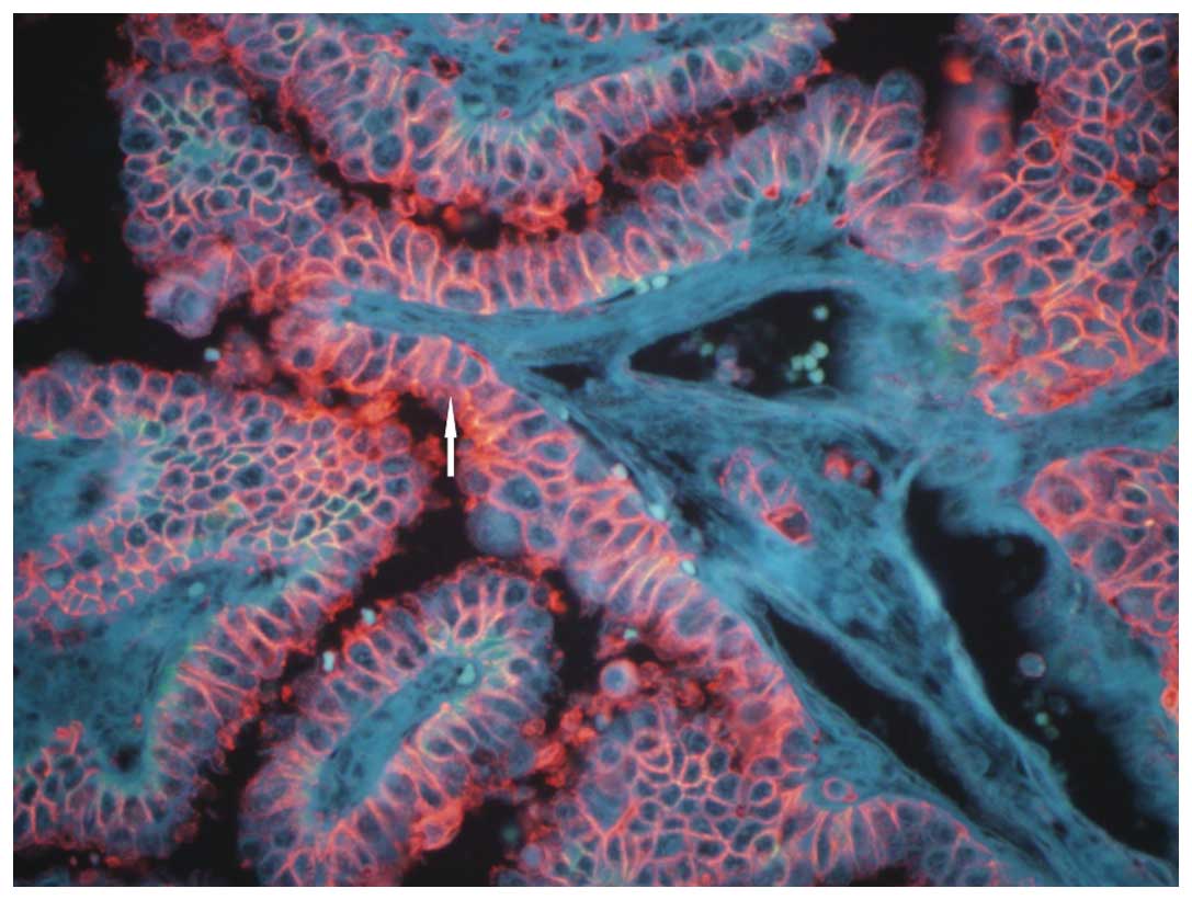
Strong and red fluorescence of EMMPRIN was observed
in the cell membrane or cytoplasm of papillary thyroid carcinoma
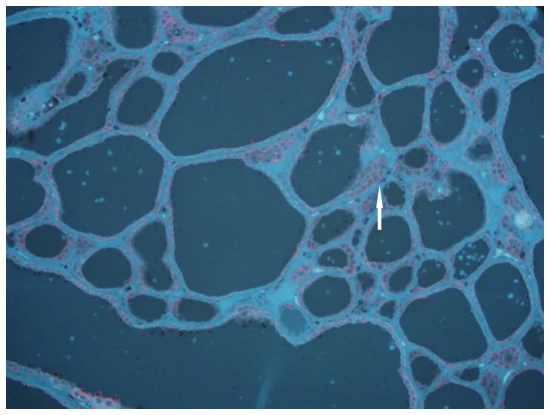
(Fig. 1). A slight red
fluorescence appeared in peri-tumor tissues; EMMPRIN protein
exhibited negative expression (Fig.
2). The positive rate of EMMPRIN protein was 75.71% in
papillary thyroid carcinoma and 20.00% in peri-tumor tissues
(P<0.05; Table I). The positive
rate of EMMPRIN protein in carcinoma tissues with LNM was
significantly higher than that in carcinoma tissues without LNM
(80.95 vs. 60.71%, P<0.05; Table
II).
 | Table IExpression of EMMPRIN and HER-2 in
thyroid papillary carcinoma and adjacent (peri-tumor) tissues. |
Table I
Expression of EMMPRIN and HER-2 in
thyroid papillary carcinoma and adjacent (peri-tumor) tissues.
| | EMMPRIN | HER-2 |
|---|
| |
|
|
|---|
| Group | n | − | + | − | + |
|---|
| Thyroid papillary
carcinoma | 70 | 17 | 53 | 38 | 32 |
| Peri-tumor
tissue | 5 | 4 | 1 | 5 | 0 |
| Table IIExpression of EMMPRIN and HER-2
protein and the association with LNM. |
Table II
Expression of EMMPRIN and HER-2
protein and the association with LNM.
| | EMMPRIN | HER-2 |
|---|
| |
|
|
|---|
| LNM | n | − | + | − | + |
|---|
| Yes | 42 | 8 | 34 | 17 | 25 |
| No | 28 | 11 | 17 | 20 | 8 |
HER-2 protein expression
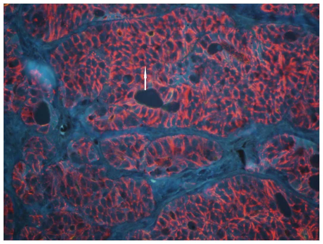
Red fluorescence was observed in the cell membrane
or cytoplasm of papillary thyroid carcinoma, which indicated
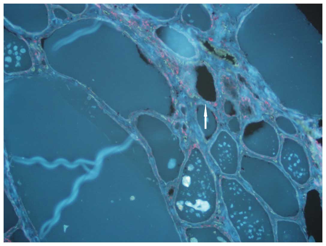
positive HER-2 protein expression (Fig. 3). A slight red fluorescence
appeared in peri-tumor tissues; HER-2 protein expression was
negative (Fig. 4). The positive
rate of HER-2 protein expression was 45.71% in papillary thyroid
carcinoma and 0% in peri-tumor tissues (P<0.05; Table I). The positive rate of HER-2
protein expression in carcinoma tissues with LNM was significantly
higher than that in carcinoma tissues without LNM (59.52 vs.
28.57%, P<0.05; Table II).
Correlation between EMMPRIN and HER-2
protein expression in papillary thyroid carcinoma
Among 70 cases of papillary thyroid carcinoma
tissues, 53 cases exhibited a positive expression of EMMPRIN
protein and 32 cases were positive for HER-2 protein. There were 17
cases negative for EMMPRIN protein and 38 cases negative for HER-2.
EMMPRIN and HER-2 had a positive correlation (r=0.375,
P=0.001).
Discussion
Biochip technology, as a biological research
technique that emerged in the 1980s, generally includes gene and
protein chips. Gene and protein chips are widely applied in
molecular/synthetic biology to improve the development of a growing
number of new functional genes and proteins. In order to further
explain their function, it is necessary to turn to histology and
histomorphological studies, which play a pivotal role in the study
of cancer and require urgent development (5).
Currently, tissue slices are widely applied in
medical studies, generally containing one (type of) tissue sample
in one wax mass, that is, a single tissue slice. In scientific
studies on large samples, a large number of tissue wax masses and
tissue slices are prepared by repetitively carrying out the same
procedure, resulting in a waste of time and resources, unavoidable
errors and poor comparability. It is, therefore, necessary to
design new multi-tissue embedding chip-making techniques, simplify
the experiment times, reduce the workload of experiments and
improve the efficiency. Tissue chips have the characteristics of
small volume and high information content, as well as providing
substantial results in a one-off experiment. In the present study,
a 150-chip papillary thyroid carcinoma tissue chip in a dual-chip
matrix was adopted. The experiment was completed with only a few
chips, saving funds and reducing work load. Data from EMMPRIN and
HER-2 protein expression in papillary thyroid carcinoma were
obtained in a short time. Compared with traditional pathological
techniques and methods, the results available are uniform, reliable
and comparable, saving time and money in addition to providing a
large quantity of information. This presents great potential for
the future application of tissue chips in medicine (6,7).
QDs, made up of Group II–IV or Group III–V elements,
are a type of semiconductor nanoparticle (diameter, 2–6 nm) capable
of producing fluorescence upon laser excitation. Due to their
special structure and unique optical properties, such as a broad
and continuously distributed excitation spectrum, and narrow and
symmetrically distributed emission spectrum, QDs have been widely
used in medical studies. The size of QD kernels can be changed to
precisely regulate the length of optical waves; thus, QDs have
become a powerful tool in biomedical tagging and optical imaging
(8). QDs, as new nanometer
fluorescence probes, are characterized by high efficiency, wide
coverage of light and stable optical properties. They are uniquely
advantageous in live action monitoring and as long-term in
vivo tracers (9). In recent
years, nanometer-sized fluorescence probes with biological
compatibility, developed on the basis of semiconductor QDs, have
been provided with a unique optical property. QDs are promising in
terms of application in the field of biomedicine, particularly in
studies of biomedical imaging technology.
Tumors have a unique disease progession, involving
invasion and metastasis. This is a complex process, with the
following steps: Firstly, tumor cells break away from the primary
tumor lesion, intrude into the mesenchyme and break down the base
membrane to infiltrate and adhere to the outside of the vessel and
intravasate into the vascular endothelium; secondly they enter the
blood circulation system through the vascular endothelium, evading
the immune response; thirdly, the tumor cells reach a new location
through circulation and pass through the vascular wall and base
membrane and complete extravasation into the extracellular matrix.
Ultimately they survive, clone and proliferate in particular organs
to form metastases.
EMMPRIN is a type of multifunctional protein,
capable of inducing the production of matrix metalloproteinase
(MMP) and promoting tumor cell invasion and metastasis (10).
The role of EMMPRIN, also known as CD147, in the
process of tumor development, invasion and metastasis is a popular
topic in the field of the cellular biology of tumors (11). The proliferative activity of tumor
cells is closely associated with tumor invasion (12), metastasis (13) and prognosis (14).
In the present study, the expression rate of EMMPRIN
in peri-tumor tissues was 20.00%, while it was 75.71% in papillary
thyroid carcinoma tissues (P<0.05). EMMPRIN is highly expressed
in papillary thyroid carcinoma tissues. The enhanced expression of
EMMPRIN in papillary thyroid carcinoma tissues indicates that it
could play a role in the occurrence and development of papillary
thyroid carcinoma (11,15).
HER-2, also referred to as c-erbB-2, is a type of
oncogene with an homologous sequence with the virus oncogene as
well as the oncogene of epidermal growth factor receptor (EGFR),
and is overexpressed in various types of tumor (16,17).
HER-2 can form compounds (such as heterodimers) with other members
of the EGFR family, such as EGFR erbB1/HER1, erbB3/HER3 and
erbB4/HER4, engage in cell proliferation signal transduction, and
finally result in cell proliferation or even carcinomatosis
(18). HER-2 can be slightly
expressed in coelomic epithelium and glandular epithelium tissues,
although without gene amplification, and its overexpression is
associated with cellular carcinomatosis. Therefore, the detection
of overexpressed HER-2 protein can indicate HER-2 oncogene
amplification indirectly (19–22).
HER-2 overexpression plays an important role in the occurrence and
development of certain forms of cancer and has effects on tumor
progression and therapy (23).
In the current study, the positive rate of EMMPRIN
protein expression in tumor tissues with LNM was found to be 80.95%
(34/42), significantly higher compared with the EMMPRIN protein
expression in tumor tissues without LNM at 60.71% (17/28)
(P<0.05). This shows that EMMPRIN protein expression is closely
associated with LNM. The result corresponds well with the
previously reported literature (24,25).
EMMPRIN shows high expression in papillary thyroid
carcinoma; however, only a few factors inducing its expression in
the development of tumors are known (26). Amphiregulin and EGF serve as
regulation factors, which are able to induce the expression of
EMMPRIN by the activation of protein tyrosine kinase of EGFR
(27). In addition, anti-EGFR
antibody can inhibit EMMPRIN expression and MMP activity (28). This demonstrates that EGFR
signaling may play a decisive role in the regulation process. In
the present study, it was found that EMMPRIN and HER-2 were
concurrently expressed in papillary thyroid carcinoma tissues; the
rate of co-expression was 45.71% (32/70). EMMPRIN is positively and
significantly associated with HER-2. EMMPRIN and HER-2 could
jointly control MMP activity and collaboratively promote the LNM of
papillary thyroid carcinoma, therefore being jointly involved in
the occurrence, development and metastasis of papillary thyroid
carcinoma. This result shows that the development of a tumor is a
multi-factorial, multi-stage process.
EMMPRIN and HER-2 exhibit positive expression in
papillary thyroid carcinoma, which is substantially associated with
LNM. Further study on EMMPRIN and HER-2 inhibitors may serve as a
new approach for inhibiting the invasion and metastasis of
papillary thyroid carcinoma and, most significantly, may improve
the prognosis of patients with papillary thyroid carcinoma.
Acknowledgements
The authors would like to thank all the individuals
who participated in this study and thank Professor Wu-Dong Cheng
for assisting in preparation of this manuscript.
References
|
1
|
Ito Y, Miyauchi A, Kobayashi K and Miya A:
Prognosis and growth activity depend on patient age in clinical and
subclinical papillary thyroid carcinoma. Endocr J. 61:205–213.
2014. View Article : Google Scholar
|
|
2
|
Dalberg K, Eriksson E, Enberg U, Kjellman
M and Bäckdahl M: Gelatinase A, membrane type 1 matrix
metalloproteinase, and extracellular matrix metalloproteinase
inducer mRNA expression: Correlation with invasive growth of breast
cancer. World J Surg. 24:334–340. 2000. View Article : Google Scholar : PubMed/NCBI
|
|
3
|
Meert AP, Martin B, Paesmans M, Berghmans
T, Mascaux C, Verdebout JM, Delmotte P, Lafitte JJ and Sculier JP:
The role of HER-2/neu expression on the survival of patients with
lung cancer: A systematic review of the literature. Br J Cancer.
89:959–965. 2003. View Article : Google Scholar : PubMed/NCBI
|
|
4
|
Delellis RA, Lloyd RV, Heitx PU and Eng C:
Pathology and Genetics of Tumors of Endocrine Organs. World Health
Organization Classification of Tumours Lyon: pp. 73–76. 2004
|
|
5
|
Kramer MW, Merseburger AS, Hennenlotter J
and Kuczyk M: Tissue microarrays in clinical urology - technical
considerations. Scand J Urol Nephrol. 41:478–484. 2007. View Article : Google Scholar
|
|
6
|
Jawhar NM: Tissue microarray: A rapidly
evolving diagnostic and research tool. Ann Saudi Med. 29:123–127.
2009. View Article : Google Scholar : PubMed/NCBI
|
|
7
|
Kallioniemi OP, Wagner U, Kononen J and
Sauter G: Tissue microarray technology for high-throughput
molecular profiling of cancer. Hum Mol Genet. 10:657–662. 2001.
View Article : Google Scholar : PubMed/NCBI
|
|
8
|
Hall M, Kazakova I and Yao YM: High
sensitivity immunoassays using particulate fluorescent labels. Anal
Biochem. 272:165–170. 1999. View Article : Google Scholar : PubMed/NCBI
|
|
9
|
Hermanson GT: Bioconjugate Techniques. 2nd
edition. Academic Press; London, UK: pp. 297–416. 1996, View Article : Google Scholar
|
|
10
|
Sienel W, Polzer B, Elshawi K, et al:
Cellular localization of EMMPRIN predicts prognosis of patients
with operable lung adenocarcinoma independent from MMP-2 and MMP-9.
Mod Pathol. 21:1130–1138. 2008. View Article : Google Scholar : PubMed/NCBI
|
|
11
|
Zheng HC, Takahashi H, Murai Y, et al:
Upregulated EMMPRIN/CD147 might contribute to growth and
angiogenesis of gastric carcinoma: A good marker for local invasion
and prognosis. Br J Cancer. 95:1371–1378. 2006. View Article : Google Scholar : PubMed/NCBI
|
|
12
|
Quemener C, Gabison EE, Naïmi B, et al:
Extracellular matrix metalloproteinase inducer up-regulates the
urokinase-type plasminogen activator system promoting tumor cell
invasion. Cancer Res. 67:9–15. 2007. View Article : Google Scholar : PubMed/NCBI
|
|
13
|
Bougatef F, Quemener C, Kellouche S, et
al: EMMPRIN promotes angiogenesis through hypoxia-inducible
factor-2alpha-mediated regulation of soluble VEGF isoforms and
their receptor VEGFR-2. Blood. 114:5547–5556. 2009. View Article : Google Scholar : PubMed/NCBI
|
|
14
|
Lu Q, Lv G, Kim A, Ha JM and Kim S:
Expression and clinical significance of extracellular matrix
metalloproteinase inducer, EMMPRIN/CD147, in human osteosarcoma.
Oncol Lett. 5:201–207. 2013.
|
|
15
|
Zheng H, Takahashi H, Murai Y, Cui Z,
Nomoto K, Miwa S, Tsuneyama K and Takano Y: Pathobiological
characteristics of intestinal and diffuse-type gastric carcinoma in
Japan: an immunostaining study on the tissue microarray. J Clin
Pathol. 60:273–277. 2007. View Article : Google Scholar
|
|
16
|
Chao WR, Lee MY, Lin WL, Chen CK, Lin JC,
Koo CL, Sheu GT and Han CP: HER2 amplification and overexpression
are significantly correlated in mucinous epithelial ovarian cancer.
Hum Pathol. 45:810–816. 2014. View Article : Google Scholar : PubMed/NCBI
|
|
17
|
Brennan PJ, Kumagai T, Berezov A, Murali R
and Greene MI: HER-2/neu: Mechanisms of
dimerization/oligomerization. Oncogene. 19:6093–6101. 2000.
View Article : Google Scholar
|
|
18
|
Lohrisch C and Piccart M: An overview of
HER2. Semin Oncol. 28(Suppl 18): 3–11. 2001. View Article : Google Scholar
|
|
19
|
Ho VW, Leung K, Hsu A, et al: A low
carbohydrate, high protein diet slows tumor growth and prevents
cancer initiation. Cancer Res. 71:4484–4493. 2011. View Article : Google Scholar : PubMed/NCBI
|
|
20
|
Debled M, Dalenc F, Mauriac L and Brain E:
Medical treatments of endocrine-sensitive Her-2 negative breast
cancers: A review. Bull Cancer. 98:655–670. 2011.(In French).
PubMed/NCBI
|
|
21
|
Menashi S, Serova M, Ma L, Vignot S,
Mourah S and Calvo F: Regulation of extracellular matrix
metalloproteinase inducer and matrix metalloproteinase expression
by amphiregulin in transformed human breast epithelial cells.
Cancer Res. 63:7575–7580. 2003.PubMed/NCBI
|
|
22
|
Auvinen P, Tammi R, Kosma VM, Sironen R,
Soini Y, Mannermaa A, Tumelius R, Uljas E and Tammi M: Increased
hyaluronan content and stromal cell CD44 associate with HER2
positivity and poor prognosis in human breast cancer. Int J Cancer.
132:531–539. 2013. View Article : Google Scholar
|
|
23
|
Papadopoulou E, Tripsianis G,
Anagnostopoulos K, Tentes I, Kakolyris S, Galazios G, Sivridis E,
Simopoulos K and Kortsaris A: The influence of serum HER-2 levels
and HER-2 codon 655 polymorphism on breast cancer outcome.
Neoplasma. 55:113–121. 2008.PubMed/NCBI
|
|
24
|
Yang Q, Liu Y, Huang Y, Huang D, Li Y, Wu
J and Duan M: Expression of COX-2, CD44v6 and CD147 and
relationship with invasion and lymph node metastasis in
hypopharyngeal squamous cell carcinoma. PLoS One. 8:e710482013.
View Article : Google Scholar : PubMed/NCBI
|
|
25
|
Huang T, Chen MH, Wu MY and Wu XY:
Correlation between expression of extracellular matrix
metalloproteinase inducer and matrix metalloproteinase-2 and
cervical lymph node metastasis of nasopharyngeal carcinoma. Ann
Otol Rhinol Laryngol. 122:210–215. 2013.PubMed/NCBI
|
|
26
|
Tan H, Ye K, Wang Z and Tang H: CD147
expression as a significant prognostic factor in differentiated
thyroid carcinoma. Transl Res. 152:143–149. 2008. View Article : Google Scholar : PubMed/NCBI
|
|
27
|
Suzuki S and Ishikawa K: Combined
inhibition of EMMPRIN and epidermal growth factor receptor prevents
the growth and migration of head and neck squamous cell carcinoma
cells. Int J Oncol. 44:912–917. 2014.PubMed/NCBI
|
|
28
|
Gialeli Ch, Theocharis AD, Kletsas D,
Tzanakakis GN and Karamanos NK: Expression of matrix macromolecules
and functional properties of EGF-responsive colon cancer cells are
inhibited by panitumumab. Invest New Drugs. 31:516–524. 2013.
View Article : Google Scholar
|