Introduction
Smoke inhalation injury (SII) mainly affects the
airways and lung parenchyma, causing severe toxic pneumonitis or
pulmonary edema. These conditions may rapidly develop into acute
lung injury (ALI) and acute respiratory distress syndrome, which
increase the morbidity and mortality rates in patients (1). Oxidative stress is an important SII
mechanism, as high-temperature smoke contains a high concentration
of strong oxidants. The resultant inflammatory response, if
uncontrolled, causes abundant inflammatory cell accumulation in the
lungs, producing excessive reactive oxygen species (ROS) and
inducing oxidative stress injury.
Previous studies have shown that hydrogen sulfide
(H2S) exerts antioxidative (2) and antifibrotic effects, and plays
important roles in vasodilation (3–6) and the
regulation of the inflammatory response (7), endocrine and reproductive systems
(8). Inhalation of 80 ppm
H2S for 6 h has been demonstrated to suppress the
systemic inflammatory response and enhance survival rates in mouse
models of endotoxin-induced ALI (9,10). In
addition, inhalation of H2S has been shown to reduce
lung damage in mouse models of hyperventilation-induced ALI by
inhibiting pulmonary inflammation and alveolar epithelial cell
apoptosis (11). In a previous
study, increased oxidative stress was observed in rats with cotton
smoke inhalation-induced lung injury (12). The aim of the present study was to
observe the effect of 80 ppm H2S inhalation for 6 h on
oxidative stress in rats with SII.
Materials and methods
Animals and grouping
A total of 24 healthy, clean, adult male
Sprague-Dawley rats (weight, 150–250 g) were provided by the
Laboratory Animal Center of the Academy of Military Medical
Sciences [SCXK-(Military)-2012-0004] and fed in the Laboratory
Animal Center of the Navy General Hospital
[SCXK-(Military)-2012-0012]. The rats were randomly divided into
four groups, including the control, H2S, smoke and smoke
+ H2S groups (n=6 in each), in accordance with the
regulations for the administration of affairs concerning
experimental animals (13). This
study was conducted in strict accordance with the recommendations
of the Guide for the Care and Use of Laboratory Animals of the
National Institutes of Health (14).
Furthermore, the animal use protocol was reviewed and approved by
the Institutional Animal Care and Use Committee of the Second
Military Medical University (Beijing, China).
Establishment of the SII model and
H2S inhalation
A rat model of SII was established by placing two
randomly selected rats from the smoke and smoke + H2S
groups into a smoke chamber. The smoke was generated by sealing 2 g
cotton (Sunny Cotton Company, Xinjiang, China) in a soldering
machine (Fudi HT-B, Hongtai Hardware and Electric Equipment
Company, Guangzhou, China) at 300°C, which was subsequently
channeled into the chamber containing the rats. The rats were left
to inhale the smoke for 2 min or until red/purple spots developed
on the plantar skin, with symptoms of restlessness, tachypnea,
mouth breathing, Kussmaul breathing and stridor (15–17). The
smoke chamber was opened to allow the rats to breathe fresh air for
7 min before the chamber was resealed. This procedure was repeated
three to five times, until the rats remained unconscious after 7
min of air inhalation. The rats in the control and H2S
groups underwent the same procedure, but without smoke inhalation.
Following the smoke or sham smoke inhalation, rats in the
H2S and smoke + H2S groups inhaled 80 ppm
H2S + 30% oxygen for 6 h, while rats in the control and
smoke groups inhaled 30% oxygen for 6 h. The rats had free access
to food and water.
Enzyme-linked immunosorbent assay
(ELISA)
Rats were euthanized with an intraperitoneal
injection of pentobarbital sodium. A double-antibody sandwich
avidin-biotin-peroxidase complex-ELISA (Jiamay Biotech Co. Ltd.,
Beijing, China) was performed to determine the levels of nitric
oxide (NO), inducible nitric oxide synthase (iNOS) and nuclear
factor (NF)-κBp65 in the lower right lung homogenate. In addition,
the concentration of malondialdehyde (MDA) was measured using
colorimetry.
Immunohistochemistry (IHC) of
NF-κBp65
The right middle lobes of the rat lungs were fixed
in 4% paraformaldehyde for 72 h, embedded in paraffin, sectioned at
3 µm and preheated at 60–65°C for 4 h. This was followed by
deparaffinization, rehydration, washing in phosphate-buffered
saline, high temperature antigen retrieval, endogenous peroxidase
blocking in 3% H2O2 and normal goat serum
blocking. The slides were incubated with 50 µl anti-NF-κBp65
primary antibody (ab16502; Abcam, Cambridge, UK) at 1:200 dilution
overnight (4°C), and a secondary horseradish peroxidase-conjugated
anti-mouse/rabbit IgG antibody (KIT-5020; Maixin-Bio, Fuzhou,
China) for 20 min at room temperature. The slides were subsequently
stained with diaminobenzidine and counterstained with hematoxylin.
For the negative control, the primary antibody was replaced with
serum. Positive expression was observed as yellow or brown
staining. Image-Pro Plus 6.0 software (Media Cybernetics, Inc.,
Rockville, MD, USA) was used for semiquantitative analysis by
randomly selecting five high-power fields (magnification, x1,000)
from each slide. Image-Pro Plus 6.0 software was used to calculate
the sum-integrated optical density (IOD) of the mean density, and
the IOD of the positive staining in each field, as well as the mean
value of these parameters.
Quantitative fluorescence-polymerase
chain reaction (qF-PCR)
Forward and reverse primers for iNOS were
synthesized by Jiamay Biotech Co. Ltd. as follows:
5-ACACCGATTCCACTCAACTA-3 and 5-ACCACCTGTTAGTTCAAGCC-3′,
respectively. The amplified products were 159 bp in length and
β-actin was used as an internal reference gene (CW0918; CWbio Co.,
Ltd., Beijing, China). Total RNA was extracted using an Ultrapure
RNA Kit (CW0581; CWbio Co., Ltd.) and analyzed (5 µl) with 1%
agarose gel electrophoresis. The total RNA was reverse transcribed
with a HiFi-MMLV-cDNA First-Strand cDNA Synthesis kit (CW0744;
CWbio Co., Ltd.) and amplified using an UltraSYBR Mixture with ROX
(CW0956; CWbio Co., Ltd.) under the following conditions: 95°C for
10 min, 40 cycles of 95°C for 15 sec and 60°C for 60 sec. qF-PCR
was performed with a LightCycler® 480 II PCR system (Roche
Diagnostics, Basel, Switzerland), and the 2−ΔΔCt method
was employed to analyze the relative changes in gene
expression.
Statistical analysis
Data were analyzed with SPSS 18.0 software (SPSS,
Inc., Chicago, IL, USA). Measurement data are presented as the mean
± standard deviation, and comparisons were performed using one-way
analysis of variance. Comparisons between groups were performed
using Fishers least significant difference test. P<0.05 was
considered to indicate a statistically significant difference.
Results
ELISA
Significantly higher concentrations of MDA, NO, iNOS
and NF-κBp65 were observed in the rat lung homogenate from the
smoke group, as compared with those in the control or smoke +
H2S groups (P<0.001). Furthermore, the H2S
group exhibited a higher concentration of iNOS than the control
group (Table I). The levels of all
the measured indicators were lower in the H2S group when
compared with those in the smoke group.
 | Table I.Concentrations of indicators in the
rat lung tissue. |
Table I.
Concentrations of indicators in the
rat lung tissue.
| Group | MDA (nmol/ml) | NO (µM/ml) | iNOS (pg/ml) | NF-κBp65
(pg/ml) |
|---|
| Control |
161.24±15.68a |
85.25±10.07a |
320.11±30.91a |
7636.77±535.48a |
| H2S |
188.29±20.44a |
78.75±6.61a |
394.11±34.95a,b |
9543.63±755.25a |
| Smoke |
332.00±52.23b |
179.00±16.04b |
603.44±50.67b |
13803.19±2196.37b |
| Smoke +
H2S |
240.38±24.26a,b |
93.09±5.33a |
406.33±52.45a,b |
8123.51±2095.33a,b |
| F-value | 34.120 | 123.124 | 46.967 | 18.729 |
| P-value | <0.001 | <0.001 | <0.001 | <0.001 |
Rat body weight and relative mRNA
expression of iNOS
The mean rat body weight was 186.68±28.79 g and was
comparable between the groups; thus, body weight was determined to
have no effect on the results (P>0.05). The relative mRNA
expression of iNOS was significantly higher in the smoke, smoke +
H2S and H2S groups when compared with the
control group (P<0.01); however, the levels were markedly lower
in the smoke + H2S and H2S groups when
compared with the smoke group (P<0.001; Table II).
| Table II.Results of immunohistochemistry in
rat lung tissue. |
Table II.
Results of immunohistochemistry in
rat lung tissue.
| Group | iNOS mRNA | p65 Density
(mean) | p65 IOD (sum) | Weight (g) |
|---|
| Control |
0.07±0.03a |
0.244±0.016a |
9275.25±1219.39a | 182.13±14.29 |
| H2S |
0.26±0.05a,b |
0.218±0.005b |
22536.16±3107.68a,b | 200.43±23.23 |
| Smoke |
2.20±0.21b |
0.219±0.009b |
32782.06±4826.13b | 195.42±47.38 |
| Smoke +
H2S |
1.04±0.24a,b |
0.218±0.010b |
25668.15±2420.81a,b | 168.75±9.80 |
| F-value | 221.670 | 8.814 | 57.700 | 1.576 |
| P-value | <0.001 | 0.001 | <0.001 | 0.226 |
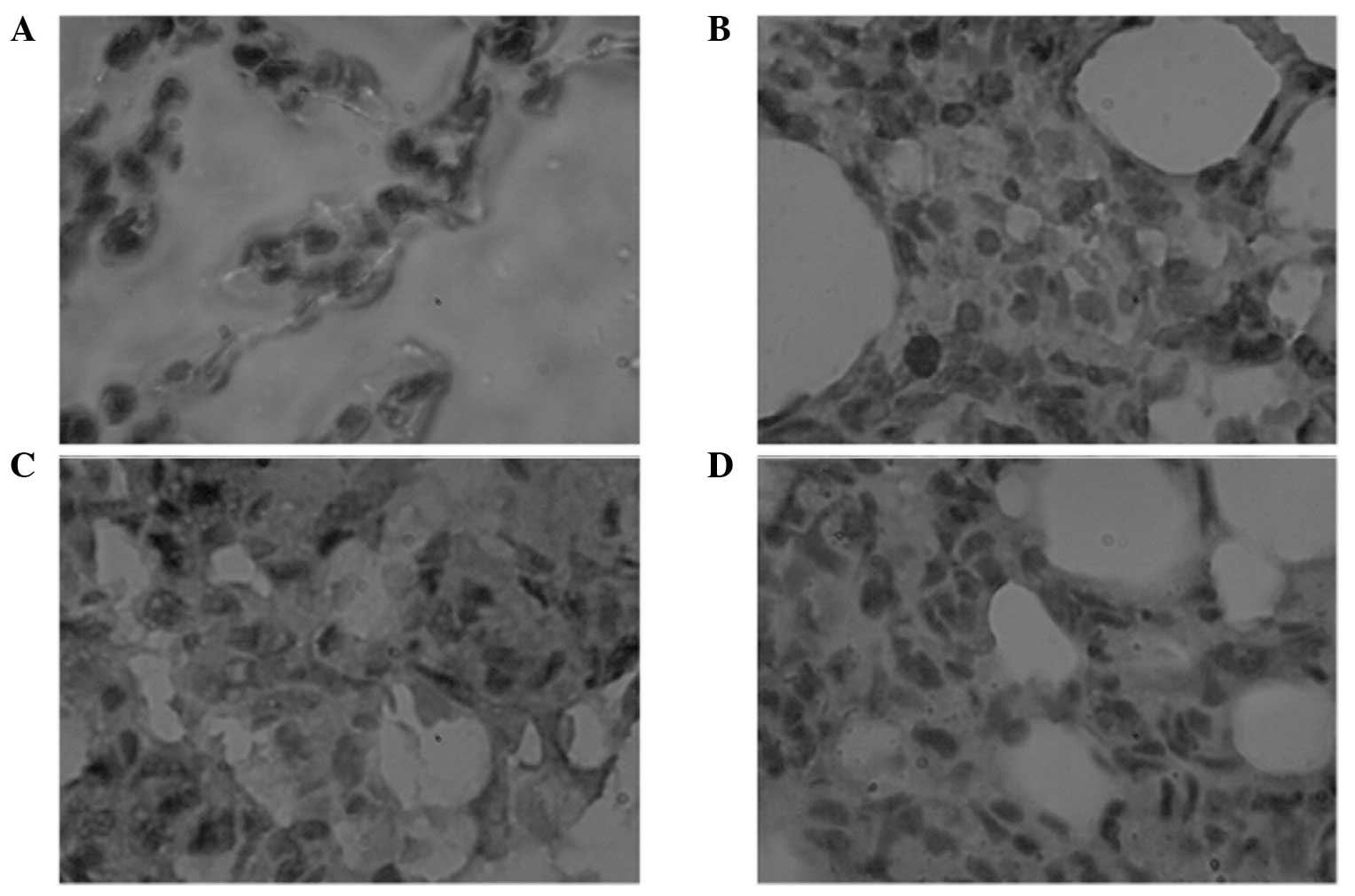
IHC of NF-κBp65
IHC results showing NF-κBp65 expression in the rat
lungs are presented in Fig. 1. The
sum-IOD of NF-κBp65 expression was higher in the smoke group when
compared with the control group (P<0.001). However, in the smoke
+ H2S and H2S groups, the sum-IOD was lower
when compared with the smoke group (P<0.01), but higher when
compared with the control group (P<0.001). The mean density
values of NF-κBp65 expression in the rat lungs were comparable in
the smoke, smoke + H2S and H2S groups, which
were all lower than the value observed in the control group
(P<0.01; Table II).
Discussion
The complex composition of smoke determines the
complex SII pathogenesis, in which oxidative stress is important
(18). Smoke inhalation can
stimulate lung macrophages, neutrophils, vascular endothelial and
smooth muscle cells to release abundant cytokines, including tumor
necrosis factor (TNF)-α, interleukin (IL)-1β, −6 and −8, which
activate NF-κB. Following NF-κB decomposition in the cytoplasm,
active fragments of NF-κBp65 are translocated to the nucleus to
promote downstream iNOS gene transcription, enhancing the synthesis
of iNOS. Arginine is subsequently metabolized, generating large
amounts of NO that react with the superoxide free radical to
synthesize peroxynitrite, leading to lipid peroxidation in the cell
membrane (19). Simultaneously, NO,
nitrous oxide, disulfur monoxide and other oxidizing particles in
smoke are strong oxidants and stimulate granulocytes to release
large quantities of oxyradicals. In addition, oxygen therapy
following hypoxia increases the production of oxyradicals, which
results in lipid peroxidation, membrane destruction and the
activation of inflammatory mediator synthesis. Subsequently, energy
metabolism is affected, causing protein denaturation and
dysfunction. These factors enhance alveolar-capillary permeability,
leading to exudation of the blood component into the alveolar space
and pulmonary edema. Moreover, SII-induced ROS causes excessive NO
synthesis, which results in vascular leakage, failure of hypoxic
pulmonary vasoconstriction and an increase in the production of
cytotoxic reactive nitrogen species (RNS), which further aggravate
pulmonary injury (20–22). A previous study confirmed that cotton
smoke inhalation for 6 h in rats induced typical lung injury
(12). The present study showed the
concentration and sum-IODs of NF-κBp65, and the relative mRNA
expression and concentration of iNOS and NO were increased in the
rat lung tissue after 6 h of smoke inhalation. In addition, there
was an increase in MDA, a peroxide produced in the reaction of free
radicals and polyunsaturated fatty acids in the cell membranes,
indicating an intensified oxidative stress response, lipid
peroxidation and tissue injury.
Early treatment for SII following smoke inhalation
is crucial. With regard to the pathogenesis of SII, interrupting
oxidative stress pathways and decreasing downstream products by
inhibiting NF-κB activation may theoretically ameliorate lung
injury. A previous study demonstrated that continuous intravenous
infusion of arginine vasopressin at a low dose can suppress the
excessive generation of NO by iNOS, significantly reducing lung
injury induced by burning and smoke inhalation (23).
H2S is a harmful gas, commonly associated
with the smell of rotten eggs, and has recently emerged as the
third gaseous signaling molecule in addition to NO and CO (24,25).
Animal experiments have confirmed that intravenous infusion of NaHS
or H2S inhalation has antioxidative, anti-inflammatory
and antiapoptotic effects in animal models of various types of lung
injury (2,9,10,26).
However, the effects of H2S inhalation on SII have not
yet been investigated. In macrophages activated in vitro by
lipopolysaccharide, H2S can inhibit the activation of
the NF-κB signaling pathway, reduce NO production and exert an
antioxidative effect (27).
Therefore, the aim of the present study was to investigate the
effects of H2S inhalation on SII.
The present study revealed that inhalation of 80 ppm
H2S for 6 h immediately after smoke inhalation markedly
reduced rat lung injury. This reduction in injury was characterized
by decreases in the concentration and sum-IOD of NF-κBp65, relative
mRNA expression of iNOS and concentrations of iNOS, NO and MDA in
the rat lung tissue, indicating that H2S inhalation may
reduce iNOS mRNA transcription, and iNOS and NO production, by
inhibiting NF-κBp65 activation. Subsequently, oxidative stress and
cotton smoke inhalation-induced lung injury were reduced. The mean
density of NF-κBp65 in the rat lungs may not be used as an
indicator, since it represents the depth of positive IHC staining
intensity, but not the total quantity of positive staining.
In the H2S group, the concentrations of
MDA, NO and NF-κBp65 were comparable to those in the control group.
However, the iNOS concentration, relative mRNA expression of iNOS
and the sum-IOD of NF-κBp65 were higher in the H2S group
compared with the control group, suggesting that inhalation of 80
ppm H2S for 6 h caused no damage to the rats, but may
activate NF-κBp65 signaling pathways to increase iNOS synthesis,
which correlated with the negative feedback.
Recently, research into the effects of
H2S in vivo has been increasing. Different cell
types or stimulations may lead to opposite results in terms of the
effects of H2S on NF-κB signaling pathways (28). For example, H2S amplifies
the IL-1β-activated NF-κB signaling pathway, increases iNOS
expression and NO production in rat vascular smooth muscle cells
(29). In addition, H2S
activates the NF-κB signaling pathway to increase the production of
proinflammatory cytokines in human monocytes pretreated with
interferon-γ (30). However,
H2S inhibits the activation of the NF-κB signaling
pathway to reduce iNOS expression in lipopolysaccharide-activated
macrophages (27) or microgliacytes
(31). NF-κB is a nuclear
transcription factor that maintains cell sensitivity to oxidative
stress, and TNF-α, IL-1β and NO can activate the signaling cascade
of the NF-κB pathway. This pathway is influenced by numerous
factors, including the status of potential sites sensitive to
oxidative stress, ROS and RNS, selectivity of signaling pathways
and cell types, which may affect the process of oxidative stress
reactions (32). Inhalation of
strong oxidant H2S disturbs the redox balance in
vivo and enhances reduction reactions, which activates the
NF-κB signaling pathway, increases iNOS synthesis and produces NO
against the reducing property of H2S to complete the
negative feedback loop, without evident injury in rats. However,
smoke inhalation increases the oxidizing reactions, which activates
the NF-κB signaling pathway, elevates iNOS expression and NO
synthesis, causing an increase in oxidative stress that cannot be
regulated by the antioxidant system (33). Therefore, H2S inhalation
provides a negative feedback system, inhibiting the activation of
the NF-κB signaling pathway and reducing iNOS expression and NO
synthesis, which subsequently decreases lung injury in rats.
H2S regulates NF-κB activity via the extracellular
signal-regulated kinase signaling pathway (29,30),
p38-mitogen-activated protein kinase signaling pathway (31), heme oxygenase-1 and heat shock
protein 70 (27). A previous study
investigating the mechanisms of H2S (34) revealed that H2S-mediated
sulfhydration (the binding of H2S to active-site
cysteine residues of target proteins to induce protein
sulfhydration and post-translational modification) may be central
to the H2S mechanism.
The present study expands the application of
H2S inhalation. Moreover, strong reductant
H2S in airways can directly neutralize oxidants,
including the superoxide anion, hydrogen peroxide, superoxide
nitrogen and hypochlorous acid (35,36), to
protect the cell membrane against free radical-induced injury.
H2S inhalation is a convenient treatment for SII;
however, further investigation is required. In the present study,
H2S inhalation was applied for only 6 h and the
long-term effects require further study. H2S can
activate ATP-sensitive potassium channels to dilate blood vessels
(6), regulate Fas/Fas ligand death
receptor pathways to reduce apoptosis, and inhibit neurogenic
inflammation and the co-interaction of the three gaseous signaling
molecules (37); these mechanisms
may play roles in the treatment of SII. However, further
confirmation is required with regard to the mechanisms underlying
the effects of H2S in SII.
In conclusion, inhalation of 80 ppm H2S
reduces iNOS mRNA transcription and iNOS and NO production by
inhibiting NF-κBp65 activation, which subsequently decreases
oxidative stress and cotton smoke inhalation-induced lung
injury.
Acknowledgements
This study was funded by the Research Project of the
‘Twelfth Five-year Plan’ for Medical Science Development of PLA
(no. CWS11J180).
References
|
1
|
Ballard-Croft C, Sumpter LR, Broaddus R,
Alexander J, Wang D and Zwischenberger JB: Ovine smoke/burn ARDS
model: a new ventilator-controlled smoke delivery system. J Surg
Res. 164:e155–e162. 2010. View Article : Google Scholar : PubMed/NCBI
|
|
2
|
Esechie A, Kiss L, Olah G, Horváth EM,
Hawkins H, Szabo C and Traber DL: Protective effect of hydrogen
sulfide in a murine model of acute lung injury induced by combined
burn and smoke inhalation. Clin Sci (Lond). 115:91–97. 2008.
View Article : Google Scholar : PubMed/NCBI
|
|
3
|
Papapetropoulos A, Pyriochou A, Altaany Z,
et al: Hydrogen sulfide is an endogenous stimulator of
angiogenesis. Proc Natl Acad Sci USA. 106:21972–21977. 2009.
View Article : Google Scholar : PubMed/NCBI
|
|
4
|
Szabó C and Papapetropoulos A: Hydrogen
sulphide and angiogenesis: mechanisms and applications. Br J
Pharmacol. 164:853–865. 2011. View Article : Google Scholar : PubMed/NCBI
|
|
5
|
Yang G, Wu L, Jiang B, et al:
H2S as a physiologic vasorelaxant: hypertension in mice
with deletion of cystathionine gamma-lyase. Science. 322:587–590.
2008. View Article : Google Scholar : PubMed/NCBI
|
|
6
|
Muzaffar S, Jeremy JY, Sparatore A, Del
Soldato P, Angelini GD and Shukla N: H2S-donating
sildenafil (ACS6) inhibits superoxide formation and gp91phox
expression in arterial endothelial cells: role of protein kinases A
and G. Br J Pharmacol. 155:984–994. 2008. View Article : Google Scholar : PubMed/NCBI
|
|
7
|
Li PC, Chen WC, Chang LC and Lin SC:
Substance P acts via the neurokinin receptor 1 to elicit
bronchoconstriction, oxidative stress, and upregulated ICAM-1
expression after oil smoke exposure. Am J Physiol Lung Cell Mol
Physiol. 294:L912–L920. 2008. View Article : Google Scholar : PubMed/NCBI
|
|
8
|
Zhu XY, Gu H and Ni X: Hydrogen sulfide in
the endocrine and reproductive systems. Expert Rev Clin Pharmacol.
4:75–82. 2011. View Article : Google Scholar : PubMed/NCBI
|
|
9
|
Tokuda K, Kida K, Marutani E, et al:
Inhaled hydrogen sulfide prevents endotoxin-induced systemic
inflammation and improves survival by altering sulfide metabolism
in mice. Antioxid Redox Signal. 17:11–21. 2012. View Article : Google Scholar : PubMed/NCBI
|
|
10
|
Faller S, Zimmermann KK, Strosing KM, et
al: Inhaled hydrogen sulfide protects against
lipopolysaccharide-induced acute lung injury in mice. Med Gas Res.
2:262012. View Article : Google Scholar : PubMed/NCBI
|
|
11
|
Faller S, Ryter SW, Choi AM, Loop T,
Schmidt R and Hoetzel A: Inhaled hydrogen sulfide protects against
ventilator-induced lung injury. Anesthesiology. 113:104–115. 2010.
View Article : Google Scholar : PubMed/NCBI
|
|
12
|
Zhang Y, Han Z, Duan Y, et al: The
influence of inhalation H2S lung in rats. Jie Fang Jun Yi Xue Yuan
Xue Bao. 35:1241–1244. 2014.
|
|
13
|
McPherson C: Regulation of animal care and
research? NIH's opinion. J Animal Sci. 51:492–496. 1980.
|
|
14
|
National Research Council (US) Committee
for the Update of the Guide for the Care and Use of Laboratory
Animals, . Guide for the Care and Use of Laboratory Animals. 8th.
National Academies Press (US); Washington (DC): 2011
|
|
15
|
Lee HM, Greeley GH, Herndon DN, Sinha M,
Luxon BA and Englander EW: A rat model of smoke inhalation injury:
influence of combustion smoke on gene expression in the brain.
Toxicol Appl Pharmacol. 208:255–265. 2005. View Article : Google Scholar : PubMed/NCBI
|
|
16
|
Huang PS, Tang GJ, Chen CH and Kou YR:
Whole-body moderate hypothermia confers protection from wood
smoke-induced acute lung injury in rats: the therapeutic window.
Crit Care Med. 34:1160–1167. 2006. View Article : Google Scholar : PubMed/NCBI
|
|
17
|
Zou YY, Lu J, Poon DJ, Kaur C, Cao Q, Teo
AL and Ling EA: Combustion smoke exposure induces up-regulated
expression of vascular endothelial growth factor, aquaporin 4,
nitric oxide synthases and vascular permeability in the retina of
adult rats. Neuroscience. 160:698–709. 2009. View Article : Google Scholar : PubMed/NCBI
|
|
18
|
Rehberg S, Maybauer MO, Enkhbaatar P,
Maybauer DM, Yamamoto Y and Traber DL: Pathophysiology, management
and treatment of smoke inhalation injury. Expert Rev Respir Med.
3:283–297. 2009. View Article : Google Scholar : PubMed/NCBI
|
|
19
|
Enkhbaatar P and Traber DL:
Pathophysiology of acute lung injury in combined burn and smoke
inhalation injury. Clin Sci (Lond). 107:137–143. 2004. View Article : Google Scholar : PubMed/NCBI
|
|
20
|
Cox RA, Jacob S, Oliveras G, et al:
Pulmonary expression of nitric oxide synthase isoforms in sheep
with smoke inhalation and burn injury. Exp Lung Res. 35:104–118.
2009. View Article : Google Scholar : PubMed/NCBI
|
|
21
|
Westphal M, Enkhbaatar P, Schmalstieg FC,
et al: Neuronal nitric oxide synthase inhibition attenuates
cardiopulmonary dysfunctions after combined burn and smoke
inhalation injury in sheep. Crit Care Med. 36:1196–1204. 2008.
View Article : Google Scholar : PubMed/NCBI
|
|
22
|
Rehberg S, Maybauer MO, Maybauer DM,
Traber LD, Enkhbaatar P and Traber DL: The role of nitric oxide and
reactive nitrogen species in experimental ARDS. Front Biosci (Schol
Ed). 2:18–29. 2010. View
Article : Google Scholar : PubMed/NCBI
|
|
23
|
Westphal M, Rehberg S, Maybauer MO, et al:
Cardiopulmonary effects of low-dose arginine vasopressin in ovine
acute lung injury. Crit Care Med. 39:357–363. 2011. View Article : Google Scholar : PubMed/NCBI
|
|
24
|
Calvert JW: The summer of hydrogen
sulfide: highlights from two international conferences. Med Gas
Res. 3:52013. View Article : Google Scholar : PubMed/NCBI
|
|
25
|
Gadalla MM and Snyder SH: Hydrogen sulfide
as a gasotransmitter. J Neurochem. 113:14–26. 2010. View Article : Google Scholar : PubMed/NCBI
|
|
26
|
Liu WL, Liu ZW, Li TS, Wang C and Zhao B:
Hydrogen sulfide donor regulates alveolar epithelial cell apoptosis
in rats with acute lung injury. Chin Med J (Engl). 126:494–499.
2013.PubMed/NCBI
|
|
27
|
Oh GS, Pae HO, Lee BS, et al: Hydrogen
sulfide inhibits nitric oxide production and nuclear factor-kappaB
via heme oxygenase-1 expression in RAW264.7 macrophages stimulated
with lipopolysaccharide. Free Radic Biol Med. 41:106–119. 2006.
View Article : Google Scholar : PubMed/NCBI
|
|
28
|
Wagner F, Asfar P, Calzia E, Radermacher P
and Szabó C: Bench-to-bedside review: Hydrogen sulfide - the third
gaseous transmitter: applications for critical care. Crit Care.
13:2132009. View
Article : Google Scholar : PubMed/NCBI
|
|
29
|
Jeong SO, Pae HO, Oh GS, et al: Hydrogen
sulfide potentiates interleukin-1beta-induced nitric oxide
production via enhancement of extracellular signal-regulated kinase
activation in rat vascular smooth muscle cells. Biochem Biophys Res
Commun. 345:938–944. 2006. View Article : Google Scholar : PubMed/NCBI
|
|
30
|
Zhi L, Ang AD, Zhang H, Moore PK and
Bhatia M: Hydrogen sulfide induces the synthesis of proinflammatory
cytokines in human monocyte cell line U937 via the ERK-NF-kappaB
pathway. J Leukoc Biol. 81:1322–1332. 2007. View Article : Google Scholar : PubMed/NCBI
|
|
31
|
Hu LF, Wong PT, Moore PK and Bian JS:
Hydrogen sulfide attenuates lipopolysaccharide-induced inflammation
by inhibition of p38 mitogen-activated protein kinase in microglia.
J Neurochem. 100:1121–1128. 2007. View Article : Google Scholar : PubMed/NCBI
|
|
32
|
Janssen-Heininger YM, Poynter ME and
Baeuerle PA: Recent advances towards understanding redox mechanisms
in the activation of nuclear factor kappaB. Free Radic Biol Med.
28:1317–1327. 2000. View Article : Google Scholar : PubMed/NCBI
|
|
33
|
LaLonde C, Nayak U, Hennigan J and Demling
R: Plasma catalase and glutathione levels are decreased in response
to inhalation injury. J Burn Care Rehabil. 18:515–519. 1997.
View Article : Google Scholar : PubMed/NCBI
|
|
34
|
Paul BD and Snyder SH: H2S
signalling through protein sulfhydration and beyond. Nat Rev Mol
Cell Biol. 13:499–507. 2012. View Article : Google Scholar : PubMed/NCBI
|
|
35
|
Whiteman M, Armstrong JS, Chu SH, et al:
The novel neuromodulator hydrogen sulfide: an endogenous
peroxynitrite scavenger? J Neurochem. 90:765–768. 2004. View Article : Google Scholar : PubMed/NCBI
|
|
36
|
Whiteman M, Cheung NS, Zhu YZ, et al:
Hydrogen sulphide: a novel inhibitor of hypochlorous acid-mediated
oxidative damage in the brain? Biochem Biophys Res Commun.
326:794–798. 2005. View Article : Google Scholar : PubMed/NCBI
|
|
37
|
Arai M, Yoshioka S, Nishimura R and Okuda
K: FAS/FASL-mediated cell death in the bovine endometrium. Anim
Reprod Sci. 151:97–104. 2014. View Article : Google Scholar : PubMed/NCBI
|