Introduction
Rotavirus (RV) infection is a common disease
worldwide. Although the infection occurs in children and adults,
children are more commonly infected. RV is currently the main
pathogen that causes severe acute diarrhea in infants (1). In developing countries, 20–50% of
hospitalized children aged <5 years are infected with RV
enteritis, which can lead to the mortality of 600,000 children
worldwide each year (2,3). RV primarily affects the epithelial
cells of the intestinal villi, resulting in induction of the immune
system, predominantly the mucosal immune response. The majority of
previous studies have focused on RV infection prevention through
humoral immunity, while there are only a few studies investigating
cellular immune responses. The majority of the intestinal
epithelium is a collection of T lymphocytes in lymph nodes; thus,
investigating the role of antiviral cell-mediated immunity in the
gut is particularly important (4).
With the increasing number of studies investigating the abnormal
immune response and the mechanism underlying RV infection in the
body, a variety of cellular immune responses associated with RV
cells and cytokines have been demonstrated to be involved in the
inflammatory response (5,6). An imbalance in the proportion of
regulatory T cells (Treg)/T helper 17 cells (Th17), and the
subsequent expression of cytokines, has become the theoretical
basis for the study of various autoimmune disorders and other
diseases (7–9). Therefore, the aim of the present study
was to examine the changes in the cytokine expression of Treg/Th17
cells in the peripheral blood of children with RV enteritis, in
order to further investigate the immunomodulatory effects and
clinical significance of the Treg/Th17 imbalance in the
pathogenesis of RV enteritis.
Subjects and methods
General data
In total, 102 children admitted to the First
Affiliated Hospital of Bengbu Medical College (Bengbu, China) due
to infectious diarrhea between December 2011 and December 2013 were
recruited for the study. The children were diagnosed with an RV
enteritis infection through polymerase chain reaction inspection,
and bowel RV (+) and bacterial culture (-) tests combined with
typical clinical manifestations. The infected children had no
previous history of disease, had good nutrition and had never used
immunosuppressants. Of the 102 children, 59 were male and 43 were
female, with an age range between 3 months and 3 years (mean age,
17.5±6.6 months). A total of 30 healthy children examined in the
hospital during the same time period were selected as the control
group. The control group included 19 males and 11 females, with an
age range between 2 months and 3 years (mean age, 18.5±6.5 months).
No statistically significant differences with regard to age, gender
and weight (P>0.05) were observed between the two groups. The
study was conducted in accordance with the Declaration of Helsinki,
and with approval from the Ethics Committee of the First Affiliated
Hospital of Bengbu Medical College. Written informed consent was
obtained from the participants' parents or guardians.
Sample collection
A fasting venous blood sample (3 ml) was collected
from each individual in the two groups in the morning and divided
into two parts. One part was used for anticoagulation density
gradient centrifugation (10,000 × g, 10 min, room temperature) to
obtain peripheral blood mononuclear cells (PBMCs), for which the
PBMC density was adjusted to 2×10⁶ cells/ml for flow
cytometry. The other part was centrifuged at 5,000 × g for 10 min,
after which the supernatant was obtained and stored at −80°C for
ELISA analysis.
Treg cell detection
A 200-µl sample of cell suspension was added to two
test tubes, which were marked as the isomorphic control and the
sample. Subsequently, 10 µl artificial antigen PerCP-CD4 mAb (BD
Biosciences, Franklin Lakes, NJ, USA) and 10 µl artificial antigen
FITC-CD25 mAb (BD Biosciences) were added to the sample test tube,
while 10 µl PerCP-CD4 mAb and 10 µl FITC-Ig-G1 mAb (BD Biosciences)
were added to the isomorphic control tube and mixed. The test tubes
were incubated in the dark at room temperature for 30 min, and flow
cytometry (FC 500; Beckman Coulter, Inc., Miami, FL, USA) was
conducted following washing and resuspension, using CXP software
(Beckman Coulter).
Th17 cell detection
A 2×10⁶ cells/ml sample of PBMC
suspension was added to a 24-well culture plate, with 1 ml in each
well. Phorbol myristate acetate (20 ng/ml; Sigma-Aldrich, St.
Louis, MO, USA), ionomycin (1 µg/ml; Sigma-Aldrich) and monensin (2
nmol/m1; Sigma-Aldrich) were added to each well and the plates were
incubated in a CO2 incubator at 37°C for 4 h.
Subsequently, the cells were collected and divided into isomorphic
control and sample test tubes, after which 10 µl PerCP-CD4 mAb was
added and the tubes were incubated in the dark at room temperature
for 30 min. Following washing with phosphate-buffered saline, the
cells were fixed with 4% paraformaldehyde. The cells were
maintained at room temperature for 20 min, parting medium (Dingguo
Biotechnology, Inc., Beijing, China) was added and the solution was
left for 10 min. Following centrifugation at 10,000 × g for 10 min
at room temperature, the supernatant was discarded and the cells
were resuspended. Next, 10 µl PE-IL-17mAb artificial antigen (BD
Biosciences) was added to the sample test tube, while 10 µl
PE-Ig-G1mAb (BD Biosciences) was added to the isomorphic control
test tube. The tubes were incubated at room temperature in the dark
for 1 h. Flow cytometry (FC 500; Beckman Coulter, Inc.) was
conducted following washing and resuspension, using CXP
software.
ELISA
With the frozen blood serum, ELISA kits were used to
detect the levels of interleukin (IL)-10, transforming growth
factor (TGF)-β, IL-17 and IL-6, according to the manufacturer's
instructions (R&D Systems, Inc., Minneapolis, MN, USA). Three
repeated wells were set for each sample and standard product, and
the optical density was measured at 492 nm using a microplate
reader (Bio-Rad 680; Bio-Rad, Inc., Hercules, CA, USA).
Statistical analysis
All data were analyzed using SPSS 13.0 statistical
software (SPSS, Inc., Chicago, IL, USA). Measurement data were
analyzed using the t-test and are expressed as the mean ±standard
deviation. The count data were analyzed using the χ2
test. P<0.05 was considered to indicate a statistically
significant difference.
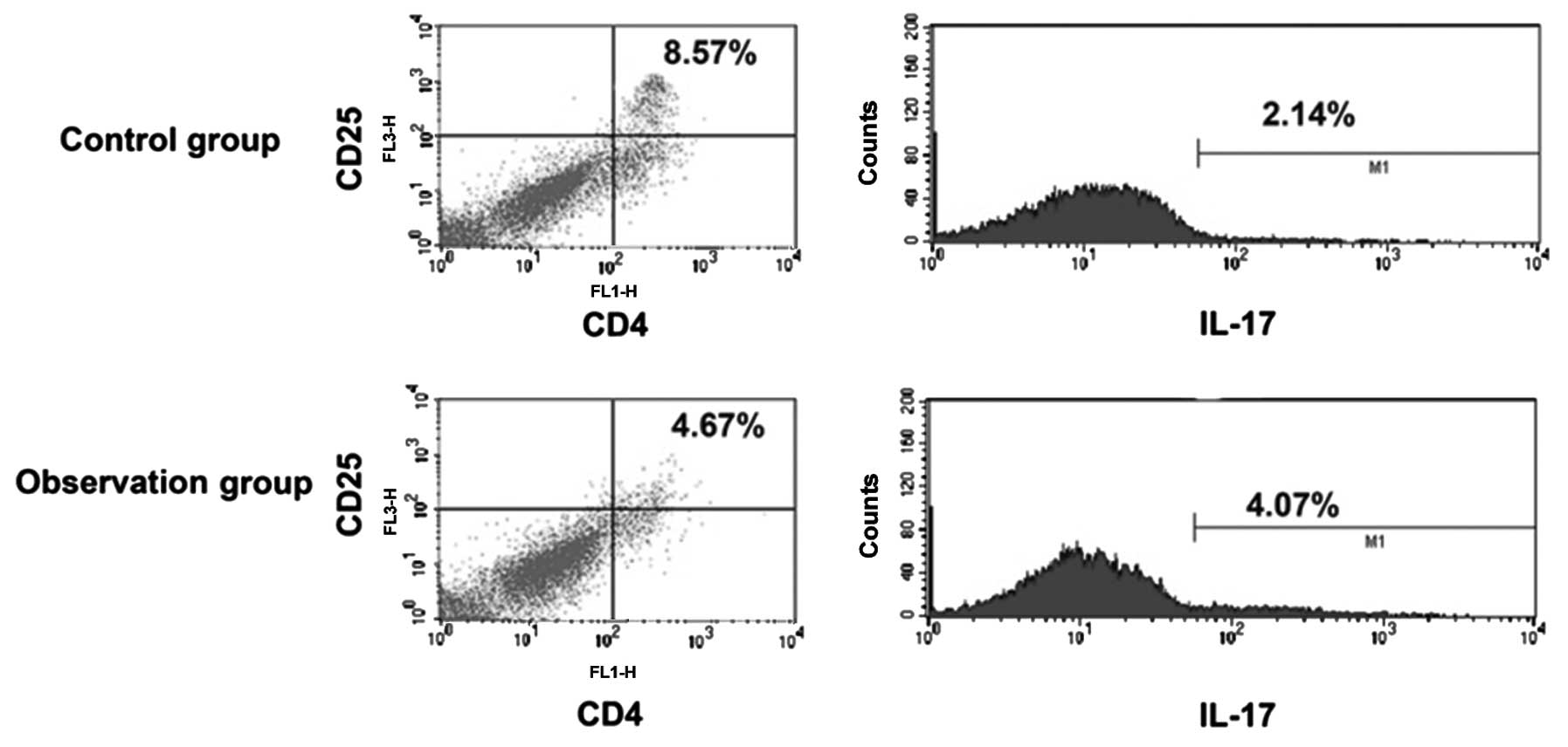
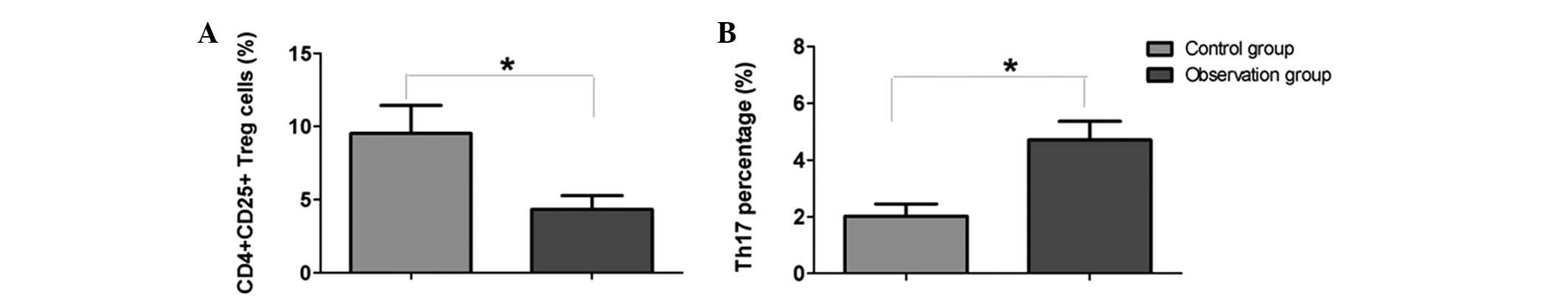
Results
Changes in the proportion of Treg and
Th17 cells in the peripheral blood of children infected with
RV
Following the detection of cell surface marker
molecules by flow cytometry, the results revealed that the
proportion of Treg cells in children with RV enteritis was
significantly decreased when compared with the control group
(P<0.05). However, the Th17 cell proportion was significantly
increased in the RV enteritis patients when compared with the
control group (P<0.05; Figs. 1
and 2).
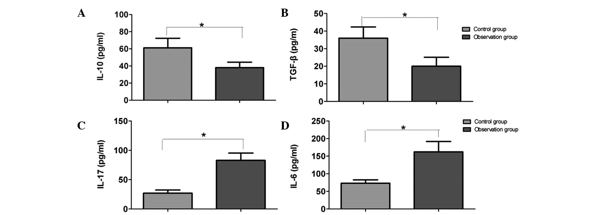
Cytokine expression changes in the
peripheral blood plasma of children infected with RV
ELISA results are shown in Fig. 3. When compared with the control
group, the levels of IL-10 and TGF-β in the peripheral blood serum
of the RV group were significantly decreased (P<0.05). However,
the levels of IL-17 and IL-6 were significantly increased in the RV
group when compared with the control group (P<0.05).
Discussion
RV enteritis is the prime cause of infectious
diarrhea in infants in China and worldwide. The high incidence
rate, acute onset and rapid change of illness have attracted the
attention of clinical professionals, and numerous clinical studies
have investigated the virological characteristics, infectious
epidemiology and clinical control of RV enteritis (10). However, there are few published
studies investigating the underlying mechanism of the immune
response to RV enteritis, and particularly on cell immunity
(11). A number of studies have
shown that balances between Thl/Th2 cells and Thl7/Treg cells exist
in the body's immune system; the imbalance of which is frequently
the basis of certain autoimmune diseases. Thl7 and Treg cells have
been identified as members of the CD4+ T cell family
(12,13). Previous studies have shown that when
the Thl/Th2 imbalance theory is unable to account for certain
diseases caused by immune system disorders, the Thl7/Treg imbalance
theory may provide a subsequent explanation (14,15). The
present study found that the proportion of Treg cells in the
peripheral blood of children infected with RV enteritis decreased
significantly, as compared with the control group, whilst the
increase in the proportion of Th17 cells was marked. These
observations indicate that a Th17/Treg cell imbalance occurs in
patients with RV enteritis, which may be one of the underlying
causes of RV.
Thl7 cells are a T cell subpopulation that has been
identified to be different from the Th1 and Th2 subtypes, which
secrete IL-17, IL-6 and other cytokines. Th17 cells play an
important role in autoimmune diseases, infections, cancer and other
diseases (16). Animal models have
confirmed their synthesis of the proinflammatory cytokines, IL-17,
IL-6 and TNF-α, which are involved in inflammatory response
(17). Studies have also
hypothesized that Th17 cells play a more important role in the
pathogenesis of several inflammatory diseases, as compared with
Th1/Th2 cells. IL-17 is a proinflammatory cytokine that can cause
the upregulation of chemokines and the invasion of inflammatory
cells in tissues (18,19). IL-6 is a pleiotropic cytokine that
functions within a variety of cell types, which adjusts a number of
aspects of innate and adaptive immunity (20). The present study demonstrated that
the expression levels of IL-17 and IL-6 in the peripheral blood of
children with RV were significantly increased when compared with
the control group.
Treg cells are a T cell subpopulation with
immunosuppressive effects that enable active and effective control
over the function of other immune cells. Changes in the number of
Treg cells or inhibition of their activity may result in the
occurrence and development of autoimmune diseases (21). Treg cells primarily secrete IL-10 and
TGF-β (22–24). Kim et al (25) revealed the impact of Tregs on the
immune response during RV infection. Furthermore, Chen et al
(26) found that detection of the
serum level of IL-10 was of great significance for RV clinical
diagnosis. Jiang et al (27)
also confirmed that IL-6, IL-10 and other cytokines play a
potential role in the pathogenesis of RV enteritis. The current
study revealed a significant decrease in the levels of IL-10 and
TGF-β in the peripheral blood of children infected with RV when
compared with the control subjects, which is in line with the
decrease in the Treg cell proportion in the peripheral blood. Nish
et al (28) found that IL-6
exhibited an inhibitory effect on Treg cells, and Hou et al
(29) showed that IL-6 and IL-17
promoted viral persistence via the inhibition of apoptosis and the
synergistic function of cytotoxic T cells. These results indicate
that a Th17 and Treg disorder destroys the basis of the immune
balance, exacerbates viral infections and promotes the occurrence
of RV enteritis.
In conclusion, an imbalance in the proportion of
Treg/Th17 cells and the expression of cytokines in the peripheral
blood of children with RV enteritis may be one of the underlying
pathogeneses. This conclusion has important clinical value with
regard to the detection of Treg/Th17 cells and cytokine expression
for the diagnosis, treatment and prognosis judgment of RV
enteritis.
References
|
1
|
Enweronu-Laryea CC, Sagoe KW, Damanka S,
Lartey B and Amrah GE: Rotavirus genotypes associated with
childhood severe acute diarrhoea in southern Ghana: A
cross-sectional study. Virol J. 10:2872013. View Article : Google Scholar : PubMed/NCBI
|
|
2
|
Turner A, Ngwira B, Witte D, Mwapasa M,
Dove W and Cunliffe N: Surveillance of rotavirus gastro-enteritis
in children in Blantyre, Malawi. Paediatr Int Child Health.
33:42–45. 2013. View Article : Google Scholar : PubMed/NCBI
|
|
3
|
Lesanu G, Becheanu CA, Vlad RM, Pacurar D,
Tincu IF and Smadeanu RE: Clinical characteristics of rotavirus
diarrhea in hospitalized Romanian infants. Pediatr Infect Dis J.
32:89–91. 2013. View Article : Google Scholar : PubMed/NCBI
|
|
4
|
Ogden KM, Snyder MJ, Dennis AF and Patton
JT: Predicted structure and domain organization of rotavirus
capping enzyme and innate immune antagonist VP3. J Virol.
88:9072–9085. 2014. View Article : Google Scholar : PubMed/NCBI
|
|
5
|
Cai HF, Lan JH and Qian LJ: Application of
clinical pathways in children with rotavirus enteritis. Zhongguo
Dang Dai Er Ke Za Zhi. 13:820–822. 2011.(In Chinese). PubMed/NCBI
|
|
6
|
Medici MC, Abelli LA, Guerra P, Dodi I,
Dettori G and Chezzi C: Case report: Detection of rotavirus RNA in
the cerebrospinal fluid of a child with rotavirus gastroenteritis
and meningism. J Med Virol. 83:1637–1640. 2011. View Article : Google Scholar : PubMed/NCBI
|
|
7
|
Li S, Li Y, Qu X, Liu X and Liang J:
Detection and significance of TregFoxP3(+) and Th17 cells in
peripheral blood of non-small cell lung cancer patients. Arch Med
Sci. 10:232–239. 2014. View Article : Google Scholar : PubMed/NCBI
|
|
8
|
Huang X, Chen Y, Zhang F, Yang Q and Zhang
G: Peripheral Th17/Treg cell-mediated immunity imbalance in
allergic rhinitis patients. Braz J Otorhinolaryngol. 80:152–155.
2014. View Article : Google Scholar : PubMed/NCBI
|
|
9
|
Huang XK, Yang QT, Chen YL, Zhang FC and
Zhang GH: Expression of peripheral blood gammadelta T cells, treg
cells and cytokines IL-17 and TGF-beta1 in patients with allergic
rhinitis. Zhonghua Er Bi Yan Hou Tou Jing Wai Ke Za Zhi.
48:544–548. 2013.(In Chinese). PubMed/NCBI
|
|
10
|
Zhou R, Xu JL, Wu D and Tang JL: Analysis
of prognostic factors for infantile rotavirus infection. Genet Mol
Res. 14:790–796. 2015. View Article : Google Scholar : PubMed/NCBI
|
|
11
|
Medici MC, Abelli LA, Martinelli M, et al:
Clinical and molecular observations of two fatal cases of
rotavirus-associated enteritis in children in Italy. J Clin
Microbiol. 49:2733–2739. 2011. View Article : Google Scholar : PubMed/NCBI
|
|
12
|
Li L, Zhang SN, Ran JH, Liu J, Li Z and Li
LB: Mechanism of immune hyporesponsiveness induced by
recipient-derived immature dendritic cells in liver transplantation
rat. Chin Med Sci J. 26:28–35. 2011. View Article : Google Scholar : PubMed/NCBI
|
|
13
|
Shen Y, Hong SL, Hu GH, Tang XY, Kou W and
Pan CK: Imbalance of Th17/Treg cell ratio in peripheral blood of
patients with nasal polyposis and its clinical significance. Xi Bao
Yu Fen Zi Mian Yi Xue Za Zhi. 27:1339–1342. 2011.(In Chinese).
|
|
14
|
Mai J, Wang H and Yang XF: Th 17 cells
interplay with Foxp3+ Tregs in regulation of
inflammation and autoimmunity. Front Biosci (Landmark Ed).
15:986–1006. 2010. View
Article : Google Scholar : PubMed/NCBI
|
|
15
|
Shen X, Du J, Guan W and Zhao Y: The
balance of intestinal Foxp3+ regulatory T cells and Th17
cells and its biological significance. Expert Rev Clin Immunol.
10:353–362. 2014. View Article : Google Scholar : PubMed/NCBI
|
|
16
|
Cosmi L, Santarlasci V, Maggi L, Liotta F
and Annunziato F: Th17 plasticity: Pathophysiology and treatment of
chronic inflammatory disorders. Curr Opin Pharmacol. 17:12–16.
2014. View Article : Google Scholar : PubMed/NCBI
|
|
17
|
Brandenberger C, Li N, Jackson-Humbles DN,
Rockwell CE, Wagner JG and Harkema JR: Enhanced allergic airway
disease in old mice is associated with a Th17 response. Clin Exp
Allergy. 44:1282–1292. 2014. View Article : Google Scholar : PubMed/NCBI
|
|
18
|
Cosmi L, Maggi L, Santarlasci V, Liotta F
and Annunziato F: T helper cells plasticity in inflammation.
Cytometry A. 85:36–42. 2014. View Article : Google Scholar : PubMed/NCBI
|
|
19
|
Han Y, Ye A, Bi L, Wu J, Yu K and Zhang S:
Th17 cells and IL-17 increase with poor prognosis in patients with
acute myeloid leukemia. Cancer Sci. 105:933–942. 2014. View Article : Google Scholar : PubMed/NCBI
|
|
20
|
Pal M, Febbraio MA and Whitham M: From
cytokine to myokine: The emerging role of interleukin-6 in
metabolic regulation. Immunol Cell Biol. 92:331–339. 2014.
View Article : Google Scholar : PubMed/NCBI
|
|
21
|
Cao Y, Li C, Zhang Q, Wang Y and Xia R:
Extracellular ubiquitin enhances the suppressive effects of
regulatory T cells on effectot T cell responses. Clin Lab.
60:1983–1991. 2014.PubMed/NCBI
|
|
22
|
Yang X, Gao T, Shi R, Zhou X, Qu J, Xu J,
Shan Z and Teng W: Effect of iodine excess on Th1, Th2, Th17, and
Treg cell subpopulations in the thyroid of NOD.H-2h4 mice. Biol
Trace Elem Res. 159:288–296. 2014. View Article : Google Scholar : PubMed/NCBI
|
|
23
|
Wen K, Li G, Yang X, et al:
CD4+ CD25− FoxP3+ regulatory cells
are the predominant responding regulatory T cells after human
rotavirus infection or vaccination in gnotobiotic pigs. Immunology.
137:160–171. 2012. View Article : Google Scholar : PubMed/NCBI
|
|
24
|
Mesa MC, Gutiérrez L, Duarte-Rey C, Angel
J and Franco MA: A TGF-beta mediated regulatory mechanism modulates
the T cell immune response to rotavirus in adults but not in
children. Virology. 399:77–86. 2010. View Article : Google Scholar : PubMed/NCBI
|
|
25
|
Kim B, Feng N, Narváez CF, He XS, Eo SK,
Lim CW and Greenberg HB: The influence of CD4+
CD25+ Foxp3+ regulatory T cells on the immune
response to rotavirus infection. Vaccine. 26:5601–5611. 2008.
View Article : Google Scholar : PubMed/NCBI
|
|
26
|
Chen SM, Ku MS, Lee MY, Tsai JD and Sheu
JN: Diagnostic performance of serum interleukin-6 and
interleukin-10 levels and clinical predictors in children with
rotavirus and norovirus gastroenteritis. Cytokine. 59:299–304.
2012. View Article : Google Scholar : PubMed/NCBI
|
|
27
|
Jiang B, Snipes-Magaldi L, Dennehy P, et
al: Cytokines as mediators for or effectors against rotavirus
disease in children. Clin Diagn Lab Immunol. 10:995–1001.
2003.PubMed/NCBI
|
|
28
|
Nish SA, Schenten D, Wunderlich T, et al:
T cell-intrinsic role of IL-6 signaling in primary and memory
responses. eLife. 3:e019492014. View Article : Google Scholar : PubMed/NCBI
|
|
29
|
Hou W, Jin YH, Kang HS and Kim BS:
Interleukin-6 (IL-6) and IL-17 synergistically promote viral
persistence by inhibiting cellular apoptosis and cytotoxic T cell
function. J Virol. 88:8479–8489. 2014. View Article : Google Scholar : PubMed/NCBI
|