Spandidos Publications Logo
  • About
    • About Spandidos
    • Aims and Scopes
    • Abstracting and Indexing
    • Editorial Policies
    • Reprints and Permissions
    • Job Opportunities
    • Terms and Conditions
    • Contact
  • Journals
    • All Journals
    • Oncology Letters
      • Oncology Letters
      • Information for Authors
      • Editorial Policies
      • Editorial Board
      • Aims and Scope
      • Abstracting and Indexing
      • Bibliographic Information
      • Archive
    • International Journal of Oncology
      • International Journal of Oncology
      • Information for Authors
      • Editorial Policies
      • Editorial Board
      • Aims and Scope
      • Abstracting and Indexing
      • Bibliographic Information
      • Archive
    • Molecular and Clinical Oncology
      • Molecular and Clinical Oncology
      • Information for Authors
      • Editorial Policies
      • Editorial Board
      • Aims and Scope
      • Abstracting and Indexing
      • Bibliographic Information
      • Archive
    • Experimental and Therapeutic Medicine
      • Experimental and Therapeutic Medicine
      • Information for Authors
      • Editorial Policies
      • Editorial Board
      • Aims and Scope
      • Abstracting and Indexing
      • Bibliographic Information
      • Archive
    • International Journal of Molecular Medicine
      • International Journal of Molecular Medicine
      • Information for Authors
      • Editorial Policies
      • Editorial Board
      • Aims and Scope
      • Abstracting and Indexing
      • Bibliographic Information
      • Archive
    • Biomedical Reports
      • Biomedical Reports
      • Information for Authors
      • Editorial Policies
      • Editorial Board
      • Aims and Scope
      • Abstracting and Indexing
      • Bibliographic Information
      • Archive
    • Oncology Reports
      • Oncology Reports
      • Information for Authors
      • Editorial Policies
      • Editorial Board
      • Aims and Scope
      • Abstracting and Indexing
      • Bibliographic Information
      • Archive
    • Molecular Medicine Reports
      • Molecular Medicine Reports
      • Information for Authors
      • Editorial Policies
      • Editorial Board
      • Aims and Scope
      • Abstracting and Indexing
      • Bibliographic Information
      • Archive
    • World Academy of Sciences Journal
      • World Academy of Sciences Journal
      • Information for Authors
      • Editorial Policies
      • Editorial Board
      • Aims and Scope
      • Abstracting and Indexing
      • Bibliographic Information
      • Archive
    • International Journal of Functional Nutrition
      • International Journal of Functional Nutrition
      • Information for Authors
      • Editorial Policies
      • Editorial Board
      • Aims and Scope
      • Abstracting and Indexing
      • Bibliographic Information
      • Archive
    • International Journal of Epigenetics
      • International Journal of Epigenetics
      • Information for Authors
      • Editorial Policies
      • Editorial Board
      • Aims and Scope
      • Abstracting and Indexing
      • Bibliographic Information
      • Archive
    • Medicine International
      • Medicine International
      • Information for Authors
      • Editorial Policies
      • Editorial Board
      • Aims and Scope
      • Abstracting and Indexing
      • Bibliographic Information
      • Archive
  • Articles
  • Information
    • Information for Authors
    • Information for Reviewers
    • Information for Librarians
    • Information for Advertisers
    • Conferences
  • Language Editing
Spandidos Publications Logo
  • About
    • About Spandidos
    • Aims and Scopes
    • Abstracting and Indexing
    • Editorial Policies
    • Reprints and Permissions
    • Job Opportunities
    • Terms and Conditions
    • Contact
  • Journals
    • All Journals
    • Biomedical Reports
      • Information for Authors
      • Editorial Policies
      • Editorial Board
      • Aims and Scope
      • Abstracting and Indexing
      • Bibliographic Information
      • Archive
    • Experimental and Therapeutic Medicine
      • Information for Authors
      • Editorial Policies
      • Editorial Board
      • Aims and Scope
      • Abstracting and Indexing
      • Bibliographic Information
      • Archive
    • International Journal of Epigenetics
      • Information for Authors
      • Editorial Policies
      • Editorial Board
      • Aims and Scope
      • Abstracting and Indexing
      • Bibliographic Information
      • Archive
    • International Journal of Functional Nutrition
      • Information for Authors
      • Editorial Policies
      • Editorial Board
      • Aims and Scope
      • Abstracting and Indexing
      • Bibliographic Information
      • Archive
    • International Journal of Molecular Medicine
      • Information for Authors
      • Editorial Policies
      • Editorial Board
      • Aims and Scope
      • Abstracting and Indexing
      • Bibliographic Information
      • Archive
    • International Journal of Oncology
      • Information for Authors
      • Editorial Policies
      • Editorial Board
      • Aims and Scope
      • Abstracting and Indexing
      • Bibliographic Information
      • Archive
    • Medicine International
      • Information for Authors
      • Editorial Policies
      • Editorial Board
      • Aims and Scope
      • Abstracting and Indexing
      • Bibliographic Information
      • Archive
    • Molecular and Clinical Oncology
      • Information for Authors
      • Editorial Policies
      • Editorial Board
      • Aims and Scope
      • Abstracting and Indexing
      • Bibliographic Information
      • Archive
    • Molecular Medicine Reports
      • Information for Authors
      • Editorial Policies
      • Editorial Board
      • Aims and Scope
      • Abstracting and Indexing
      • Bibliographic Information
      • Archive
    • Oncology Letters
      • Information for Authors
      • Editorial Policies
      • Editorial Board
      • Aims and Scope
      • Abstracting and Indexing
      • Bibliographic Information
      • Archive
    • Oncology Reports
      • Information for Authors
      • Editorial Policies
      • Editorial Board
      • Aims and Scope
      • Abstracting and Indexing
      • Bibliographic Information
      • Archive
    • World Academy of Sciences Journal
      • Information for Authors
      • Editorial Policies
      • Editorial Board
      • Aims and Scope
      • Abstracting and Indexing
      • Bibliographic Information
      • Archive
  • Articles
  • Information
    • For Authors
    • For Reviewers
    • For Librarians
    • For Advertisers
    • Conferences
  • Language Editing
Login Register Submit
  • This site uses cookies
  • You can change your cookie settings at any time by following the instructions in our Cookie Policy. To find out more, you may read our Privacy Policy.

    I agree
Search articles by DOI, keyword, author or affiliation
Search
Advanced Search
presentation
Experimental and Therapeutic Medicine
Join Editorial Board Propose a Special Issue
Print ISSN: 1792-0981 Online ISSN: 1792-1015
Journal Cover
August-2015 Volume 10 Issue 2

Full Size Image

Sign up for eToc alerts
Recommend to Library

Journals

International Journal of Molecular Medicine

International Journal of Molecular Medicine

International Journal of Molecular Medicine is an international journal devoted to molecular mechanisms of human disease.

International Journal of Oncology

International Journal of Oncology

International Journal of Oncology is an international journal devoted to oncology research and cancer treatment.

Molecular Medicine Reports

Molecular Medicine Reports

Covers molecular medicine topics such as pharmacology, pathology, genetics, neuroscience, infectious diseases, molecular cardiology, and molecular surgery.

Oncology Reports

Oncology Reports

Oncology Reports is an international journal devoted to fundamental and applied research in Oncology.

Experimental and Therapeutic Medicine

Experimental and Therapeutic Medicine

Experimental and Therapeutic Medicine is an international journal devoted to laboratory and clinical medicine.

Oncology Letters

Oncology Letters

Oncology Letters is an international journal devoted to Experimental and Clinical Oncology.

Biomedical Reports

Biomedical Reports

Explores a wide range of biological and medical fields, including pharmacology, genetics, microbiology, neuroscience, and molecular cardiology.

Molecular and Clinical Oncology

Molecular and Clinical Oncology

International journal addressing all aspects of oncology research, from tumorigenesis and oncogenes to chemotherapy and metastasis.

World Academy of Sciences Journal

World Academy of Sciences Journal

Multidisciplinary open-access journal spanning biochemistry, genetics, neuroscience, environmental health, and synthetic biology.

International Journal of Functional Nutrition

International Journal of Functional Nutrition

Open-access journal combining biochemistry, pharmacology, immunology, and genetics to advance health through functional nutrition.

International Journal of Epigenetics

International Journal of Epigenetics

Publishes open-access research on using epigenetics to advance understanding and treatment of human disease.

Medicine International

Medicine International

An International Open Access Journal Devoted to General Medicine.

Journal Cover
August-2015 Volume 10 Issue 2

Full Size Image

Sign up for eToc alerts
Recommend to Library

  • Article
  • Citations
    • Cite This Article
    • Download Citation
    • Create Citation Alert
    • Remove Citation Alert
    • Cited By
  • Similar Articles
    • Related Articles (in Spandidos Publications)
    • Similar Articles (Google Scholar)
    • Similar Articles (PubMed)
  • Download PDF
  • Download XML
  • View XML
Article Open Access

Expression of glucocorticoid receptor-α in the epidermis of patients with psoriasis vulgaris

  • Authors:
    • Xiaowen Pang
    • Ping Zhang
    • Sainan Zhu
    • Guangjin Guo
  • View Affiliations / Copyright

    Affiliations: Department of Dermatology, Air Force General Hospital of People's Liberation Army, Beijing 100142, P.R. China, Department of Statistics, The First Hospital of Peking University, Beijing 100030, P.R. China
    Copyright: © Pang et al. This is an open access article distributed under the terms of Creative Commons Attribution License.
  • Pages: 419-422
    |
    Published online on: May 26, 2015
       https://doi.org/10.3892/etm.2015.2522
  • Expand metrics +
Metrics: Total Views: 0 (Spandidos Publications: | PMC Statistics: )
Metrics: Total PDF Downloads: 0 (Spandidos Publications: | PMC Statistics: )
Cited By (CrossRef): 0 citations Loading Articles...

This article is mentioned in:


Abstract

Expression of glucocorticoid receptor (GR)-α is observed in almost all tissues and cells of the body; thus, investigating the expression of the receptor in the epidermis of patients with psoriasis vulgaris. The aim of the present study was to investigate GR‑α expression in the epidermis of psoriasis vulgaris patients The study population consisted of 26 patients with psoriasis vulgaris and 10 normal control cases. None of the patients had received any prior treatment with glucocorticoids. Epithelial tissue samples were detected using a streptavidin peroxidation‑enzymatic method for biopsy. Non‑lesional tissue samples from within a range of 3 cm of lesions formed the non‑lesional group, and lesional tissue samples formed the lesional group. Pathological image analysis system (named CMIAS) was used to convert image signals into numeric values, according to the optical density per unit area. GR‑α expression was observed within the nucleus in the normal control group; however, cytoplasmic expression was observed in the lesions of the psoriatic group. The optical density values were significantly lower in the psoriatic group when compared with the normal control group, indicating a statistically significant difference in GR‑α expression between the two groups (P<0.001), and this decreasing correlation was unaffected by the administration of steroids for 6 months. Therefore, decreased expression of GR‑α may play an important role in the degeneration of keratinocytes in patients with psoriasis vulgaris.

Introduction

The glucocorticoid receptor (GR) is a hormone-dependent transcription factor and a member of the nuclear receptor superfamily. The functions of the receptor include the transcription and regulation of a variety of genes. Glucocorticoids (GCs) usually target GR-α, which is expressed in almost all tissues and cells of the body (1). GR-α is not only involved in signal transduction, but has also been demonstrated to exert multiple functions, including anti-inflammatory, antiproliferative and immunosuppressive effects (1).

Psoriasis is a form of chronic inflammatory dermatosis, which is characterized by the excessive proliferation and abnormal differentiation of epidermal keratinocytes (2). The epidermal tissue has been reported to be one of the target tissues for GCs. Thus, GR-α was hypothesized to be involved in the overproliferation and abnormal differentiation of epidermal keratinocytes (2) underlying the pathogenesis of psoriasis. In the present study, immunohistochemistry was used to detect the expression of GR-α in normal control subjects, and lesional and non-lesional samples from patients with psoriasis. The aim of the present study was to investigate the function of GR-α in the molecular pathogenesis underlying psoriasis.

Materials and methods

Study population

The study population was composed of 26 patients with psoriasis vulgaris and 10 healthy control subjects, who were recruited from the Air Force General Hospital of the People's Liberation Army (Beijing, China) in 2013. The patients had been undergoing consultations at least once a week with a physician with regard to their skin condition. However, prior to sampling, none of the patients had received treatment with GCs, immunosuppressants or vitamin A acid drugs, and had only used Vaseline or other moisturizing creams. With regard to the effect of treatment with GCs, studies were performed prior to and after using it in the 6 months follow-up survey independently.

Institutional Ethics Board approval was obtained from the Medical Ethics Committee of the Air Force General Hospital of People's Liberation Army (Beijing, China). All the patients had previously provided written, informed consent to having their clinical and pathological information used for this research.

Sample collection

The psoriasis area and severity index (PASI) was used to detect and score the skin lesions of the 26 psoriasis cases. The present study included three groups, the lesional group contained samples from typical lesions of psoriasis cases, while the non-lesional group consisted of samples from non-lesional tissue within the range of 3 cm. In addition, normal control samples were collected from the normal control group.

In the two psoriasis groups (lesional and non-lesional), five specimens were obtained from the torso and 21 specimens were collected from the arms or legs. With regard to the control group with normal skin tissue, five specimens were collected from the head tissue, two specimens were obtained from the torso and four specimens were collected from the arms or legs. Every epidermis sample had a diameter of 3 mm and was obtained using a trephine. All the samples were fixed in 10% formalin and embedded in paraffin, immediately.

Section staining

Paraffin sections (4 µm) from the three groups were analyzed using a streptavidin peroxidase-enzymatic (SP) method. Two processes were utilized for sample preparation, which all samples underwent. Firstly, after 2 h incubation at 60°C, the paraffin sections were dewaxed in xylene and hydrated in graded ethanol. Subsequently, the sections were blocked with 3% hydrogen peroxide for 5 min at 25°C. In the second process of sample preparation, the sections were subjected to a high temperature of 140°C for 2 min in citrate buffer. At the end of each process, the samples were washed three times with phosphate-buffered saline (PBS) for 2 min at 25°C.

GR-α expression was detected using a rabbit anti-human GR-α primary antibody (1:300, EH-1657; Beijing Biosynthesis Biotechnology Co., Ltd., Beijing, China). Subsequently, samples were analyzed using an anti-rabbit Ultra-Sensitive Immunohistochemistry Kit (#7770; Fuzhou Maixin Biotechnology Development, Co., Ltd., Fuzhou, China), following the manufacturer's instruction. The chromogenic reaction was observed using a 3,3′-diaminobenzidine (DAB) kit (#0017; Maixin Biotechnology Development, Co., Ltd.), following the manufacturer's protocol. The primary antibody was incubated overnight under 4°C. The secondary and horse radish peroxidase-conjugated tertiary antibodies were incubated for 20 min at 37°C. The secondary and tertiary antibodies were provided with the kit. DAB was used as a substrate to visualize the reaction.

Samples were clarified using xylene and counterstained with hematoxylin, in order to produce clearer and identifiable images. PBS was used as a negative control. The specific brown granules in the tissues and cells were observed using an Olympus BX60 optical microscope (Olympus Corporation, Tokyo, Japan).

Quantification of GR-α expression

Using five typical fields of view under the optical microscope, images of the epidermis, excluding the stratum corneum, were collected. A pathological image analysis system, known as CMIAS (jointly developed by the Air Force General Hospital and Beijing University of Aeronautics and Astronautics, Beijing, China) (3), was used to convert the image signal into a numeric value, according to the optical density per unit area, which was measured using a Nikon DS-SMC-UI system (Nikon Corporation, Tokyo, Japan) (4,5). The mean value of the specimens was calculated.

Reagents

The polyclonal rabbit anti-human GR-α antibody was purchased from Beijing Biosynthesis Biotechnology Co, Ltd. (Beijing, China). The highly sensitive SP immunohistochemical kit was obtained from Beijing Hantian Biotechnology Co., Ltd. (Beijing, China), and the DAB chromogenic kit was obtained from Fuzhou Maxim Biotechnology Co., Ltd. (Fuzhou, China).

Statistical analysis

All the data are represented as the mean ± standard deviation of three or more independent experiments. If the data exhibited homogeneity, analysis of variance, the Student-Newman-Keuls test and Pearson's correlation analysis were performed. If the data exhibited heterogeneity, the Kruskal-Wallis test, Games-Howell test and Spearman's correlation analysis were performed. All statistical analyses were conducted using SPSS 17.0 software (SPSS, Inc, Chicago, IL, USA), and P<0.05 was considered to indicate a statistically significant difference.

Results

Population characteristics

Of the 26 psoriasis cases, 19 individuals were male and 7 patients were female, with an average male-to-female gender ratio of 2.714. The average age was 36.0±11.2 months (range, 19–57 months), and the average PASI value was 21.9±6.1 (range, 11.6–31.8). In the normal control group, 7 subjects were male and 3 were female, with an average male-to-female gender ratio of 2.333. The average age of the subjects was 35.5±14.0 months (range, 10–50 months). There were no statistically significant differences with regard to patient gender and age between the groups.

Expression of GR-α

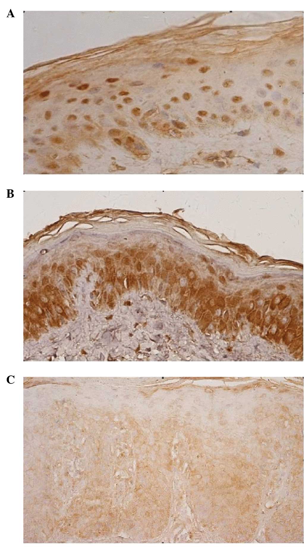
GR-α expression within the nucleus was observed in the normal control group, while cytoplasmic expression was observed in the lesions of the psoriatic group (Fig. 1A and C), excluding the stratum corneum. However, in the non-lesional psoriasis samples, GR-α expression was observed in the nucleus and cytoplasm (Fig. 1B). Thus, the microscopic observations revealed that GR-α was differentially expressed among the three groups.

Figure 1.

Expression of glucocorticoid receptor (GR)-α in epidermal cells (magnification, ×400; pathological sections treated with streptavidin peroxidase). (A) Intranuclear expression of GR-α in the normal control group. (B) Intranuclear and intracytoplasmic expression of GR-α in the non-lesional psoriatic epidermis group. (C) Intracytoplasmic expression of GR-α in the lesions of the psoriatic epidermis group.

The results from the quantitative image analysis (Table I) revealed that there was no statistically significant difference in the expression levels of GR-α between the normal control group and the non-lesional psoriasis samples (P=0.677). However, the expression levels of GR-α in the psoriasis lesional group were significantly lower when compared with the other two groups (P<0.001).

Table I.

Analysis of GR-α expression.

Table I.

Analysis of GR-α expression.

GroupCases (n)Optical density
Normal epidermis control10 0.166±0.032
Non-lesional psoriatic epidermis26 0.161±0.033
Lesional psoriatic epidermis26 0.093±0.025a

{ label (or @symbol) needed for fn[@id='tfn1-etm-0-0-2522'] } Results are expressed as the mean ± standard deviation.

a P<0.001, vs. normal control group. GR, glucocorticoid receptor.

According to the 6-month follow-up survey, administration of GCs was not shown to affect GR-α expression. There was no statistically significant difference prior to and following the administration of GCs (P=0.300).

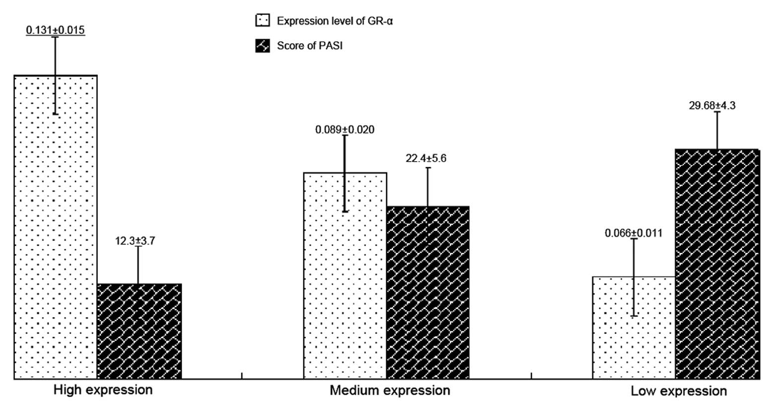
The association between GR-α expression and psoriasis severity, according to the PASI score, is illustrated in Fig. 2. According to the GR-α expression level, the samples were divided into high, medium and low expression groups. A higher PASI score was observed in those with lower expression levels of GR-α. In addition, Pearson's correlation analysis revealed that there was a negative correlation between the average optical density of GR-α and the PASI score in 26 patients with psoriasis (r=-0.627, P=0.007).

Figure 2.

Negative correlation between the expression of GR-α and the PASI score in the high, medium and low GR-α expression groups. GR, glucocorticoid receptor; PASI, psoriasis area and severity index.

Discussion

Chrousos and Kino (6) suggested that the expression of 20% of genes in the human genome was influenced by GCs. Thus, GCs were hypothesized to participate in the regulation of a number physiological and pathological processes, such as cell growth and differentiation, metabolism, immunity and inflammation, through activating the corresponding GR-α in cells (6). The inactive form of GR-α, which is not combined with GC, has been demonstrated to primarily exist in the cytoplasm. By contrast, the active form of GR-α, which is combined with GC, can be rapidly transported into the nucleus, where the receptor is able to recognize and bind to the GC reactive element, which is located upstream of the target gene (6,7). As a transcription factor, activated GR-α is able to activate or inhibit the expression of the target gene by directly or indirectly regulating the transcription of the target gene, to subsequently produce various biological effects, including antiproliferation, anti-inflammation and immunosuppression (7). In the study by Fan et al, the level of serum GC in patients with psoriasis was demonstrated to be higher than normal, while the mRNA expression level of GR in peripheral blood leukocytes was significantly lower than normal. These results indicated that the decreased expression of GR in psoriasis patients may prevent GC from effectively exerting an anti-inflammatory effect, subsequently inducing or aggravating the psoriasis immune inflammatory reaction (8).

The results of the present study demonstrated that GR-α was expressed in the nucleus, where the receptor played a role in the cell proliferation and differentiation of normal human epidermal keratinocytes. Abnormal expression of GR-α was observed in the psoriatic epidermis, where the receptor was abundantly expressed in the cytoplasm; however, the expression level of GR-α in the non-lesional psoriasis epidermis did not decrease. These results indicated that the translocation of GR-α into the nucleus was blocked. In the psoriasis lesions, the expression level of GR-α decreased significantly; thus, GR-α failed to enter the nucleus, resulting in complete malfunction with regard to gene regulation.

Results from Pearson's correlation analysis revealed that there was a negative correlation between the psoriasis severity and the expression of GR-α in the psoriasis lesions. This observation indicates that the more serious the lesions of the psoriasis, the lower the expression of GR-α in the epidermis.

A previous study found that the decreasing expression of GR was the result of decreased levels of GC (9). However, the results of the present study indicate that reduced expression of GR-α was not associated with long-term exposure to GC. To eliminate this hypothesis, in the present study, the psoriasis patients were divided into two groups. One group did not receive GC treatment, while the other group received partial GC treatment (no third GC administration) over 6 months. The results demonstrated that there was no statistically significant difference in the expression levels of GR-α in the psoriasis lesions between the two groups, indicating that partial GC administration was not the fundamental reason for the abnormal expression of GR-α in the psoriasis epidermis.

In conclusion, the present study demonstrated that psoriasis epidermal keratinocytes exhibit decreased expression and abnormal localization of GR-α. Thus, GR-α may play an important role in the excessive proliferation and abnormal differentiation observed in the epidermal cells of patients with psoriasis. However, the factors contributing to this phenomenon remain unclear. These factors may be crucially involved in the pathogenesis of psoriasis vulgaris and require further investigation. In summary, the results of the present study provides insights relevant to the investigation of the mechanisms underlying the pathogenesis of psoriasis vulgaris, and may aid the development of novel therapies.

Abbreviations:

GR-α

glucocorticoid receptor-α

SP

streptavidin peroxidase

GC

glucocorticoid

PASI

psoriasis area and severity index

DAB

3,3′-diaminobenzidine

PBS

phosphate-buffered saline

References

1 

Luft FC: Glucocorticoid receptor function and clinical medicine. J Mol Med Berl. 80:267–269. 2002. View Article : Google Scholar : PubMed/NCBI

2 

Serres M, Viac J and Schmitt D: Glucocorticoid receptor localization in human epidermal cells. Arch Dermatol Res. 288:140–146. 1996. View Article : Google Scholar : PubMed/NCBI

3 

Yu P, Bu H, Wang H, Zhao G, Zhang J and Zhou Q: Comparative study on image analysis and manual counting of immunohistochemistry. Sheng Wu Yi Xue Gong Cheng Xue Za Zhi. 20:288–290. 2003.(In Chinese). PubMed/NCBI

4 

Ying C, Chunmin Y, Qingsen L, Mingzhou G, Yunsheng Y, Gaoping M and Ping W: Effects of simulated weightlessness on tight junction protein occludin and Zonula Occluden-1 expression levels in the intestinal mucosa of rats. J Huazhong Univ Sci Technolog Med Sci. 31:26–32. 2011. View Article : Google Scholar : PubMed/NCBI

5 

Li CH, Yang ZW, Yin ZR, Jin Z, Xing DG, Piao LH, Kim YC and Xu WX: Relationship between atrial natriuretic peptide-immunoreactive cells and microvessels in rat gastric mucosa. Acta Pharmacol Sin. 27:205–211. 2006. View Article : Google Scholar : PubMed/NCBI

6 

Chrousos GP and Kino T: Intracellular glucocorticoid signaling: A formerly simple system turns stochastic. Sci STKE. 304:pe482005.

7 

Pujols L, Mullol J, Roca-Ferrer J, et al: Expression of glucocorticoid receptor alpha- and beta-isoforms in human cells and tissues. Am J Physiol Cell Physiol. 283:C1324–C1331. 2002. View Article : Google Scholar : PubMed/NCBI

8 

Fan JY, Yang XQ, Zhang LJ and Liu XQ: Determination of level of glucocorticoid and its receptor gene mRNA in the blood of psoriatic patients and its significance. Di 4 Jun Yi Da Xue Xue Bao. 22:2091–2093. 2001.(In Chinese).

9 

Yu B, He W, Zhang B, Yang L and Wu J: Effects of dexamethasone on the expression of glucocorticoid receptor α in human keratinocyte HaCaT cell line. Lin Chuang Pi Fu Ke Za Zhi. 37:216–218. 2008.(In Chinese).

Related Articles

  • Abstract
  • View
  • Download
  • Twitter
Copy and paste a formatted citation
Spandidos Publications style
Pang X, Zhang P, Zhu S and Guo G: Expression of glucocorticoid receptor-α in the epidermis of patients with psoriasis vulgaris. Exp Ther Med 10: 419-422, 2015.
APA
Pang, X., Zhang, P., Zhu, S., & Guo, G. (2015). Expression of glucocorticoid receptor-α in the epidermis of patients with psoriasis vulgaris. Experimental and Therapeutic Medicine, 10, 419-422. https://doi.org/10.3892/etm.2015.2522
MLA
Pang, X., Zhang, P., Zhu, S., Guo, G."Expression of glucocorticoid receptor-α in the epidermis of patients with psoriasis vulgaris". Experimental and Therapeutic Medicine 10.2 (2015): 419-422.
Chicago
Pang, X., Zhang, P., Zhu, S., Guo, G."Expression of glucocorticoid receptor-α in the epidermis of patients with psoriasis vulgaris". Experimental and Therapeutic Medicine 10, no. 2 (2015): 419-422. https://doi.org/10.3892/etm.2015.2522
Copy and paste a formatted citation
x
Spandidos Publications style
Pang X, Zhang P, Zhu S and Guo G: Expression of glucocorticoid receptor-α in the epidermis of patients with psoriasis vulgaris. Exp Ther Med 10: 419-422, 2015.
APA
Pang, X., Zhang, P., Zhu, S., & Guo, G. (2015). Expression of glucocorticoid receptor-α in the epidermis of patients with psoriasis vulgaris. Experimental and Therapeutic Medicine, 10, 419-422. https://doi.org/10.3892/etm.2015.2522
MLA
Pang, X., Zhang, P., Zhu, S., Guo, G."Expression of glucocorticoid receptor-α in the epidermis of patients with psoriasis vulgaris". Experimental and Therapeutic Medicine 10.2 (2015): 419-422.
Chicago
Pang, X., Zhang, P., Zhu, S., Guo, G."Expression of glucocorticoid receptor-α in the epidermis of patients with psoriasis vulgaris". Experimental and Therapeutic Medicine 10, no. 2 (2015): 419-422. https://doi.org/10.3892/etm.2015.2522
Follow us
  • Twitter
  • LinkedIn
  • Facebook
About
  • Spandidos Publications
  • Careers
  • Cookie Policy
  • Privacy Policy
How can we help?
  • Help
  • Live Chat
  • Contact
  • Email to our Support Team