Introduction
Idiopathic pulmonary fibrosis (IPF) is an
interstitial lung disease that is characterized by the pathological
conditions of common interstitial pneumonia (1). The etiology of IPF remains unknown,
although the disease primarily occurs in adults, with pathological
changes limited to the lungs and a progressive development
(2). Due to the unknown pathogenesis
and the challenges associated with diagnosis, there is a lack of an
effective treatment and the prognosis is poor; the average survival
time is only 2.8–4.2 years following a diagnosis of IPF (1). Therefore, investigations into the
etiology and pathogenesis of IPF are necessary in order to identify
an effective treatment.
Previous studies have demonstrated that interleukin
(IL)-17A is a proinflammatory cytokine that is involved in chronic
inflammation and autoimmune diseases (3). In the study by Mi et al
(3), components of the IL-17A
signaling pathway were hypothesized to become new potential
molecular therapy targets in the treatment of pulmonary
fibrosis.
The aim of the present study was to investigate the
role of IL-17A in the pathogenesis of pulmonary fibrosis, and lay a
solid foundation for the further exploration of new
molecularly-targeted therapeutic agents in the treatment of IPF.
Bleomycin (BLM) is a glycopeptide antitumor antibiotic and
antiviral drug produced by bacterium, which was initially produced
in 1966 (4). As an anticancer agent,
BLM is used to treat Hodgkin's lymphoma, squamous cell carcinomas
and testicular cancer. The most serious complication of BLM is
pulmonary fibrosis and impaired lung function (5); therefore, BLM was used in the present
study to induce pulmonary fibrosis and impair lung function.
Materials and methods
Materials
The present study was approved by the ethics
committee of the Affiliated Hospital of Luzhou Medical College
(Luzhou, China). A total of 20 female Wistar rats (weight, 180–200
g) were purchased from Chongqing Teng Xin Bill Experimental Animal
Sales Co. Ltd. (Chongqing, China) for use in the study. The
reagents used in the experiment were purchased from the following
companies. Pentobarbital sodium was obtained from Jiangsu Hengrui
Medicine Co. Ltd. (Lianyungang, China), while bleomycin A5 was
purchased from Harbin Pharmaceutical Co. Ltd, (Shanghai, China). A
rabbit anti-IL-17 immunohistochemistry kit was purchased from
Beijing Boosen Biological Technology Co. Ltd. (Beijing, China), and
the IL-17A ELISA kit was obtained from Bio-Rad Laboratories, Inc.
(Hercules, CA, USA). A total RNA extraction kit was obtained from
Tiangen Biotech Co. Ltd. (Beijing, China), while improved RPMI-1640
culture medium, 1X Dulbecco's modified Eagle's medium (DMEM) in
high glucose medium and fetal bovine serum were all purchased from
HyClone (GE Healthcare, Logan, UT, USA). Primers for polymerase
chain reaction (PCR) were purchased from Shanghai Sangon Biological
Engineering Co. Ltd. (Shanghai, China), and the RNA reverse
transcription (RT) and PCR kits were purchased from Chengdu
FOREGENE Biotechnology Co. Ltd. (Chengdu, China).
Establishment of an animal model of
pulmonary fibrosis
A total of 20 female Wistar rats were randomly
divided into a control group (normal saline, NS) and a model group
(BLM). Model establishment was conducted according to previously
described methods (6). The rats in
the BLM group were anesthetized by an intraperitoneal injection of
pentobarbital sodium (40 mg/kg body weight) and the trachea was
exposed according to a blunt separation method during aseptic
surgery. The trachea was filled with 0.3 ml NS and BLM A5 (5
mg/kg), after which the rats were quickly erected and spun to
distribute the liquid in the lung evenly. With regard to the NS
group, the same methods were applied, but with an injection of the
same volume of NS in the rat tracheas.
Specimens and pathological analysis of
the lung tissue
Experimental animals from the two groups were
sacrificed with 10% chloral hydrate (3 ml/kg) and right ventricular
blood sampling was performed following intratracheal instillation
of NS or BLM at week one (day 7, n=10) and week four (day 28,
n=10). Collection of the lung tissue and bronchoalveolar lavage
fluid (BALF) was conducted according to previously described
methods (7). A total of 40 g/l
paraformaldehyde was injected into the left pulmonary tissue for
fixation. Following paraffin embedding, three sections collected
from different parts of the lung tissue were sliced to a thickness
of 5 µm. Hematoxylin and eosin and Masson's trichrome staining were
used to evaluate the pathological changes (alveolitis and pulmonary
fibrosis) in the pulmonary tissue, with the specific criterion
predominantly formed according to previous studies (8,9).
Briefly, pathological changes were graded according to the
Edmondson-Steiner grading method. Grade I, highly differentiated
cancer cells, mucleocytoplasmic ratio is close to normal; grade II,
moderately differentiated cancer cells, mucleocytoplasmic ratio
increases with deeper nuclear staining; grade III, highly
differentiated cancer cells, mucleocytoplasmic ratio is increases
with marked heterogenous nuclear division; and grade IV, highest
differentiation of cancer cells, reduced cytoplasm, densely stained
chromatin, irregular cell shape and loose arrangement.
Immunohistochemical staining for IL-17
in the lung tissue
Lung tissue slices were subjected to dewaxing,
hydration, antigen repair and serum blocking. The primary antibody
used was a rabbit anti-IL-17 polyclonal antibody (dilution, 1:100;
cat. no. sc-7927; Santa Cruz Biotechnology, Inc., Dallas, TX, USA),
and the secondary antibody used was a goat anti-rabbit antibody
(dilution 1:100; cat. no. sc-45101; Santa Cruz Biotechnology, Inc.)
with the Dako REAL™ Envision™ kit (Dako, Glostrup, Denmark). The
marker used was horseradish peroxidase, while 3,3′-diaminobenzidine
solution (Beijing Solarbio Science & Technology Co., Ltd.,
Beijing, China) was used for visualization. Other steps included
hematoxylin counterstaining, dehydration using an ascending
concentration of ethanol followed by treatment with xylene and
mounting. The control group was treated with phosphate-buffered
saline instead of the primary antibody. The image results of the
immunohistochemical staining were acquired with a Leica system
(Leica DMi8; Leica Microsystems, Inc., Buffalo Grove, IL, USA) and
analysis was performed using Image-Pro Plus software (version 6.0;
Media Cybernetics, Inc., Rockville, MD, USA). The average
absorbance of the positive staining was calculated through randomly
selected, stained lung tissue areas of five high power fields at
two time points using ImageJ 1.48u software (National Institutes of
Health, Bethesda, MD, USA).
Counting and classification of BALF
cells
According to previously described methods (10), BALF cells were counted and
classified. A total of 10 µl BALF cell suspension liquid was
inserted into a cell counting plate (Costar, Corning Life Sciences,
Tewkesbury, MA, USA), and the number of cells was counted in the
four large squares at the four corners at a low magnification
(IX83; Olympus Corporation, Tokyo, Japan). A sedimentation smear
was taken and Wright's stain (Sigma-Aldrich, St. Louis, MO, USA)
cell sorting was used to select 100 white blood cells to count
according to their morphological characteristics. The percentage of
the various white blood cell types was calculated. In addition, the
absolute value of the various white blood cell types in the BALF
were determined by counting the total cell number. The formula was
as follows: Cell number/ml = N/4 × 10 × C × 106, where N
was the total number of white blood cells in four large squares and
C was the dilution ratio.
Using ELISA to detect the levels of
IL-17A in the BALF
Levels of IL-17A in the BALF were detected using an
ELISA kit, according to the manufacturer's instructions.
Separation, purification and
cultivation of alveolar macrophages (AMs)
BALF was collected using a right pulmonary
bronchoalveolar lavage, and the cell suspension was mixed with
RPMI-1640 medium containing 100 ml/l calf serum, which was followed
by incubation and purification for 3 h at 37°C in a 50 ml/l
CO2 cell culture box (Thermo Scientific 3913 incubator;
Thermo Fisher Scientific, Inc., Waltham, MA, USA). Subsequently,
the culture fluid was discarded and the purified and adherent AMs
were cultured in DMEM. Two groups of AMs (day 7 and day 28) were
cultivated at a density of 5×10⁵cells/well in six-well
plates. Following culture for 12, 24 and 48 h, the cells and the
culture supernatant were collected. The supernatant was used for
the detection of the IL-17A concentration, while the cells were
used for the detection of IL-17A mRNA expression.
Detecting the expression of IL-17A
mRNA in AMs using RT-PCR
Using the sequence of rat IL-17A mRNA, as listed in
the PubMed database (US National Library of Medicine, National
Institutes of Health), IL-17A primers were designed and synthesized
by Shanghai Sangon Biological Engineering Co. Ltd. Glyceraldehyde
3-phosphate dehydrogenase (GAPDH) was used as the internal
reference. The primer sequences were as follows: IL-17A (188 bp)
upstream primer, 5′-CTA CCT CAA CCG TTC CAC T-3′ and downstream
primer, 5′-TTC TCA GGC TCC CTC TTC-3′; GAPDH (450 bp) upstream
primer, 5′-ACC ACA GTC CAT GCC ATC AC-3′ and downstream primer,
5′-TCC ACC ACC CTG TTG CTG TA-3′. A TRIzol® kit was used to extract
the total RNA from the AMs. The synthesis of cDNA by RT and the
RT-PCR assay were carried out in strict accordance with the
manufacturer's instructions. The PCR cycling conditions were as
follows: Denaturation at 90°C for 5 min, amplification and
quantification at 90°C for 30 sec, 60°C for 30 sec and 72°C for 30
sec for 30 cycles, followed by a final extension step at 72°C for 7
min. The odds ratios for the samples, where GAPDH was used as the
reference gene, were used to determine the expression quantity of
IL-17A mRNA.
Statistical analysis
Data are presented as the mean ±standard deviation,
and were analyzed using SPSS software (version 13.0; SPSS, Inc.,
Chicago, IL, USA). The statistical significance of differences was
assessed using one-way analysis of variance and Dixon's Q test,
where P<0.05 was considered to indicate a statistically
significant difference.
Results
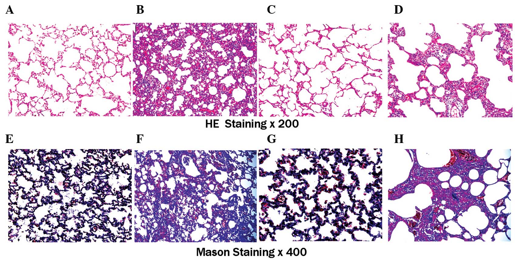
Pathological changes in the lung
tissues from the two groups of rats
Inflammatory cell infiltration was observed in the
alveolar cavity and pulmonary interstitium of the experimental
group rats at day 7, and alveolar septal thickening and a small
amount of collagen fiber organization was observed in the lung
interstitium. At day 28, the alveolar exudate had decreased and the
alveolar structure was destroyed. Furthermore, the interstitial gap
had widened significantly and hyperplasia of the extracellular
matrix was evident. Fibroblast proliferation was also observed in
the pulmonary interstitium, and the accumulation of collagen fibers
had formed extensive fibrosis. When compared with the NS group, the
alveolitis was evident in the BLM group at day 7. By day 28, the
alveolitis was relieved; however, the degree of pulmonary fibrosis
remained high (Fig. 1, Table I).
 | Table I.Comparison of pulmonary alveolitis and
fibrosis in the rats from the two groups (n=5). |
Table I.
Comparison of pulmonary alveolitis and
fibrosis in the rats from the two groups (n=5).
|
| Alveolitis score | Pulmonary fibrosis
score |
|---|
|
|
|
|
|---|
| Group | Day 7 | Day 28 | Day 7 | Day 28 |
|---|
| NS | 1.00±0.00 | 1.00±0.00 | 1.00±0.00 | 1.00±0.00 |
| BLM |
3.60±0.55a |
1.40±0.55a |
1.60±0.55a |
3.80±0.45a |
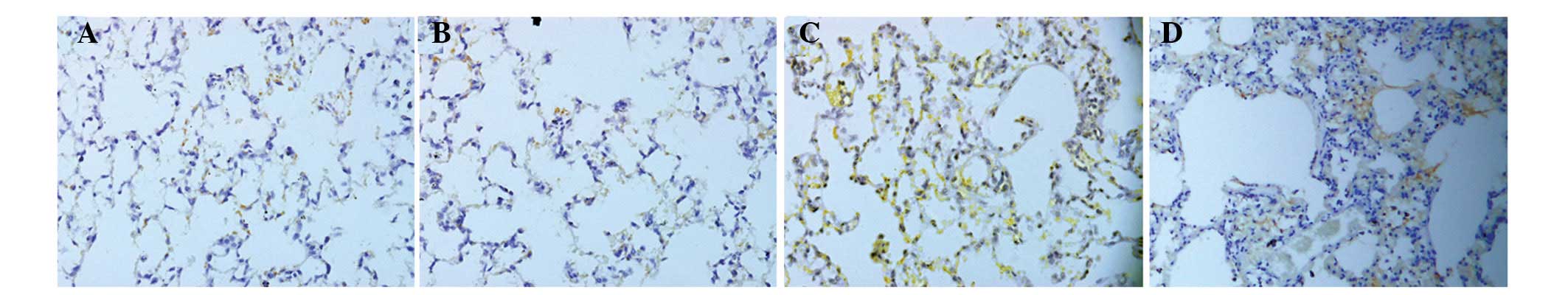
Immunohistochemical detection of the
protein expression of IL-17A in the lung tissue
Weak expression of IL-17A was observed in the lung
tissues from the NS rats; the expression levels were 23.67±3.19 and
24.26±3.35 at days 7 and 28, respectively. However, the expression
of IL-17A in the BLM group was significantly increased at days 7
(103.48±9.49) and 28 (87.42±7.64). When compared with the NS group
at day 7, the difference was statistically significant (P<0.05),
although expression levels decreased at day 28 compared with day 7
(Fig. 2).
Analysis of the BALF cell number and
type
At day 7, the total number of BALF cells in the NS
group was 9.45±0.76, while the number of BALF cells in the BLM
group was 35.28±2.76. When compared with the NS group, the total
number of BALF cells increased significantly in the BLM group and
the difference was statistically significant (P<0.05). At day
28, the total number of BALF cells in the NS group was 9.52±0.67,
while the number in the BLM group was 9.71±0.80; thus, no
statistically significant difference was observed (P>0.05). When
compared with the NS group, the AM ratio of BALF at day 7 was
smaller in the BLM group. In addition, the percentages of
neutrophils and lymphocytes were significantly increased, with the
differences statistically significant (P<0.05). When compared
with the NS group, the classification percentage of BALF cells in
the BLM group at day 28 was not significantly different (P>0.05;
Table II), which demonstrated that
the cell types had returned to normal.
| Table II.Cell types in the bronchoalveolar
lavage fluid at different time points. |
Table II.
Cell types in the bronchoalveolar
lavage fluid at different time points.
| Group | Cell type | Day 7 (%) | Day 28 (%) |
|---|
| NS | Macrophage | 90.20±1.64 | 90.40±1.14 |
|
| Neutrophil | 3.00±0.71 | 3.20±0.84 |
|
| Lymphocyte | 6.80±1.10 | 6.40±0.54 |
| BLM | Macrophage |
71.40±2.70a | 89.40±1.14 |
|
| Neutrophil |
17.60±3.05a | 3.00±0.71 |
|
| Lymphocyte |
11.00±0.71a | 7.20±0.84 |
Detection of IL-17A in the BALF
IL-17A levels in the BALF of the NS group were
57.45±6.68 pg/ml at day 7 and 55.49±6.06 pg/ml at day 28, while the
levels of IL-17A in the BLM group were 278.37±13.99 and
213.26±11.62 pg/ml at days 7 and 28, respectively. When compared
with the NS group, the IL-17A levels were significantly increased
in the BLM group and the difference was statistically significant
(P<0.01). The level of IL-17A at day 28 was marginally lower
compared with those on day 7; however, the difference was not
statistically significant.
Changes in the concentration of IL-17A
in the supernatant at different culture stages of the AMs
Compared with the NS group, the IL-17A concentration
in the supernatant of the BLM group at days 7 and 28 increased
significantly prior to AM culturing at 12, 24 and 48 h. The
differences between the two groups were statistically significant
(P<0.05, Table III).
| Table III.Changes in the supernatant
concentration (pg/ml) of IL-17A at different culturing stages of
the alveolar macrophages at day 7 and day 28. |
Table III.
Changes in the supernatant
concentration (pg/ml) of IL-17A at different culturing stages of
the alveolar macrophages at day 7 and day 28.
|
| Day 7 | Day 28 |
|---|
|
|
|
|
|---|
| Group | 12 h | 24 h | 48 h | 12 h | 24 h | 48 h |
|---|
| NS | 0.34±0.05 | 0.67±0.05 | 0.96±0.04 | 0.35±0.06 | 0.66±0.06 | 0.95±0.05 |
| BLM |
0.94±0.05a |
1.67±0.09a |
2.13±0.08a |
0.85±0.08a |
1.42±0.05a |
1.92±0.06a |
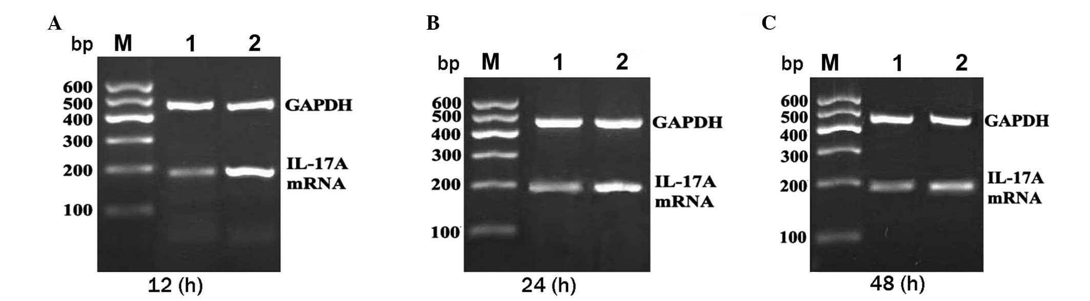
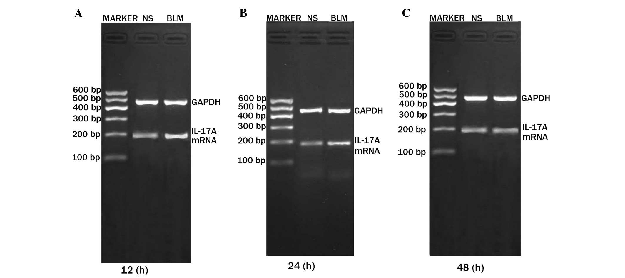
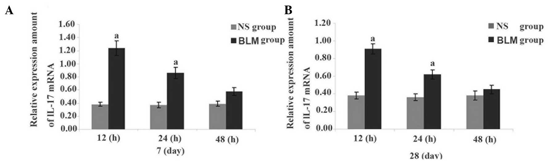
Expression of IL-17A mRNA in the
AMs
Expression levels of IL-17A mRNA at days 7 and 28
were higher in the BLM group when compared with the NS group, and
the differences were statistically significant (P<0.05). The
difference in expression was particularly notable in the first 12
h, decreasing after 24 and 48 h (Figs.
3, 4 and 5).
Discussion
The pathogenesis of IPF is generally considered to
result from the abnormal proliferation of epithelial cells and
re-epithelialization repair, which leads to pulmonary fibrosis
following alveolar epithelial cell damage due to inflammation
(1). Previous studies have
demonstrated that the overexpression of IL-17A, an important
proinflammatory cytokine, may lead to chronic inflammation and
autoimmune diseases (11,12), while antibodies targeting anti-IL-17A
and the anti-IL-17A receptor have demonstrated a particular effect
for the treatment of immune diseases, such as ankylosing
spondylitis and psoriasis, by inhibiting IL-17A or IL-17A receptors
(13–15). Therefore, anti-IL-17A antibodies may
have potential for the treatment of IPF.
In the present study, the expression of IL-17A in
the lung tissue of rats with pulmonary fibrosis was analyzed under
pulmonary inflammatory conditions. The results revealed that the
expression of IL-17A in the lung tissue and the secretion of IL-17A
in the AMs were increased during the occurrence and development of
pulmonary fibrosis. IL-17A expression was more evident during the
alveolar inflammation stage, indicating that IL-17A may contribute
to the formation of alveolitis and subsequently result in pulmonary
fibrosis. The results provide a theoretical basis for the targeted
therapy of IPF in future research.
IL-17A is an important proinflammatory cytokine that
belongs to the IL-17 family. The present view considers IL-17A to
play an important role in the process of resistance to the invasion
of certain pathogens, in addition to the induction and maintenance
of chronic inflammation (3). Wilson
et al (16) demonstrated that
BLM-induced pulmonary fibrosis was mediated by IL-17A, while
IL-17A-induced pulmonary fibrosis was dependent on transforming
growth factor-β. Large-dose injections of IL-17A in the airway have
been shown to induce pulmonary fibrosis, indicating that IL-17A and
other cytokines may exert a synergistic effect on the occurrence
and development of pulmonary fibrosis. Gasse et al (17) also found that the expression of
IL-17A was increased during the early stages of BLM-induced lung
injury, and confirmed that IL-17A and the IL-17A signaling
transduction pathway were involved in the early alveolar
inflammation induced by BLM in gene-deficient mice and neutralizing
antibody-treated mice. Neutralizing the activity of IL-17A can
improve the lung function of mice with pulmonary fibrosis (18). The results of the present study
revealed that the total number of BALF cells in the BLM group
increased significantly at day 7 when compared with the NS group.
In addition, the ratio of neutrophils and lymphocytes increased
significantly in the BLM group, while the ratio of AMs decreased
significantly. In the BLM group, the expression levels of IL-17A in
the BALF and the lung tissue were increased significantly at days 7
and 28. These results demonstrated that IL-17A, as an inflammatory
cell factor, may have a stronger chemotactic aggregation role in
neutrophil cells and other inflammatory cell types, which may also
play a very important role in the process of alveolitis and
pulmonary fibrosis. These observations were consistent with the
results of Mi et al (3),
Wilson et al (16) and Gasse
et al (17).
Abnormal AM activation can result in the abnormal
expression of cytokines, which has an important role in the
regulation of lung inflammation, and may affect the development of
pulmonary fibrosis through a variety of mechanisms (19). In the present study, the application
of ELISA was used to detect the concentration of IL-17A in the
supernatant of the AMs from different culture periods. The results
revealed that the expression levels of IL-17A in the culture
supernatant of the AMs were increased significantly at days 7 and
28 in the BLM group, as compared with the NS group. The difference
in expression was particularly notable in the first 12 h, and
decreased after 24 and 48 h, which confirmed that AMs are able to
secrete IL-17A, which may be involved in the formation of pulmonary
fibrosis. When compared with the BLM group at day 7, the expression
of IL-17A in the culture supernatant of the AMs was higher compared
with the levels observed at day 28, which also demonstrated that
IL-17A may play an important role in the formation and development
of pulmonary fibrosis. In addition, the role of IL-17A may be more
significant in the alveolar inflammation stage, as compared with
the pulmonary fibrosis stage. Using RT-PCR to detect the mRNA
expression levels of IL-17A in the AMs, the results revealed that
the mRNA expression levels of IL-17A in the AMs at different
culture periods were significantly increased in the BLM group at
days 7 and 28, particularly in the first 12 h, when compared with
the NS group. The expression of IL-17A was shown to gradually
weaken after 24 and 48 h, indicating that the expression of IL-17A
mRNA gradually decreased due to the culture of AMs in vitro
without the participation of an inducer. This further confirmed
that the secretion of IL-17A was induced by AMs, and a substantial
secretion was observed after stimulation, indicating that IL-17A is
involved in the formation of pulmonary fibrosis. Furthermore, the
results of the current study revealed that the mRNA expression of
IL-17A in the AM medium of the BLM group at day 7 was higher
compared with the levels at day 28, which further confirmed that
IL-17A may play an important role in the pathological process and
development of pulmonary fibrosis, particularly in the alveolitis
stage. Therefore, neutralization of IL-17A may be a promising
therapeutic measure for the treatment of IPF, and future studies
should focus on this research direction.
In conclusion, the present study indicated that
IL-17A was able to promote the development of pulmonary
inflammation in lung tissue, thus suggesting it may have an
important role in the development of pulmonary fibrosis.
Neutralization of IL-17A may be used to treat IPF; however, further
research is required.
Acknowledgements
The study was supported by a grant from the Science
and Technology Bureau of Luzhou City (no. 12102).
References
|
1
|
Tanaka K, Ishihara T, Azuma A, Kudoh S,
Ebina M, Nukiwa T, Sugiyama Y, Tasaka Y, Namba T, Ishihara T, Sato
K, Mizushima Y and Mizushima T: Therapeutic effect of lecithinized
superoxide dismutase on bleomycin-induced pulmonary fibrosis. Am J
Physiol Lung Cell Mol Hysiol. 298:L348–L360. 2010. View Article : Google Scholar
|
|
2
|
Raghu G, Collard HR, Egan JJ, Martinez FJ,
Behr J, et al: ATS/ERS/JRS/ALAT Committee on Idiopathic Pulmonary
Fibrosis: An official ATS/ERS/JRS/ALAT statement: Idiopathic
pulmonary fibrosis: Evidence-based guidelines for diagnosis and
management. Am J Respir Crit Care Med. 183:788–824. 2011.
View Article : Google Scholar : PubMed/NCBI
|
|
3
|
Mi S, Li Z, Yang HZ, Liu H, Wang JP, Ma
YG, Wang XX, Liu HZ, Sun W and Hu ZW: Blocking IL-17A promotes the
resolution of pulmonary inflammation and fibrosis via TGF-beta
1-dependent and -independent mechanisms. J Immunol. 187:3003–3014.
2011. View Article : Google Scholar : PubMed/NCBI
|
|
4
|
Umezawa H, Maeda K, Takeuchi T and Okami
Y: New antibiotics, bleomycin A and B. J Antibiot (Tokyo).
19:200–209. 1966.PubMed/NCBI
|
|
5
|
Hoshino T, Okamoto M, Sakazaki Y, Kato S,
Young HA and Aizawa H: Role of proinflammatory cytokines IL-18 and
IL-1beta in bleomycin-induced lung injury in humans and mice. Am J
Respir Cell Mol Biol. 41:661–670. 2009. View Article : Google Scholar : PubMed/NCBI
|
|
6
|
Huang CL, Wang WJ, Zhu HL, Fan XM, Chen
JP, Zhan XQ and Li YY: Effect of andrographolide on expression of
hydroxyproline and PDGF in lung tissue of bleomycin-induced rat
pulmonary fibrosis. Shizhen Guo Yi Guo Yao. 23:904–907. 2012.(In
Chinese).
|
|
7
|
Huang C, Li Y, Fan X, Wang W and Zhan X:
Effects of combination of Salvia and Ligustrazine on TNF-α and
TGF-β1 in serum and BALF of rats with pulmonary fibrosis. Xi Bao Yu
Fen Zi Mian Yi Xue Za Zhi. 29:673–676. 2013.(In Chinese).
PubMed/NCBI
|
|
8
|
Tabata C, Kubo H, Tabata R, Wada M, Sakuma
K, Ichikawa M, Fujita S, Mio T and Mishima M: All-trans retinoic
acid modulates radiation-induced proliferation of lung fibroblasts
via IL-6/IL-6R system. Am J Physiol Lung Cell Mol Physiol.
290:L597–L606. 2006. View Article : Google Scholar : PubMed/NCBI
|
|
9
|
Lee KY, Ito K, Hayashi R, Jazrawi EP,
Barnes PJ and Adcock M: NF-kappa B and activator protein 1 response
elements and the role of histone modifications in IL-1 beta-induced
TGF-beta 1 gene transcription. J Immunol. 176:603–615. 2006.
View Article : Google Scholar : PubMed/NCBI
|
|
10
|
Liu JP, Di NL, Fan XM, Li SC and Zhang SP:
The expressions of STAT1 and PDGF in lung tissue of rats with
pulmonary fibrosis induced by bleomycin. Shan Dong Yi Yao.
31:16–18. 2008.(In Chinese).
|
|
11
|
Halwani R, Al-Muhsen S and Hamid Q: T
helper 17 cells in airway diseases: From laboratory bench to
bedside. Chest. 143:494–501. 2013. View Article : Google Scholar : PubMed/NCBI
|
|
12
|
Truchetet ME, Mossalayi MD and Boniface K:
IL-17 in the rheumatologist's line of sight. Biomed Res Int.
2013:2951322013. View Article : Google Scholar : PubMed/NCBI
|
|
13
|
Baeten D, Baraliakos X, Braun J, Sieper J,
Emery P, et al: Anti-interleukin-17A monoclonal antibody
secukinumab in treatment of ankylosing spondylitis: A randomised,
double-blind, placebo-controlled trial. Lancet. 382:1705–1713.
2013. View Article : Google Scholar : PubMed/NCBI
|
|
14
|
Leonardi C, Matheson R, Zachariae C,
Cameron G, Li L, Edson-Heredia E, Braun D and Banerjee S:
Anti-interleukin-17 monoclonal antibody ixekizumab in chronic
plaque psoriasis. N Engl J Med. 366:1190–1199. 2012. View Article : Google Scholar : PubMed/NCBI
|
|
15
|
Papp KA, Leonardi C, Menter A, Ortonne JP,
Krueger JG, Kricorian G, Aras G, Li J, Russell CB, Thompson EH and
Baumgartner S: Brodalumab, an anti-interleukin-17-receptor antibody
for psoriasis. N Engl J Med. 366:1181–1189. 2012. View Article : Google Scholar : PubMed/NCBI
|
|
16
|
Wilson MS, Madala SK, Ramalingam TR,
Gochuico BR, Rosas IO, Cheever AW and Wynn TA: Bleomycin and
IL-1β-mediated pulmonary fibrosis is IL-17A dependent. J Exp Med.
207:535–552. 2010. View Article : Google Scholar : PubMed/NCBI
|
|
17
|
Gasse P, Riteau N, Vacher R, Michel ML,
Fautrel A, di Padova F, Fick L, Charron S, Lagente V, Eberl G, Le
Bert M, Quesniaux VF, Huaux F, Leite-de-Moraes M, Ryffel B and
Couillin I: IL-1 and IL-23 mediate early IL-17A production in
pulmonary inflammation leading to late fibrosis. PLoS One.
6:e231852011. View Article : Google Scholar : PubMed/NCBI
|
|
18
|
Liu H, Li Z, Lv XX and Mi S:
Neutralization of IL-17A activity can improve pulmonary function in
mice with pulmonary fibrosis. Hang Tian Yi Xue Yu Yi Xue Gong
Cheng. 26:38–42. 2013.(In Chinese).
|
|
19
|
Fan XM, Zhang SB, Liu CT, Xiong B and Wang
ZL: STAT1 antisense oligonucleotides inhibit secretion of
TNF-alpha, IL-8 and NO in alveolar macrophages of rats suffering
from interstitial pulmonary fibrosis. Xi Bao Yu Fen Zi Mian Yi Xue
Za Zhi. 22:487–489. 2006.(In Chinese). PubMed/NCBI
|