Introduction
Telomeres are protective structures at the ends of
eukaryotic chromosomes, which consist of tandem arrays of hexameric
repeats (TTAGGG) bound to specific proteins. Telomeres play a
critical role in the maintenance of the structural integrity of the
genome, as they can prevent chromosomes from nucleolytic decay,
fusion and atypical recombination (1). In human somatic cells, telomeres
shorten by 30–200 bp with each mitotic cell division (2). Thus, telomere length is expected to be
an effective indicator of the mitotic history of a cell, since it
reflects the cumulative effect of cell divisions occurring in
vitro upon stimulation and in vivo due to aging
(3,4). Telomere length is also a determining
factor for the residual life span of normal somatic cells, since
further cell division is not possible in these cells when telomeres
shorten to a critical length (5,6). For
this reason, telomere length has long been proposed as a biomarker
of aging (7).
The functional activity of the immune system depends
on the degree of lymphocyte proliferation and clonal expansion
during development, activation and differentiation (8). Telomere maintenance is of paramount
importance for dynamic cellular systems, such as the immune system.
The thymus is a primary lymphoid organ responsible for T-lymphocyte
differentiation and maturation, which provides an inductive
microenvironment in which bone marrow-derived progenitors undergo
proliferation. Thymocytes differentiate into mature T cells that
are essential for the immune response against infection (9–11). It
has also been suggested that intrathymic maturation and selection
of T cells are associated with telomerase activity, which allows T
cells to acquire telomere sequences that are long enough to undergo
several rounds of replication (12).
A variety of environmental and cellular factors have
been shown to affect the rate of telomere attrition and telomerase
activity, as well as the number of cell divisions (history of
division), such as low temperature, dry, strong ultraviolet
radiation, smoking, diet and psychological stress (13–16). In
recent years, the effect of hypoxia on telomere length has become a
topic of increasing interest and debate. Previous studies have
indicated that the telomere length varies considerably among
hypoxia levels and cell types (17–19), and
thus telomere length in lymphocytes may indicate the extent to
which hypoxia affects the immune system.
Our previous study detected telomere length of
leukocytes in rats exposed to different hypoxic altitudes. The
results showed that the leukocyte telomeres were significantly
elongated in a mildly hypoxic environment, as opposed to a highly
hypoxic environment. To the best of our knowledge, there are no
previous studies investigating the impact of different hypoxic
altitudes on thymocyte telomere length and telomerase reverse
transcriptase (TERT) expression.
In the present study, the telomere length and
telomerase reverse transcriptase (TERT) of rat thymocytes exposed
to different altitudes (10, 2,260 and 5,000 m) was measured after
different periods of time (1, 3, 7, 15 and 30 days), and their
effect on the rat immune system was investigated, with a focus on
the dynamic behavior of telomere length and TERT in thymocytes.
Materials and methods
Animal experiments
All animal experiments were approved by the
Institutional Animal Ethics Committee of the Medical College of
Qinghai University (Xining, China). A total of 110 male Wistar rats
of the same age (6 weeks) and weight (50–180 g) were used in the
present study. The rats were maintained in a controlled environment
at a temperature of 18–20°C on a 12-h light/dark cycle with
unlimited access to food and water, and were randomly assigned to
one of the following three groups: i) Sea level (SL) group, in
which 10 rats were maintained at an altitude of 10 m in Nanjing,
China, and immediately sacrificed on day 1; ii) moderate altitude
(MA) group, in which 50 rats were maintained at an altitude of
2,260 m in Xining, China, and then randomly sacrificed on days 1,
3, 7, 15 or 30 (n=10 each); and iii) simulated high altitude (SHA)
group, in which the remaining 50 rats were maintained in an 8×3×3-m
hypobaric chamber (DYC-3000; Guizhou Fenglei Aviation Armament Co.,
Ltd., Anshun, China) with a simulated altitude of 5,000 m in
Xining, China, and were then randomly sacrificed on days 1, 3, 7,
15 or 30 (n=10 each). The rats were sacrificed under Nembutal
anesthesia (40 mg/kg, intraperitoneally; H. Lundbeck A/S,
Copenhagen, Denmark), and their thymus glands were excised and
weighed. Thymus specimens (0.3 cm3) were fixed for
histopathological analysis as described in a subsequent section,
and the remainder of the thymus was fresh-frozen and stored in
liquid nitrogen at −80°C for molecular analysis.
Peripheral blood lymphocyte count and
percentage
The peripheral blood lymphocyte count and percentage
were measured using an automatic blood cell analyzer (BC-2300;
Mindray Medical International Ltd., Wuhan, China) following the
manufacturer's instructions.
Thymus index and histopathological
analysis
The rats were anesthetized using Nembutal (40 mg/kg,
intraperitoneally; H. Lundbeck A/S), and their thymuses were
excised and weighed. The thymus index was expressed as the thymus
weight relative to the body weight. A 0.3-cm3 sample of
the thymus was fixed in 4% paraformaldehyde, routinely embedded in
paraffin, sectioned, and stained with hematoxylin and eosin
(Nanjing Biological Co., Jiangsu, China) for histopathological
analysis.
Measurement of telomere length using
quantitative polymerase chain reaction (qPCR)
The thymus tissues were ground and passed through a
200-mesh sieve, centrifuged at 400 × g for 10 min at 4°C, washed
twice with phosphate-buffered saline and counted using a microscope
(Olympus Corporation, Tokyo, Japan). The genomic DNAs were isolated
from 106−107 cells using a DNA extraction kit
(Tiangen Biotech Co., Ltd., Beijing, China) following the
manufacturer's instructions, and dissolved in Tris-EDTA buffer (pH
8.0). The concentration was measured using a DU 800
spectrophotometer (Beckman Coulter, Inc., Fullerton, CA, USA). The
ratio of OD260 to OD280 was 1.8–2.0 and the DNA concentration was
50 ng/µl.
qPCR reactions were performed using SYBR® Select
Master Mix with an ABI 7500 system (Applied Biosystems, Carlsbad,
CA, USA) in a total volume of 20 µl containing 2 µl DNA template.
The PCR plate was incubated at 95°C for 30 sec, followed by 40
cycles of 95°C for 5 sec and 60°C for 34 sec. All samples were
tested in triplicate, and the amplification efficiency was compared
with that of the angiotensin type 1 (AT1) receptor reference gene
and was found to be similar. The primers used for the amplification
of telomeres were as follows: Tel-1, forward (270 nmol) 5′GGT TTT
TGA GGG TGAGGGTGAGGGTGAGGGTGAGGGT-3′ and reverse (900 nmol)
5′-TCCCGACTATCCCTATCCCTATCCCTATCCCTATCCCTA-3′; AT1, forward (400
nmol) 5′-ACGTGTTCTCAGCATCGACCGCTACC-3′; and reverse (400 nmol)
5′-AGAATGATAAGGAAAGGGAACAAGAAGCCC-3′. Primers were synthesized by
Invitrogen (Grand Island, NY, USA).
The relative telomere length was expressed as the
T/S ratio, where T is the amount of telomere and S the amount of a
single gene copy of the AT1 receptor. The 2−∆∆Ct method
was used to calculate the T/S ratio.
Protein and mRNA expression levels of
TERT
Total RNA was extracted from the thymocytes and
purified using TRIzol® reagent (Invitrogen Life Technologies)
following the manufacturer's instructions. The RNA concentration
was measured using the DU 800 spectrophotometer. cDNA synthesis was
performed with 2 µg total RNA, and this cDNA was used as a template
in qPCR. PCR reactions were performed using SYBR® Select Master Mix
with an ABI 7500 system in a total volume of 20 µl. The PCR plate
was incubated at 50°C for 2 min and 95°C for 2 min, followed by 40
cycles of 95°C for 15 sec and 60°C for 1 min. The amplification
efficiency of TERT was compared with that of the 18S rRNA reference
gene and was found to be similar. The primers used for the
amplification of TERT were as follows: TERT forward (400 nmol),
5′-GAC ATG GAG AAC AAG CTG TTT GC-3′, and reverse (400 nmol),
5′-ACA GGG AAG TTC ACC ACT GTC-3′; 18S rRNA forward (400 nmol),
5′-AGT GATCCC CGA GAA GTTT-3′, and reverse (400 nmol), 5′-GCT TTC
CTC AAC ACC ACAT-3′ (Invitrogen). The 2−∆∆Cq method was
used to calculate the relative expression levels.
Total protein of rat thymocyte cell suspension was
extracted and measured by the BCA Protein Assay kit (Pierce
Biotechnology, Inc., Rockford, IL, USA) and the protein levels of
TERT were detected using an ELISA kit (Cloud-Clone Corp., Houston,
TX, USA) following the manufacturer's instructions.
Statistical analysis
Data were analyzed using SPSS software, version 17.0
(SPSS, Inc., Chicago, IL, USA). All data are presented as the mean
± standard deviation. Statistical analysis was performed using
one-way analysis of variance followed by a least significant
difference post-hoc test. P<0.05 was considered to indicate a
statistically significant difference.
Results
Hematological changes
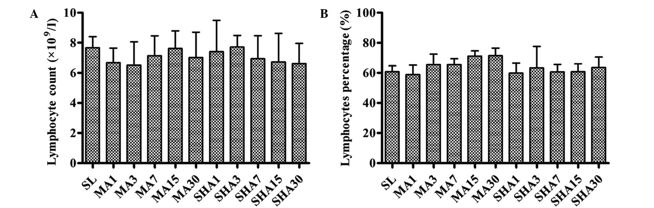
As shown in Fig. 1,
no statistically significant changes were observed in the
peripheral blood lymphocyte count and percentage in rats exposed to
different altitudes for different periods of time (P>0.05). The
thymus is the primary lymphoid organ of cellular immunity
responsible for T-lymphocyte choice and output. No changes means
that the body cellular immunity has not been obviously damaged, the
rat thymus still plays the effective immune function under hypoxic
conditions.
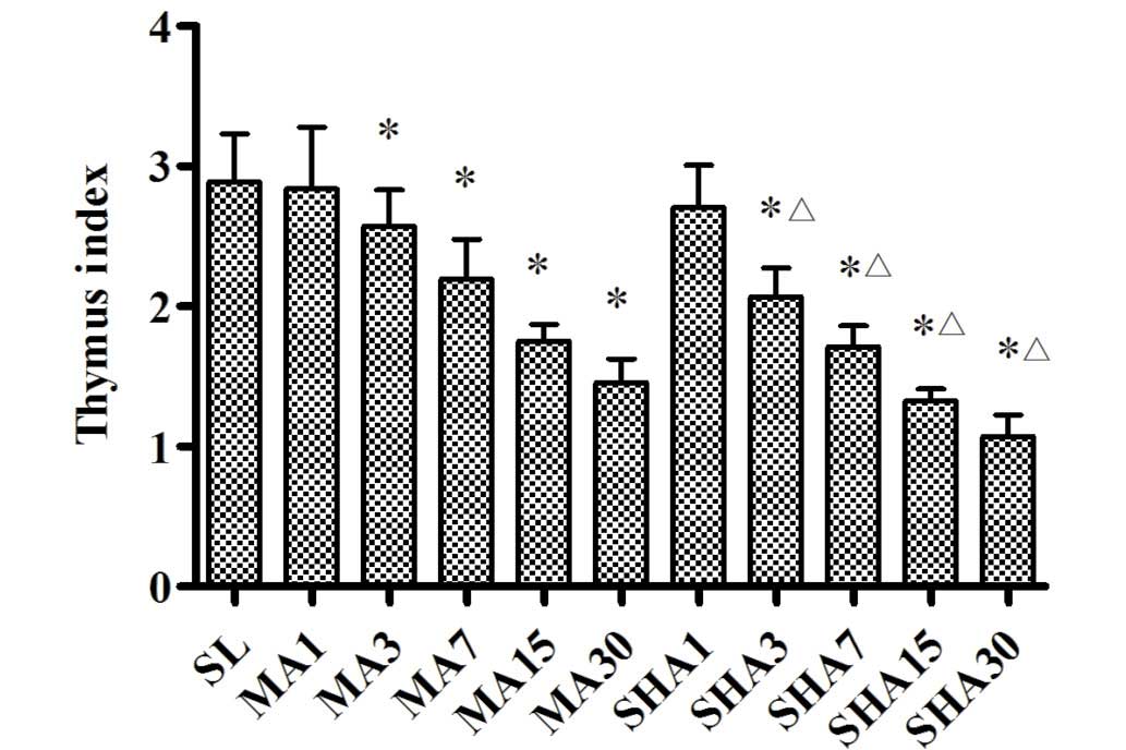
Morphological changes of thymus
It is known that the degree of hypoxia is associated
with altitude, higher altitude results in increased levels of
hypoxia. According to the international standard, the moderate
altitude used in the preset study (2,260 m) is associated with mild
hypoxia, while simulated high altitude (5,000 m) is associated with
severe hypoxia. Fig. 2 shows that
the thymus index of rats exposed to hypoxic conditions decreased
with increasing exposure time (P<0.05). This decrease in the
thymus index appeared to be more pronounced in the SHA group
compared with that in the MA group (P<0.05). The decreased
thymus index indicated that the thymocyte number reduction and
thymus tissue atrophy occurred as a result of the hypoxic
conditions.
As shown in Fig. 3A,
in the SL group, the cortex was thick and distinct corticomedullary
demarcation was observed. The lymphocytes were small and densely
clustered in the cortex, with deeply stained nuclei. Thymocyte
apoptosis or epithelial cell hyperplasia were not observed, and
there was no abnormal distribution or hyperemia of mesenchymal
vessels; however, thymic corpuscles were observed. In the MA group,
the medulla expanded, and the cortex became thin with distinct
corticomedullary demarcation. The thymocytes were small and densely
clustered in the cortex, with deeply stained nuclei. Thymocyte
apoptosis was rarely observed in the MA group. Hyperplastic and
hypertrophic epithelial cells were present in the cortex and
medulla, while thymic corpuscles were also identified. Furthermore,
the mesenchymal vessels were found to be expanded and hyperemic,
with small patches of hemorrhage. The parenchyma of thymic lobules
was found to be atrophied and infiltrated by adipose tissue. The
mild tissue damage and thymocyte apoptosis observed as a result of
moderate altitude was not evidently exacerbated following an
extension of hypoxic exposure time, and there were no significant
differences between the five times.
 | Figure 3.Histological changes (hematoxylin and
eosin stain; magnification, ×400). (A) In the SL group, the
lymphocytes were small and densely clustered in the cortex with
deeply stained nuclei. No thymocyte apoptosis was observed. In the
MA group on days (B) 1, (C) 3, (D) 7, (E) 15 and (F) 30, the cortex
became thin, and the thymocytes were small and densely clustered in
the cortex. Thymocyte apoptosis was rarely observed. The parenchyma
of thymic lobules was atrophied and infiltrated by adipose tissue.
In the SHA group on days (G) 1 (H) 3 (I) 7 (J) 15 (K) 30, the
cortex was thin, and the thymocytes in the cortex were large.
Significant thymocyte apoptosis and focal loss of thymocytes were
observed, and loose plaque formed in the sub-capsule and cortex.
Certain thymus tissues had atrophied and were replaced by adipose
tissue. |
In the SHA group, the medulla expanded, the cortex
became thin with distinct corticomedullary demarcation, and the
thymocytes were large with lightly stained nuclei. The chromatin
was thick, and significant thymocyte apoptosis was observed. Focal
loss of thymocytes was identified, and loose plaque was found in
the subcapsule and cortex. Furthermore, hyperplastic and
hypertrophic epithelial cells were present in the cortex and
medulla and thymic corpuscles were also observed. Mesenchymal
vessels were found to be expanded and hyperemic, with small patches
of hemorrhage. Certain thymus tissues were found to be atrophied
and to have been replaced by adipose tissue. As a result of the
extension of hypoxic exposure time, the apoptosis and focal loss of
thymocyte under simulated high altitude was gradually
exacerbated.
In conclusion, hypoxia induced significant
morphological changes and apoptosis in thymocytes, as well as
thymus tissue atrophy, and this effect was more apparent in highly
hypoxic conditions rather than in mild hypoxic conditions
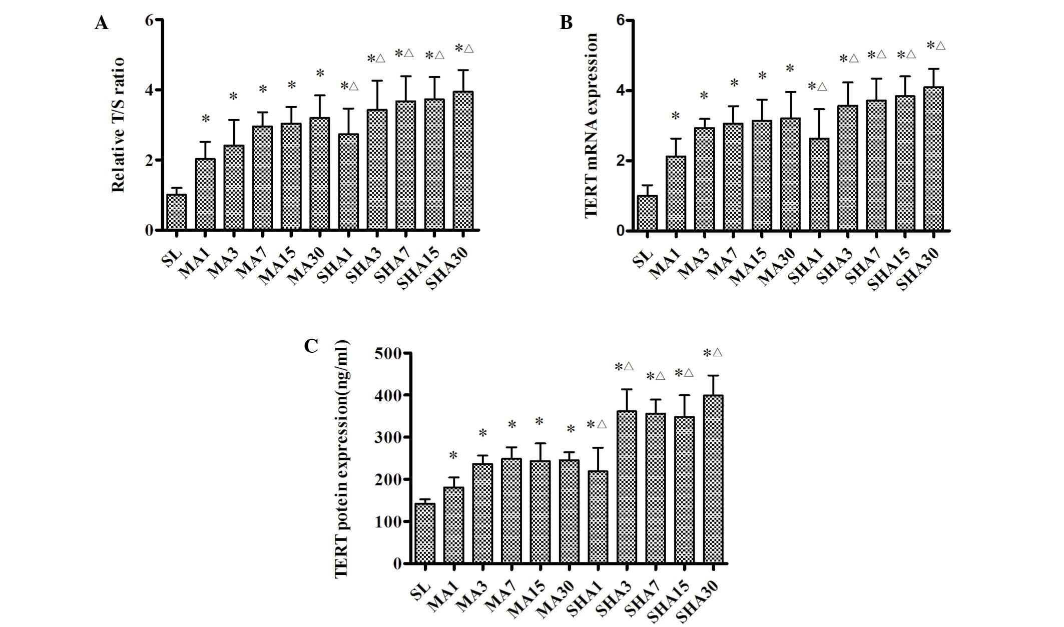
Telomere length
The relative T/S ratios for each group are shown in
Fig. 4A. It was evident that
telomere length was significantly increased during the first 7 days
of exposure to moderate or simulated high altitude (P<0.05), but
with a less significant increase over the next few weeks. Again,
this effect tended to be more pronounced in the SHA group than in
the MA group (P<0.05). The results suggested that thymocyte
telomere length was elongated gradually in response to an acute
hypoxic exposure period, and elongated no further as a result of
the chronic hypoxic exposure period.
TERT mRNA and protein expression
levels
Fig. 4B and C shows
that TERT mRNA and protein expression changed in a manner similar
to that observed for the telomere length. TERT mRNA and protein
expression were significantly increased in response to hypoxic
exposure during the first week (P<0.05), but remained stable
over the next few weeks. It was also noted that this increase was
more pronounced in the SHA group than it was in the MA group
(P<0.05). Telomere length was closely associated with TERT
expression levels, and was correlated with TERT expression under
hypoxic conditions.
Discussion
In the present study, hypoxia was found to induce
morphological changes and apoptosis in thymocytes, as well as
atrophy of the thymus tissue, and this effect became more
pronounced with the increase in hypoxia. In addition, telomere
length was significantly increased in response to hypoxic exposure,
while TERT mRNA and protein expression also changed in a similar
manner.
Telomerase is a ribonucleoprotein that maintains
telomere length in germline and stem cells by adding hexameric
TTAGGG repeats to the telomeres, thereby compensating for the
progressive loss of telomeric sequences with each cell division
(20,21). The protein catalytic subunit of
telomerase, known as TERT, adds telomere repeats to the ends of
chromosomes through the reverse transcription of an RNA template
into DNA (22), and is thus
responsible for the maintenance of telomere DNA length, chromosomal
stability and cellular immortality (23). It has been shown that the
introduction of TERT gene into somatic diploid cells resulted in a
significant increase in telomerase activity and telomere length
(24,25), which suggests that it has a
protective function against genome instability-promoting events
(26).
The thymus is the primary lymphoid organ responsible
for T-lymphocyte differentiation and maturation. The thymus gland
is composed of structural components, including peripheral blood,
vascular bed, nervous fibers and connective tissues. During their
differentiation, thymocytes interact with antigen presenting cells,
such as macrophages and dendritic cells, as well as epithelial
cells localized in specific thymic areas. Intrathymic selection and
maturation of T cells is known to be associated with telomerase
activity, allowing thymocytes to acquire telomere sequences that
are long enough to undergo several rounds of replication (12). It has been recently demonstrated that
the decrease in lymphocyte function accelerated telomere shortening
in individuals with prolonged psychological stress (27,28). The
shortening of a telomere is accompanied by a host of phenotypic and
gene expression changes that result in altered functions (29). Thus, thymocyte function depends, to a
great extent, on the telomere length and TERT expression. The
telomere length of newly generated T cells is determined by the
telomere length of hematopoietic stem cells and the efficacy of
telomere elongation in thymocytes (30).
In the present study, hypoxia induced morphological
changes and apoptosis in thymocytes. Notably, no significant
changes were observed in the total number of peripheral blood
lymphocytes; however, the telomere length, as well as the TERT mRNA
and protein expression levels, increased with the increase in
altitude. These results suggest that the thymus plays an active
role in the selection and development of T cells under hypoxic
conditions. Previous studies have investigated the possible
molecular mechanisms underlying hypoxia-induced TERT expression,
and have indicated that TERT expression is upregulated following
the induction of HIF-1α during exposure to hypoxia (31). Furthermore, HIF-1α functioned as a
key transactivator for the induction of TERT transcription, thereby
resulting in elongation of telomeres (32). During exposure to hypoxia, the thymus
function per thymocyte unit may remain constant; however, a
decrease is observed in the total number of thymocyte units. The
hypoxia-associated loss of thymic function, therefore, appears to
only be quantitative and not qualitative.
In conclusion, hypoxia was found to induced
thymocyte apoptosis and thymus tissue atrophy in rats, resulting in
a significant increase in telomere length, as well as in the TERT
mRNA and protein expression levels. It was also noted that this
effect became more apparent with the increase in hypoxia. All these
findings suggest that the hypoxia-associated loss of thymic
function appears to only be quantitative and not qualitative. In
addition, the thymus may be able to maintain its immune function
even under hypoxic conditions. Further studies are required to
determine the precise underlying mechanism.
Acknowledgements
This study was supported by the National Basic
Research Program of China (grant no. 2012CB518200), the Program of
International Science and Technology Cooperation of China (grant
no. 2011DFA32720) and the National Natural Science Foundation of
China (grant no. 31160219).
References
|
1
|
Stewart SA and Weinberg RA: Telomeres:
Cancer to human aging. Annu Rev Cell Dev Biol. 22:531–557. 2006.
View Article : Google Scholar : PubMed/NCBI
|
|
2
|
Shay JW and Wright WE: Telomerase activity
in human cancer. Curr Opin Oncol. 8:66–71. 1996. View Article : Google Scholar : PubMed/NCBI
|
|
3
|
Lu W, Zhang Y, Liu D, Songyang Z and Wan
M: Telomeres - structure, function, and regulation. Exp Cell Res.
319:133–141. 2013. View Article : Google Scholar : PubMed/NCBI
|
|
4
|
Artandi SE: Telomeres, telomerase, and
human disease. N Engl J Med. 355:1195–1197. 2006. View Article : Google Scholar : PubMed/NCBI
|
|
5
|
Johnson TE: Recent results: Biomarkers of
aging. Exp Gerontol. 41:1243–1246. 2006. View Article : Google Scholar : PubMed/NCBI
|
|
6
|
Cherkas LF, Aviv A, Valdes AM, Hunkin JL,
Gardner JP, Surdulescu GL, Kimura M and Spector TD: The effects of
social status on biological aging as measured by white-blood-cell
telomere length. Aging Cell. 5:361–365. 2006. View Article : Google Scholar : PubMed/NCBI
|
|
7
|
Hunt SC, Chen W, Gardner JP, Kimura M,
Srinivasan SR, Eckfeldt JH, Berenson GS and Aviv A: Leukocyte
telomeres are longer in African Americans than in whites: The
national heart, lung and blood institute family heart study and the
bogalusa heart study. Aging Cell. 7:451–458. 2008. View Article : Google Scholar : PubMed/NCBI
|
|
8
|
Zhao Wushu: Immune balance study and its
clinical significance. Beijing: Science Press. 2005.12–18
|
|
9
|
Anderson G and Jenkinson EJ: Lymphostromal
interactions in thymic development and function. Nat Rev Immunol.
1:31–40. 2001. View
Article : Google Scholar : PubMed/NCBI
|
|
10
|
Madhok AB, Chandrasekran A, Parnell V,
Gandhi M, Chowdhury D and Pahwa S: Levels of recent thymic emigrant
cells decrease in children undergoing partial thymectomy during
cardiac surgery. Clin Diagn Lab Immunol. 12:563–565.
2005.PubMed/NCBI
|
|
11
|
Moore SE, Collinson AC, Tamba N, Gom P,
Aspinall R and Prentice AM: Early immunological development and
mortality from infectious disease in later life. Proc Nutr Soc.
65:311–318. 2006. View Article : Google Scholar : PubMed/NCBI
|
|
12
|
Brousset P, al Saati T, Zenou RC and
Delsol G: Telomerase activity might persist in the human thymus
throughout life. Mol Pathol. 51:170–173. 1998. View Article : Google Scholar : PubMed/NCBI
|
|
13
|
Lung FW, Chen NC and Shu BC: Genetic
pathway of major depressive disorder in shortening telomeric
length. Psychiatr Genet. 17:195–199. 2007. View Article : Google Scholar : PubMed/NCBI
|
|
14
|
Bekaert S, De Meyer T, Rietzschel ER, De
Buyzere ML, De Bacquer D, Langlois M, Segers P, et al: Telomere
length and cardiovascular risk factors in a middle-aged population
free of overt cardiovascular disease. Aging Cell. 6:639–647. 2007.
View Article : Google Scholar : PubMed/NCBI
|
|
15
|
von Zglinicki T: Oxidative stress shortens
telomeres. Trends Biochem Sci. 27:339–344. 2002. View Article : Google Scholar : PubMed/NCBI
|
|
16
|
Mirabello L, Huang WY, Wong JY, Chatterjee
N, Reding D, Crawford ED, De Vivo I, Hayes RB and Savage SA: The
association between leukocyte telomere length and cigarette
smoking, dietary and physical variables and risk of prostate
cancer. Aging Cell. 8:405–413. 2009. View Article : Google Scholar : PubMed/NCBI
|
|
17
|
Minamino T, Mitsialis SA and Kourembanas
S: Hypoxia extends the life span of vascular smooth muscle cells
through telomerase activation. Mol Cell Biol. 21:3336–3342. 2001.
View Article : Google Scholar : PubMed/NCBI
|
|
18
|
Guan JZ, Guan WP, Maeda T and Makino N:
Alteration of telomere length and subtelomeric methylation in human
endothelial cell under different levels of hypoxia. Arch Med Res.
43:15–20. 2012. View Article : Google Scholar : PubMed/NCBI
|
|
19
|
Davy P and Allsopp R: Hypoxia Are stem
cells in it for the long run? Cell Cycle. 10:206–211. 2011.
View Article : Google Scholar : PubMed/NCBI
|
|
20
|
Feng J, Funk WD, Wang SS, Weinrich SL,
Avilion AA, Chiu CP, Adams RR, Chang E, Alisopp RC, Yu J, et al:
The RNA component of human telomerase. Science. 269:1236–1241.
1995. View Article : Google Scholar : PubMed/NCBI
|
|
21
|
Nakamura TM, Morin GB, Chapman KB,
Weinrich SL, Andrews WH, Lingner J, Harley CB and Cech TB:
Telomerase catalytic subunit homologs from fission yeast and human.
Science. 277:955–959. 1997. View Article : Google Scholar : PubMed/NCBI
|
|
22
|
Harley CB: Telomerases. Pathol Biol
(Paris). 42:342–345. 1994.PubMed/NCBI
|
|
23
|
Cheung AL and Deng W: Telomere
dysfunction, genome instability and cancer. Front Biosci.
13:2075–2090. 2008. View
Article : Google Scholar : PubMed/NCBI
|
|
24
|
Fujiki T, Udono M, Kadooka K, Yamashita S,
Miura T, Shirahata S and Katakura Y: Regulatory mechanisms of human
and mouse telomerase reverse transcriptase gene transcription:
Distinct dependency on c-Myc. Cytotechnology. 62:333–339. 2010.
View Article : Google Scholar : PubMed/NCBI
|
|
25
|
Wang Z, Xu J, Geng X and Zhang W: Analysis
of DNA methylation status of the promoter of human telomerase
reverse transcriptase in gastric carcinogenesis. Arch Med Res.
41:1–6. 2010. View Article : Google Scholar : PubMed/NCBI
|
|
26
|
Wyatt HD, West SC and Beattie TL: In TERT
preting telomerase structure and function. Nucleic Acids Res.
38:5609–5622. 2010. View Article : Google Scholar : PubMed/NCBI
|
|
27
|
Effros RB: Telomere/telomerase dynamics
within the human immune system: Effect of chronic infection and
stress. Exp Gerontol. 46:135–140. 2011. View Article : Google Scholar : PubMed/NCBI
|
|
28
|
Jergović M, Tomičević M, Vidović A,
Bendelja K, Savić A, Vojvoda V, Rac D, Lovrić-Čavar D, Rabatić S,
Jovanovic T and Sabioncello A: Telomere shortening and immune
activity in war veterans with posttraumatic stress disorder. Prog
Neuropsychopharmacol Biol Psychiatry. 54:275–283. 2014. View Article : Google Scholar : PubMed/NCBI
|
|
29
|
Effros RB: Replicative senescence in the
immune system: Impact of the Hayflick limit on T-cell function in
the elderly. Am J Hum Genet. 62:1003–1007. 1998. View Article : Google Scholar : PubMed/NCBI
|
|
30
|
Bertho JM, Demarquay C, Moulian N, Van Der
Meeren A, Berrih-Aknin S and Gourmelon P: Phenotypic and
immunohistological analyses of the human adult thymus: Evidence for
an active thymus during adult life. Cell Immunol. 179:30–40. 1997.
View Article : Google Scholar : PubMed/NCBI
|
|
31
|
Coussens M, Davy P, Brown L, Foster C,
Andrews WH, Nagata M and Allsopp R: RNAi screen for telomerase
reverse transcriptase transcriptional regulators identifies
HIF1alpha as critical for telomerase function in murine embryonic
stem cells. Proc Natl Acad Sci USA. 107:13842–13847. 2010.
View Article : Google Scholar : PubMed/NCBI
|
|
32
|
Yang K, Zheng Deng X, Bai L, Xu Y and Cong
YS: Lysophosphatidic acid activates telomerase in ovarian cancer
cells through hypoxia-inducible factor-1alpha and the pi3k pathway.
J Cell Biochem. 105:1194–1201. 2008. View Article : Google Scholar : PubMed/NCBI
|