Open Access

Quantitative analysis of contrast-enhanced ultrasonography in acute radiation-induced liver injury: An animal model

  • Authors:
    • Jun Feng
    • Shu-Bo Chen
    • Shu-Jun Wu
    • Ping Sun
    • Tian-You Xin
    • Ying-Zhen Chen
  • View Affiliations

  • Published online on: September 22, 2015     https://doi.org/10.3892/etm.2015.2764
  • Pages: 1807-1811
  • Copyright: © Feng et al. This is an open access article distributed under the terms of Creative Commons Attribution License.

Metrics: Total Views: 0 (Spandidos Publications: | PMC Statistics: )
Total PDF Downloads: 0 (Spandidos Publications: | PMC Statistics: )


Abstract

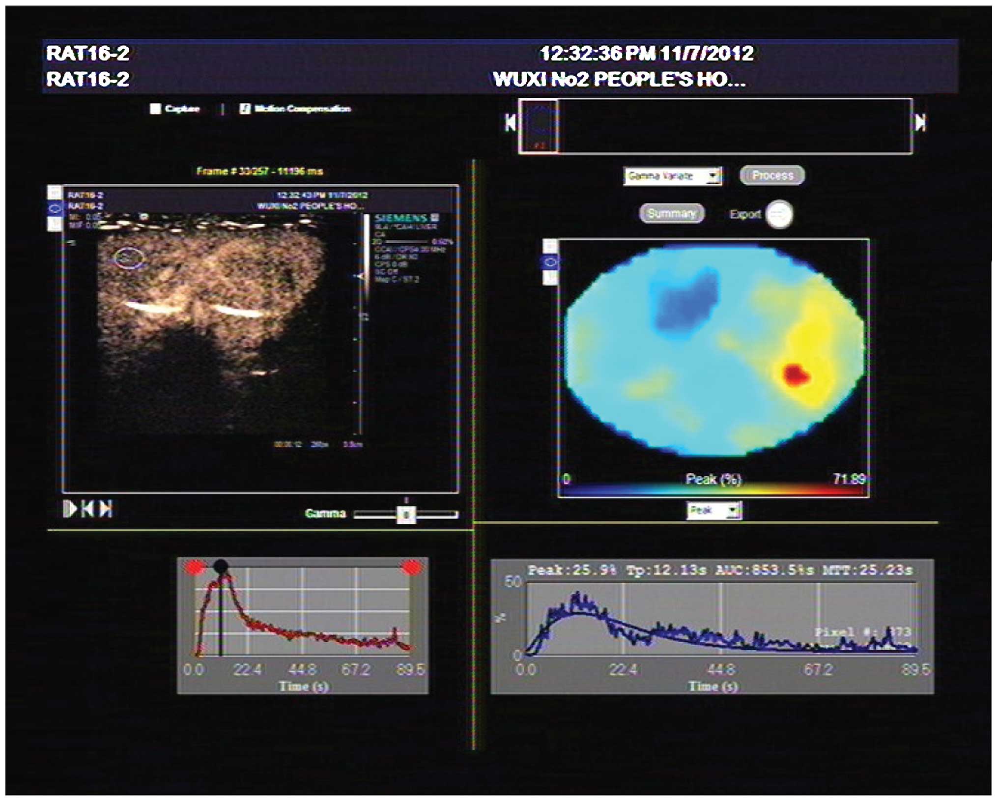
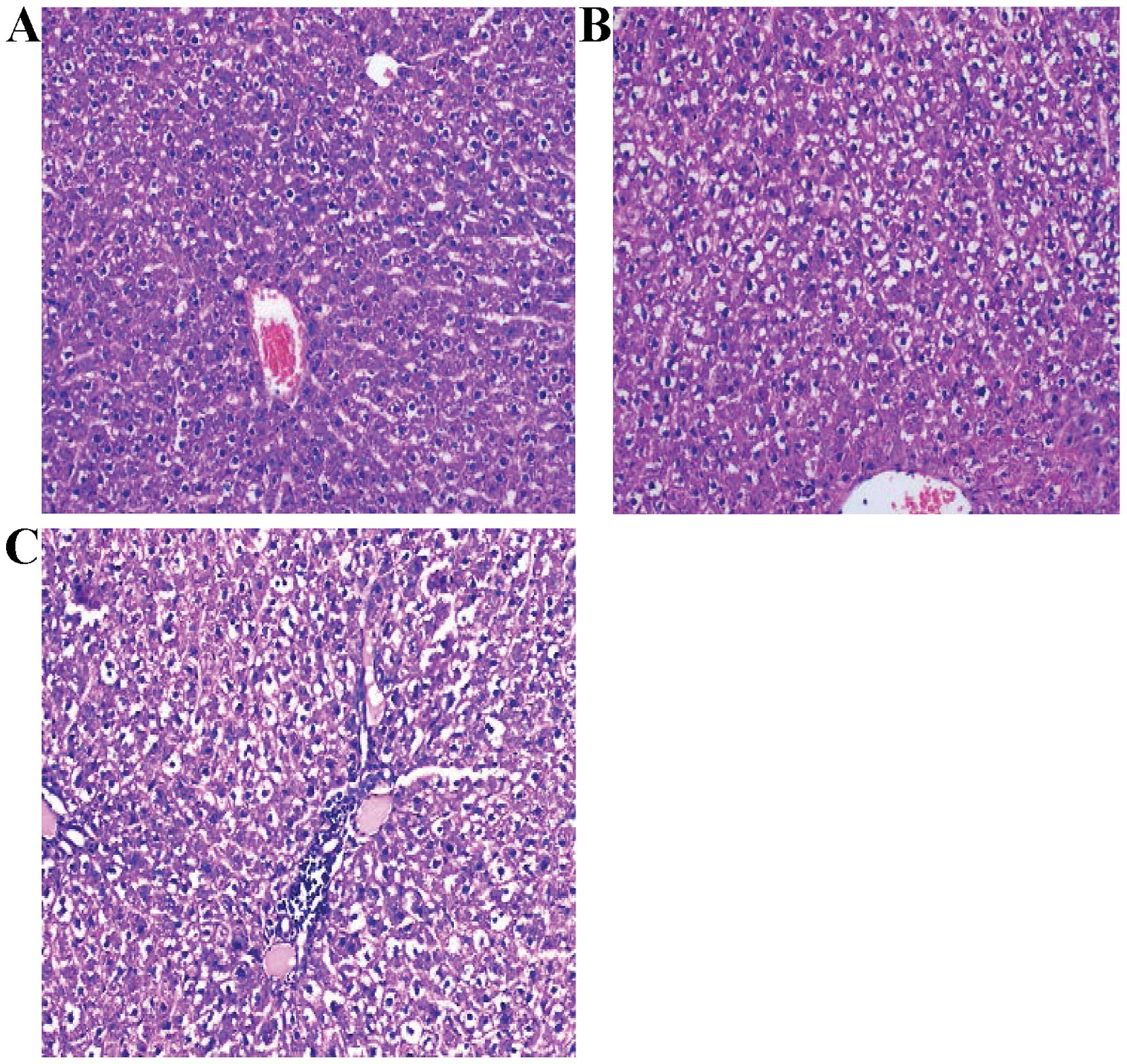
The aim of the present study was to examine and assess contrast-enhanced ultrasound in the early diagnosis of acute radiation-induced liver injury in a rat model. Sixty female rats were used, with 50 rats being utilized to produce an animal model of liver injury with a single dose of stereotactic X-ray irradiation of 20 Gy. Ten rats from the injury group and 2 rats from the control group were randomly selected on days 3, 7, 14, 21 and 28, and examined by contrast-enhanced ultrasound and histopathology of liver specimens. The rats were divided into four groups: the normal control group, mild, moderate, and severe radioactive liver injury groups based on the histopathological examination results. Hepatic artery arriving time (HAAT) and hepatic vein arriving time (HVAT) were recorded, and hepatic artery to vein transit time (HA-HVTT) was calculated. The time-intensity curve of liver parenchyma, the time to peak (TTP) and peak intensity (PI) were also obtained. Significant differences were observed between liver injury and control groups for PI and HA-HVTT (P<0.05). PI and HA-HVTT were shorter in the severe liver injury group compared to the mild and moderate liver injury groups (P<0.05). Compared to the control group, higher TTP was recorded in all the liver injury groups (P<0.05), and the highest TTP level was observed in the severe liver injury group compared to the mild or moderate group (P<0.05). However, no significant difference was observed between the mild and moderate groups for PI, HA-HVTT and TTP. In conclusion, the results showed that contrast-enhanced ultrasonography is useful for an earlier diagnosis in a rat model of acute radiation-induced liver injury.
View Figures
View References

Related Articles

Journal Cover

November-2015
Volume 10 Issue 5

Print ISSN: 1792-0981
Online ISSN:1792-1015

Sign up for eToc alerts

Recommend to Library

Copy and paste a formatted citation
x
Spandidos Publications style
Feng J, Chen S, Wu S, Sun P, Xin T and Chen Y: Quantitative analysis of contrast-enhanced ultrasonography in acute radiation-induced liver injury: An animal model. Exp Ther Med 10: 1807-1811, 2015
APA
Feng, J., Chen, S., Wu, S., Sun, P., Xin, T., & Chen, Y. (2015). Quantitative analysis of contrast-enhanced ultrasonography in acute radiation-induced liver injury: An animal model. Experimental and Therapeutic Medicine, 10, 1807-1811. https://doi.org/10.3892/etm.2015.2764
MLA
Feng, J., Chen, S., Wu, S., Sun, P., Xin, T., Chen, Y."Quantitative analysis of contrast-enhanced ultrasonography in acute radiation-induced liver injury: An animal model". Experimental and Therapeutic Medicine 10.5 (2015): 1807-1811.
Chicago
Feng, J., Chen, S., Wu, S., Sun, P., Xin, T., Chen, Y."Quantitative analysis of contrast-enhanced ultrasonography in acute radiation-induced liver injury: An animal model". Experimental and Therapeutic Medicine 10, no. 5 (2015): 1807-1811. https://doi.org/10.3892/etm.2015.2764