Introduction
Rectal cancer is a commonly occurring
gastrointestinal cancer, which has a high mortality rate of ~50%
(1). Successful early detection of
rectal cancer is rare, resulting in a poor prognosis (2). The incidence of rectal cancer is
increasing year by year (3), and
poses a serious threat to human health. Therefore, it is important
to identify novel tumor markers for the early diagnosis and
prognosis of rectal cancer.
FERM domain-containing 4A (FRMD4A), located on human
chromosome 10, is a cancer stem cell marker that belongs to the
FERM superfamily, all the members of which contain a FERM domain
(4). The FERM domain has been
demonstrated to play an important role in the cytoskeleton, the
maintenance of cell morphology, protein localization and tumor cell
migration (5–10). Levels of FRMD4A have been found to be
upregulated in skin cancer and squamous cell carcinoma, and are
closely associated with recurrence and metastasis (11). Genome-wide association analyses have
shown that FRMD4A is highly associated with drug dependence
(12,13). FERMD4A regulates the expression of
cytoskeletal proteins and the connections between mammary
epithelial cells through the activation of ADP ribosylation factor
6 (14). However, the expression of
FRMD4A in rectal cancer remains unclear.
In this study, immunohistochemistry, western
blotting and reverse transcription-quantitative polymerase chain
reaction (RT-qPCR) experiments were performed to detect the
expression of FRMD4A in rectal cancer. The correlation between
FRMD4A and cancer development was then studied, with the aim of
providing experimental evidence concerning the functions of FRMD4A
in rectal cancer.
Materials and methods
Patients
A total of 78 consecutive patients diagnosed with
rectal cancer at Zhejiang Cancer Hospital (Zhejiang, China) between
September 2013 and December 2014 were enrolled in this study. The
study was approved by the Ethics Review Board of Zhejiang Cancer
Hospital (Hangzhou, China). Prior written and informed consent was
obtained from every patient. General information about the patients
is shown in Table I. Fresh rectal
cancer and corresponding normal adjacent tissues (distance from
tumor edge, ≥5 cm) were collected, and stored at −80°C. The average
age of these patients was 53.70 years, with an age range of 32–71
years. There were 42 male patients and 36 female patients. Thirty
healthy individuals were used as the control group. The patients
underwent categorization for lymph node metastases as follows: N0
group, no evidence of lymph node metastasis; N1 group, metastasis
to lymph node.
 | Table I.Correlation between FRMD4A expression
in rectal cancer patients and cancer development. |
Table I.
Correlation between FRMD4A expression
in rectal cancer patients and cancer development.
|
|
| Immunohistochemical
scoring index
| χ2
| Correlation
|
|---|
| Characteristic | Cases (n=78) | 0–3 (n=16) | 4–6 (n=49) | 9 (n=13) |
χ2-value | P-value | Pearson's r | P-value |
|---|
| Age (years) |
|
|
|
| 1.354 | 0.852 |
|
|
|
<40 | 5 | 2 | 2 | 1 |
|
|
|
|
|
41–65 | 43 | 8 | 28 | 7 |
|
|
|
|
|
>66 | 30 | 6 | 19 | 5 |
|
|
|
|
| Gender |
|
|
|
| 2.385 | 0.184 |
|
|
| Male | 42 | 9 | 29 | 4 |
|
|
|
|
|
Female | 36 | 7 | 20 | 9 |
|
|
|
|
| Tumor size |
|
|
|
| 0.111 | 0.946 |
|
|
| <5
cm | 26 | 5 | 17 | 4 |
|
|
|
|
| ≥5
cm | 52 | 11 | 32 | 9 |
|
|
|
|
| Differentiation
status |
|
|
|
| 18.902 | 0.001 | 0.339 | 0.002 |
| High | 16 | 9 | 7 | 0 |
|
|
|
|
|
Moderate | 33 | 5 | 20 | 8 |
|
|
|
|
| Poor | 29 | 2 | 22 | 5 |
|
|
|
|
| Depth of
invasion |
|
|
|
|
|
|
|
|
| T2 | 5 | 2 | 3 | 0 | 11.128 | 0.025 | 0.352 | 0.002 |
| T3 | 56 | 13 | 37 | 6 |
|
|
|
|
| T4 | 17 | 1 | 9 | 7 |
|
|
|
|
| Dukes' stage |
|
|
|
|
|
|
|
|
| B | 46 | 12 | 30 | 4 | 10.805 | 0.029 | 0.308 | 0.006 |
| C | 25 | 4 | 16 | 5 |
|
|
|
|
| D | 7 | 0 | 3 | 4 |
|
|
|
|
| Lymph node
metastasis |
|
|
|
|
|
|
|
|
| N0 | 30 | 11 | 15 | 4 | 7.802 | 0.020 | 0.257 | 0.023 |
| N1 | 48 | 5 | 34 | 9 |
|
|
|
|
Reagents
Rabbit anti-human FRMD4A polyclonal antibody
(ab122475), rabbit anti-human epithelial cadherin (E-cadherin)
polyclonal antibody (ab15148) and mouse anti-human
glyceraldehyde-3-phosphate dehydrogenase (GAPDH) monoclonal
antibody (ab8245) were all purchased from Abcam (Cambridge, UK).
Secondary antibodies horseradish peroxidase (HRP)-conjugated goat
anti-mouse IgG (ab6789) and HRP-conjugated goat anti-rabbit IgG
(ab6721) were also purchased from Abcam. An EnVision
Immunohistochemistry Detection kit was purchased from Dako
(Glostrup, Denmark). Easyspin whole blood RNA rapid extraction kit
(centrifugal column) was purchased from Biomed (Beijing, China). A
reverse transcription kit was purchased from Boruike Biotech
(Chengdu, China). SYBR Green Real-Time PCR reagents were purchased
from Kapa Biosystems (Wilmington, MA, USA).
RT-qPCR
Total RNA was extracted from peripheral blood
samples, using the whole blood RNA extraction kit according to the
manufacturer's protocol. The quality of the RNA was detected by
electrophoresis and the spectrophotometric determination of the
ratio of optical density at 260 and 280 nm. The RNA was reverse
transcribed into cDNA using the reverse transcription kit according
to the manufacturer's protocol. qPCR was then conducted using an
ABI StepOne Plus Real-Time PCR system (Thermo Fisher Scientific,
Foster City, CA, USA). GAPDH was used as an internal control. The
relative amount of FRMD4A transcript was expressed as a ratio
relative to the level of GAPDH mRNA. The experiments were repeated
independently ≥3 times. The primers for FRMD4A were
5′-GATTCTTCGGATGCGTAA-3′ and 5′-CTGGCTCACAACATAGTC-3′. The primers
for GAPDH were 5′-ATGCTGGCGCTGAGTACGTC-3′ and
5′-GGTCATGAGTCCTTCCACGATA-3′.
Immunohistochemistry
Rectal cancer tissues were fixed with 4% neutral
formalin, followed by embedding in paraffin. Then 4-µm serial
sections were treated with citrate buffer for antigen retrieval at
95°C for 10 min. The primary rabbit anti-human FRMD4A polyclonal
antibody or rabbit anti-human E-cadherin polyclonal antibody
(dilution, 1:200) was added and the sections were incubated for 60
min at 37°C. The solution from the EnVision kit was then added and
the sections were incubated for 30 min at 37°C. The sections were
developed with diaminobenzidine chromogenic reagent and observed
under a microscope (Olympus BX51; Olympus Corp., Tokyo, Japan).
Cells with brown yellow staining were defined as FRMD4A-positive or
E-cadherin-positive cells. Ten fields at high magnification (×200)
were randomly taken from each section. The intensity of staining
and the proportion of positive cells were determined, and each was
scored on a scale of 0–3. For staining intensity: Score 0, no
staining; score 1, yellow staining; score 2, brown yellow staining;
and score 3, brown staining. For the positive rate: Score 0, 0%
positive cells; score 1, <10% positive cells; score 2, 10–50%
positive cells; and score 3, >50% positive cells. The FRMD4A and
E-cadherin staining indices were calculated by multiplying the
score for the intensity of staining by the score for the proportion
of positive cells, and graded as follows: Grade 1, score 0–3; grade
2, score 4–6; and grade 3, score 7–9. At least 200 cells were
counted in each field.
Western blotting
Total proteins were harvested from 100 mg rectal
cancer tissues and separated on 10% sodium dodecyl
sulfate-polyacrylamide gel electrophoresis gels. Then, the proteins
were transferred to a polyvinylidene difluoride (PVDF) membrane
(Thermo Fisher Scientific, Inc, Vilnius, Lithuania). After blocking
with 5% non-fat milk, the primary antibodies against FRMD4A
(dilution, 1:800) and GAPDH (dilution, 1:4,000) were added and the
membrane was incubated overnight at 4°C. The secondary
HRP-conjugated IgG antibodies (goat anti-mouse, 1:5,000; goat
anti-rabbit, 1:2,000) were added and the samples were incubated for
1 h at room temperature. The bound antibodies were detected using
an enhanced chemiluminescence system (Beyotime Institute of
Biotechnology, Jiangsu, China). The protein expression levels were
analyzed using Quantity One software, version 4.6.2 (Bio-Rad
Laboratories, Hercules, CA, USA)
Statistical analysis
Statistical analysis software (SPSS, version 10.0;
SPSS, Inc., Chicago, IL, USA) was used to conduct χ2
tests. Spearman's correlation was used to test the association
between two variables. P<0.05 was considered to indicate a
statistically significant result.
Results
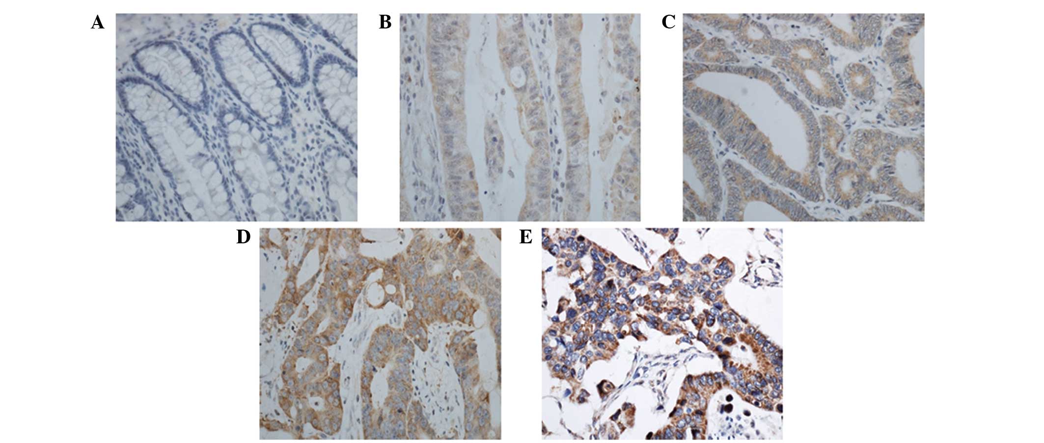
Expression of FRMD4A is upregulated in
rectal cancer tissues
To evaluate the expression of FRMD4A in rectal
cancer, immunohistochemical analysis of tissue samples taken from
patients was performed. Representative results are shown in
Fig. 1. Cells with brown and/or
yellow staining were FRMD4A-positive. The FRMD4A-positive rate in
the rectal cancer tissues was significantly higher than that in the
normal adjacent tissues (87.0 vs. 25.3%; P<0.05). These results
indicate that the expression level of FRMD4A is increased in rectal
cancer.
Correlation between the expression of
FRMD4A and clinicopathological features in rectal cancer
To determine the correlation between the expression
of FRMD4A in rectal cancer and clinicopathological features,
Spearman correlation analysis was performed. As shown in Table I, there were no statistically
significant differences in FRMD4A expression among the patients
according to age, gender or tumor size (P>0.05). However, the
expression of FRMD4A was found to be negatively correlated with the
degree of differentiation, depth of invasion, Dukes' stage and
lymph node metastasis (Table I).
These results indicate that positive correlations exist between
FRMD4A and certain clinicopathological features of patients with
rectal cancer, namely the degree of differentiation, depth of
invasion, Dukes' stage and lymph node metastasis.
Negative correlation between FRMD4A
and E-cadherin in rectal cancer patients
To determine the correlation between FRMD4A and
E-cadherin in rectal cancer, immunohistochemical analysis was used
to detect the expression of the two proteins in rectal cancer
tissue samples (Fig. 2). Spearman
correlation analysis was performed to analyze the correlation
between FRMD4A and E-cadherin expression in rectal cancer. In the
tissue sections, FRMD4A expression was found to be negatively
correlated with E-cadherin expression (r=–0.410, P<0.01),
indicating that a negative correlation exists between these two
proteins in rectal cancer.
Expression of FRMD4A protein is
increased in rectal cancer
To determine the expression of FRMD4A protein in
rectal cancer, western blotting was performed. As shown in Fig. 3, the relative expression level of
FRMD4A protein in rectal cancer tissues (3.97±0.20) was
significantly increased when compared with that in the normal
adjacent tissues (P<0.05). The relative expression level of
FRMD4A protein in the N1 group (2.76±0.31) was significantly
increased when compared with that in the N0 group (normalized value
1; P<0.05). The expression level of FRMD4A protein in cancers of
Dukes' stage C and D (1.78 ± 0.24) was significantly higher
compared with that in cancers of Dukes' stages A and B (normalized
value 1; P<0.05). The correlation between the FRMD4A expression
level and the degree of differentiation of the rectal cancer was
observed to be negative. These results indicate that the expression
of FRMD4A protein is increased in rectal cancer tissue. FRMD4A may
be associated with the invasion and metastasis of rectal cancer,
and negatively correlates with the degree of differentiation.
Expression of FRMD4A mRNA is increased
in the peripheral blood of rectal cancer patients
To determine the expression of FRMD4A mRNA in the
peripheral blood of rectal cancer patients, RT-qPCR analysis was
performed. As shown in Fig. 4, the
expression of FRMD4A mRNA in the peripheral blood of patients with
rectal cancer (3.24±0.13) was significantly increased compared with
that in healthy individuals (P<0.05). The expression of FRMD4A
mRNA in the N1 group (4.29±0.20) was significantly increased
compared with that in the N0 group (2.31±0.28) and the control
(P<0.05). Expression levels of FRMD4A mRNA in cancers of Dukes'
stages C and D were significantly increased compared with those in
cancers of Dukes' stages A and B (P<0.05). These results
indicate that the expression of FRMD4A mRNA in peripheral blood is
increased in patients with rectal cancer. The expression of FRMD4A
mRNA was increased as the Dukes' stage of the cancer increased.
Discussion
It has been reported that the expression of FRMD4A
is changed in some tumors and has an association with tumor
invasion and metastasis (11,15). For
example, Goldie et al found that FRMD4A expression levels
are increased in squamous cell carcinoma, and demonstrated that
FRMD4A-knockdown reduces tumor growth and metastasis (11). In the present study,
immunohistochemistry and western blotting assays were performed to
detect the expression of FRMD4A in rectal cancer tissue. The
results showed that the expression of FRMD4A in rectal cancer
tissue was significantly increased when compared with that in the
normal adjacent tissues (P<0.05). Positive staining of FRMD4A
protein was observed mainly in the cytoplasm, and partly in
nucleus. The level of expression of FRMD4A increased as the Dukes'
stage and the degree of differentiation decreased in the rectal
cancer. The expression of FRMD4A in patients with lymph node
metastasis was significantly increased compared with that in
patients without lymph node metastasis (P<0.05), indicating that
FRMD4A might be closely associated with the development, invasion
and metastasis of rectal cancer.
E-cadherin is a marker of epithelial-mesenchymal
transition (EMT) and has a close association with tumor invasion
and metastasis (16,17). It has been reported that erythrocyte
membrane protein band 4.1 like 5 (EPB41L5), which contains the FERM
domain, promotes EMT through the regulation of cadherin and
integrin expression (18). As
mentioned earlier, the FRMD4A protein also contains the FERM
domain. Thus, it is possible that FRMD4A is associated with EMT.
Therefore, the correlation between FRMD4A and E-cadherin was
examined in the present study. Immunohistochemical analysis showed
that the expression of FRMD4A was negatively correlated with the
expression of E-cadherin in rectal cancer. This suggests that
FRMD4A may be involved in EMT and cell adhesion and may promote the
invasion and metastasis of rectal cancer. The results of RT-qPCR
analysis showed that the expression level of FRMD4A mRNA in the
peripheral blood of patients with rectal cancer was significantly
increased compared with that in patients in the control group
(P<0.05). The expression levels of FRMD4A mRNA in the peripheral
blood of Dukes' stage C and D subgroups were significantly
increased compared with those in Dukes' stage A and B subgroups
(P<0.05). Since Dukes' stages and lymph node metastasis are
important indicators for the prognosis of patients with rectal
cancer (19), increased expression
levels of FRMD4A may indicate a poor prognosis for patients with
rectal cancer.
In conclusion, the present study indicates that the
expression of FRMD4A is closely associated with the development,
invasion and metastasis of rectal cancer. Therefore, FRMD4A may
play an important role in the early diagnosis and prognosis of
patients with rectal cancer. In addition, FRMD4A may be a potential
therapeutic target for rectal cancer.
Acknowledgements
This study was supported by the Natural Science
Foundation of Zhejiang Province (grant no. Y207427) and Medicine
and Health Science Research Fund of Zhejiang Province (grant no.
2008B019).
References
|
1
|
Cui C, Zhang M, Tian L, Jiang W, Zeng Z
and Li L: Survival implications of pretreatment pelvic CT in rectal
cancer patients after neoadjuvant chemoradiotherapy and surgery.
Int J Clin Exp Med. 8:12801–12809. 2015.PubMed/NCBI
|
|
2
|
Maffione AM, Chondrogiannis S, Capirci C,
Galeotti F, Fornasiero A, Crepaldi G, Grassetto G, Rampin L,
Marzola MC and Rubello D: Early prediction of response by (18)F-FDG
PET/CT during preoperative therapy in locally advanced rectal
cancer, A systematic review. Eur J Surg Oncol. 40:1186–1194. 2014.
View Article : Google Scholar : PubMed/NCBI
|
|
3
|
Probst CP, Becerra AZ, Aquina CT, Tejani
MA, Hensley BJ, González MG, Noyes K, Monson JR and Fleming FJ:
Watch and wait? - Elevated pretreatment CEA is associated with
decreased pathological complete response in rectal cancer. J
Gastrointest Surg Nov. 6:2015.(Epub ahead of print).
|
|
4
|
Strizzi L, Hardy KM, Margaryan V, Hillman
DW, Seftor EA, Chen B, Geiger XJ, Thompson EA, Lingle WL, et al:
Potential for the embryonic morphogen Nodal as a prognostic and
predictive biomarker in breast cancer. Breast Cancer Res.
14:R752012. View
Article : Google Scholar : PubMed/NCBI
|
|
5
|
Murata K, Nunomura W, Takakuwa Y and Cherr
GN: Two different unique cardiac isoforms of protein 4.1R in
zebrafish, Danio rerio, and insights into their cardiac
functions as related to their unique structures. Dev Growth Differ.
52:591–602. 2010. View Article : Google Scholar : PubMed/NCBI
|
|
6
|
Clucas J and Valderrama F: ERM proteins in
cancer progression. J Cell Sci. 127:267–275. 2014. View Article : Google Scholar : PubMed/NCBI
|
|
7
|
Fiévet B, Louvard D and Arpin M: ERM
proteins in epithelial cell organization and functions. Biochim
Biophys Acta. 1773:653–660. 2007. View Article : Google Scholar : PubMed/NCBI
|
|
8
|
Tepass U: FERM proteins in animal
morphogenesis. Curr Opin Genet Dev. 19:357–367. 2009. View Article : Google Scholar : PubMed/NCBI
|
|
9
|
Bennett V and Baines AJ: Spectrin and
ankyrin-based pathways: Metazoan inventions for integrating cells
into tissues. Physiol Rev. 81:1353–1392. 2001.PubMed/NCBI
|
|
10
|
Yu H, Zhang Y, Ye L and Jiang WG: The FERM
family proteins in cancer invasion and metastasis. Front Biosci
(Landmark Ed). 16:1536–1550. 2011. View
Article : Google Scholar : PubMed/NCBI
|
|
11
|
Goldie SJ, Mulder KW, Tan DW, Lyons SK,
Sims AH and Watt FM: FRMD4A upregulation in human squamous cell
carcinoma promotes tumor growth and metastasis and is associated
with poor prognosis. Cancer Res. 72:3424–3436. 2012. View Article : Google Scholar : PubMed/NCBI
|
|
12
|
Johnson C, Drgon T, Liu QR, Zhang PW,
Walther D, Li CY, Anthony JC, Ding Y, Eaton WW and Uhl GR: Genome
wide association for substance dependence, Convergent results from
epidemiologic and research volunteer samples. BMC Med Genet.
9:1132008. View Article : Google Scholar : PubMed/NCBI
|
|
13
|
Yoon D, Kim YJ, Cui WY, Van der Vaart A,
Cho YS, Lee JY, Ma JZ, Payne TJ, Li MD and Park T: Large-scale
genome-wide association study of Asian population reveals genetic
factors in FRMD4A and other loci influencing smoking initiation and
nicotine dependence. Hum Genet. 131:1009–1021. 2012. View Article : Google Scholar : PubMed/NCBI
|
|
14
|
Ikenouchi J and Umeda M: FRMD4A regulates
epithelial polarity by connecting Arf6 activation with the PAR
complex. Proc Natl Acad Sci USA. 107:748–753. 2010. View Article : Google Scholar : PubMed/NCBI
|
|
15
|
Zheng XH, Zhao JJ, Jia B, Pan J, Chen J,
Qiu XL, Han JS and Chu HX: The influence of inactive FRMD4A gene on
the biological behavior of human tongue cancer CAL-27 cell. Shi
Yong Yi Xue Za Zhi. 10:99–103. 2014.(In Chinese).
|
|
16
|
Heerboth S, Housman G, Leary M, Longacre
M, Byler S, Lapinska K, Willbanks A and Sarkar S: EMT and tumor
metastasis. Clin Transl Med. 4:62015. View Article : Google Scholar : PubMed/NCBI
|
|
17
|
Steinestel K, Eder S and Schrader AJ: andS
teinestel J: Clinical significance of epithelial-mesenchymal
transition. Clin Transl Med. 3:172014. View Article : Google Scholar : PubMed/NCBI
|
|
18
|
Hirano M, Hashimoto S, Yonemura S, Sabe H
and Aizawa S: EPB41L5 functions to post-transcriptionally regulate
cadherin and integrin during epithelial-mesenchymal transition. J
Cell Biol. 182:1217–1230. 2008. View Article : Google Scholar : PubMed/NCBI
|
|
19
|
Radovanović Z, Radovanović D, Breberina M,
Petrović T, Golubović A and Bokorov B: The value of endorectal
ultrasonography in rectal cancer staging. Med Pregl. 61:557–561.
2008.(In Serbian). View Article : Google Scholar : PubMed/NCBI
|