Introduction
Bone metabolism is coordinated by two types of
functional cells, osteoblasts and osteoclasts (1). The former cells are responsible for
bone formation, whilst the latter cells are responsible for bone
resorption. Thus, bone mass is sufficiently maintained by
osteoclastic bone resorption followed by osteoblastic bone
formation, a process termed bone remodeling (1). Disordered bone remodeling causes an
imbalance in the rates of bone resorption vs. bone formation, which
may result in metabolic bone diseases, including osteoporosis
(1). It is at present recognized
that AMP-activated protein kinase (AMPK) functions as a central
regulator of cellular energy homeostasis (2,3). AMPK
has been identified as an enzyme that catalyzes the phosphorylation
of acetyl-CoA carboxylase, which regulates lipid synthesis
(3). AMPK activity is increased by
the elevation of the AMP/ATP ratio in response to physiological and
pathological stress, resulting in the restoration of the cellular
enzyme balance through the activation of ATP-generating pathways
and the suppression of ATP-utilizing pathways (4). Therefore, evidence indicates that AMPK
regulates metabolic homeostasis throughout the body (3). With respect to AMPK in bone metabolism,
it has been revealed that AMPK activation stimulates bone formation
and bone mass (5). We have
previously demonstrated that vascular endothelial growth factor
synthesis, which is induced by basic fibroblast growth factor, is
positively regulated by AMPK in osteoblast-like MC3T3-E1 cells
(6). Conversely, AMPK limits the
interleukin (IL)-1-stimulated synthesis of IL-6 via the inhibitor
of κB/nuclear factor-κB (NF-κB) signaling pathway in MC3T3-E1 cells
(7). However, the roles of AMPK in
osteoblasts remain undefined.
Prostaglandins are important autocrine/paracrine
modulators in bone metabolism (8),
and include prostaglandin E1 (PGE1), which
has been identified as a potent bone-resorptive agent (9). It is currently accepted that
osteoblasts are vital in the regulation of bone resorption through
activation of receptor activator of NF-κB ligand (RANKL) (10). Osteoprotegerin is a glycoprotein
secreted from osteoblasts and is a member of the tumor necrosis
factor receptor superfamily, in addition to RANK (11). Osteoprotegerin binds to RANKL as a
decoy receptor, thus preventing RANKL from binding to RANK. This
results in the inhibition of osteoclastogenesis and the activation
of osteoclasts (11). Furthermore,
RANKL knockout mice reportedly develop severe osteopetrosis
(12), suggesting that RANKL is an
important regulator of osteoclastogenesis. The
RANK/RANKL/osteoprotegerin axis is currently recognized as a major
regulatory system for the formation and activation of osteoclasts
(13). Our recent study demonstrated
that PGE1 stimulates osteoprotegerin synthesis via the
activation of p38 mitogen-activated protein (MAP) kinase and
stress-activated protein kinase/c-Jun N-terminal kinase (SAPK/JNK),
but not via the activation of p44/p42 MAP kinase, in
osteoblast-like MC3T3-E1 cells (14). However, the molecular mechanism
underlying the PGE1-stimulated synthesis of
osteoprotegerin in osteoblasts has yet to be elucidated.
In the present study, the involvement of AMPK in
PGE1-induced osteoprotegerin synthesis was investigated
in osteoblast-like MC3T3-E1 cells. The present study revealed that
AMPK acts as a positive regulator in PGE1-stimulated
osteoprotegerin synthesis in MC3T3-E1 cells.
Materials and methods
Materials
PGE1 was purchased from Sigma-Aldrich
(St. Louis, MO, USA) and compound C was obtained from Calbiochem
(cat. no. 171260; Merck Millipore, La Jolla, CA, USA). A mouse
osteoprotegerin enzyme-linked immunosorbent assay (ELISA) kit (cat.
no. MOP00) was purchased from R&D Systems, Inc. (Minneapolis,
MN, USA). Rabbit anti-phosphorylated (p)-AMPKα (Thr-172; cat. no.
2531), AMPKβ (Ser-108; cat. no. 4181), acetyl-CoA carboxylase (cat.
no. 3661) polyclonal antibodies, rabbit anti-p-p38 MAP kinase (cat.
no. 4511) and p-SAPK/JNK (cat. no. 4668) monoclonal antibodies, and
rabbit anti-p38 MAP kinase (cat. no. 9212) and SAPK/JNK (cat. no.
9252) polyclonal antibodies, were purchased from Cell Signaling
Technology, Inc. (Danvers, MA, USA). Rabbit
anti-glyceraldehyde-3-phosphate dehydrogenase (GAPDH) polyclonal
antibody (sc-25778) was obtained from Santa Cruz Biotechnology,
Inc. (Santa Cruz, CA, USA). Peroxidase-conjugated goat anti-rabbit
immunoglobulin G (IgG; cat. no. 074-1506) was purchased from KPL,
Inc. (Gaithersburg, MD, USA). An enhanced chemiluminescence western
blotting detection system was purchased from GE Healthcare Life
Sciences (Chalfont, UK). All other materials and chemicals were
obtained from commercial sources.
PGE1 was dissolved in ethanol (Wako Pure
Chemical Industries, Ltd., Osaka, Japan) and compound C was
dissolved in dimethyl sulfoxide (Sigma-Aldrich). The maximum
concentration of ethanol or dimethyl sulfoxide used was 0.1% to
avoid interference with the assay for osteoprotegerin or western
blot analysis.
Cell culture
Cloned osteoblast-like MC3T3-E1 cells derived from
newborn mouse calvaria in a previous study (15) were maintained as previously described
(16). Briefly, MC3T3-E1 cells were
cultured in α-minimum essential medium (α-MEM) supplemented with
10% fetal bovine serum (FBS) at 37°C in a humidified atmosphere of
5% CO2/95% air. The cells were seeded into 35-mm
diameter dishes (5×10⁴ cells/dish) or 90-mm diameter dishes (2×10⁵
cells/dish) in α-MEM supplemented with 10% FBS. After 5 days, the
medium was exchanged for α-MEM supplemented with 0.3% FBS. The
cells were used for experiments following a 48-h incubation period
at 37°C.
Assay for osteoprotegerin
The cultured cells were pretreated with 0.1, 1, 3 or
10 µM compound C for 60 min. The cells were stimulated with 10 µM
PGE1 or vehicle [phosphate-buffered saline (PBS)
supplemented with 0.01% bovine serum albumin (Sigma-Aldrich)
containing 0.1% ethanol] in 1 ml α-MEM supplemented with 0.3% FBS,
and then incubated for 48 h. The conditioned medium was collected
by aspiration subsequent to incubation, followed by measurement
with a mouse osteoprotegerin ELISA kit, according to the
manufacturer's protocol.
Western blot analysis
The cultured cells were pretreated with 0.1, 1, 3 or
10 µM of compound C for 60 min, and then stimulated with 10 µM
PGE1 or vehicle in α-MEM supplemented with 0.3% FBS for
0, 1, 3, 5, 10, 20, 30 or 60 min. The cells were washed twice with
PBS and then lysed, homogenized and sonicated in 900 µl lysis
buffer containing 62.5 mM Tris/HCl (pH 6.8), 2% sodium dodecyl
sulfate (SDS), 50 mM dithiothreitol and 10% glycerol. Protein
samples were obtained from the lysed cells using a cell scraper,
after which equal quantities of protein (10 µl) were separated by
SDS-polyacrylamide gel electrophoresis, according to a previous
study (17), on 10% polyacrylamide
gels. The electrophoresis was run at 100 V for the separating gel
and 250 V for the stacking gel, until the dye front ran off the
bottom of the gel. The protein was fractionated and transferred to
an Immun-Blot polyvinylidene difluoride (PVDF) membrane (Bio-Rad
Laboratories, Hercules, CA, USA). Western blot analysis was
performed as previously described (18). Briefly, the membranes were blocked
with 5% fat-free dry milk in Tris-buffered saline containing
Tween-20 (TBST; 20 mM Tris/HCl, pH 7.6, 137 mM NaCl, 0.1% Tween-20)
for 1 h prior to incubation with the primary antibodies. The
membranes were incubated overnight at 4°C with the rabbit
anti-p-AMPK, anti-acetyl-CoA carboxylase, anti-GAPDH, anti-p-p38
MAP kinase, anti-p38 MAP kinase, anti-p-SAPK/JNK and anti-SAPK/JNK
primary antibodies at a dilution of 1:1,000 in 5% milk containing
TBST. Subsequently, the membranes were washed three times for 5 min
each with TBST, and then incubated for 1 h at room temperature with
peroxidase-conjugated goat anti-rabbit IgG at a dilution of 1:1,000
in 5% milk containing TBST. Peroxidase activity on the PVDF
membrane was visualized on X-ray film (Super RX; FUJIFILM,
Kanagawa, Japan) by means of the enhanced chemiluminescence western
blotting detection system.
Densitometric analysis
Densitometric analysis of the western blots was
performed using a scanner (GT-F600; Seiko Epson Corporation,
Nagano, Japan) and Image J image analysis software program (version
1.48; National Institutes of Health, Bethesda, MD, USA). The
background-subtracted signal intensity for each phosphorylation
signal was normalized to the respective total protein signal, and
plotted as the fold increase in comparison to control cells that
did not receive stimulation.
Reverse transcription-quantitative
polymerase chain reaction (RT-qPCR)
The cultured cells were pretreated with 10 µM
compound C or vehicle for 60 min, and then stimulated with 10 µM of
PGE1 or vehicle in α-MEM supplemented with 0.3% FBS for
3 h. Total RNA was isolated and reverse transcribed into cDNA using
TRIzol reagent (Invitrogen; Thermo Fisher Scientific, Inc.) and an
Omniscript Reverse Transcriptase kit (Qiagen, Inc., Valencia, CA,
USA), respectively. RT-qPCR was performed in capillaries using a
LightCycler® 480 system with the LightCycler FastStart
DNA Master SYBR Green I (Roche Diagnostics, Basel, Switzerland) and
1 ml cDNA. The primer sequences were as follows: Osteoprotegerin
forward, 5′-CAATGGCTGGCTTGGTTTCATAG-3′ and reverse,
5′-CTGAACCAGACATGACAGCTGGA-3′ (Takara Bio, Inc., Otsu, Japan); and
GAPDH forward, 5′-AACGACCCCTTCATTGAC-3′ and reverse,
5′-TCCACGACATACTCAGCAC-3′ (Sigma-Aldrich). The reaction mixtures
were incubated at 95°C for 10 min, followed by 40 cycles at 60°C
for 5 sec and 72°C for 7 sec. The amplified products were confirmed
by a melting curve analysis, according to the system protocol. The
osteoprotegerin mRNA expression levels were normalized to those of
GAPDH mRNA using PASW Statistics software, version 18 (SPSS, Inc.,
Chicago, IL, USA) and the relative mRNA expression levels were
determined from the basis of a standard curve, which was created
automatically with the LightCycler software in each run. The
results are the average of three independent experiments.
Statistical analysis
The data were analyzed by analysis of variance
followed by the Bonferroni method for multiple comparisons between
pairs. P<0.05 was considered to indicate a statistically
significant difference. Statistical analyses were conducted using
Microsoft Office Excel 2013 for Windows (Microsoft, Redmond, WA,
USA) Data are presented as the means ± standard error of the mean
of triplicate determinations from three independent cell
preparations.
Results
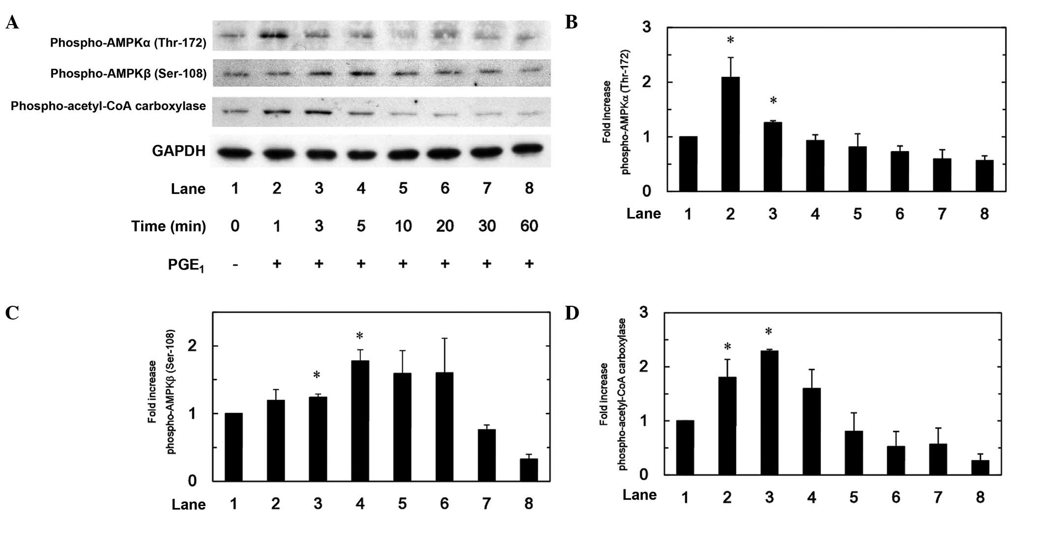
PGE1 induces the
phosphorylation of AMPK and acetyl-CoA carboxylase in MC3T3-E1
cells
It is firmly established that the phosphorylation of
AMPK is necessary for its activation (19). Therefore, to determine whether AMPK
is activated by PGE1 in osteoblast-like MC3T3-E1 cells,
the present study initially examined the effect of PGE1
on the phosphorylation of AMPK (Fig.
1A). PGE1 significantly induced the phosphorylation
of AMPKα (Thr-172; P<0.05; Fig.
1B) and AMPKβ (Ser-108; P<0.05; Fig. 1C). The effects of PGE1 on
the phosphorylation of AMPKα and AMPKβ reached peak levels at 1 and
5 min after stimulation, respectively; and decreased thereafter.
Subsequent to this, as acetyl-CoA carboxylase is a direct substrate
of AMPK (3), the effect of
PGE1 on the phosphorylation of acetyl-CoA carboxylase in
MC3T3-E1 cells was examined. PGE1 significantly
stimulated the phosphorylation of acetyl-CoA carboxylase,
displaying peak levels 3 min after stimulation (Fig. 1D).
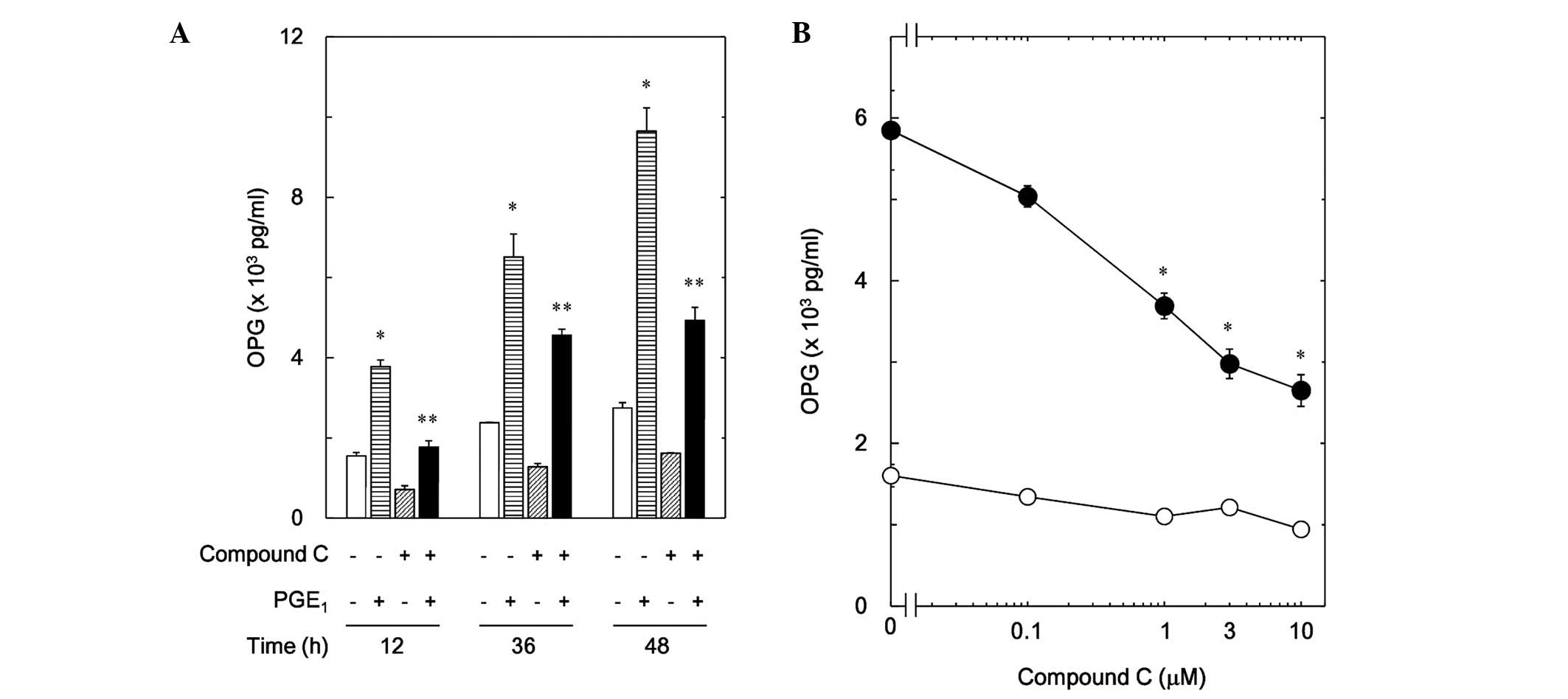
Compound C suppresses
PGE1-stimulated osteoprotegerin release in MC3T3-E1
cells
Our previous study recently revealed that
PGE1 stimulates osteoprotegerin synthesis in
osteoblast-like MC3T3-E1 cells (14). In order to investigate the
involvement of AMPK in PGE1-induced synthesis of
osteoprotegerin in the aforementioned cells, the effect of compound
C, an AMPK inhibitor (20), on
PGE1-stimulated release of osteoprotegerin was examined.
Compound C significantly suppressed PGE1-stimulated
osteoprotegerin release compared with cells treated with
PGE1 alone (P<0.05; Fig.
2A). The inhibitory effect of compound C was dose-dependent
between 0.1 and 10 mM (Fig. 2B). The
maximum suppressive activity of compound C was observed at 10 µM,
producing 60% inhibition of the effects of PGE1.
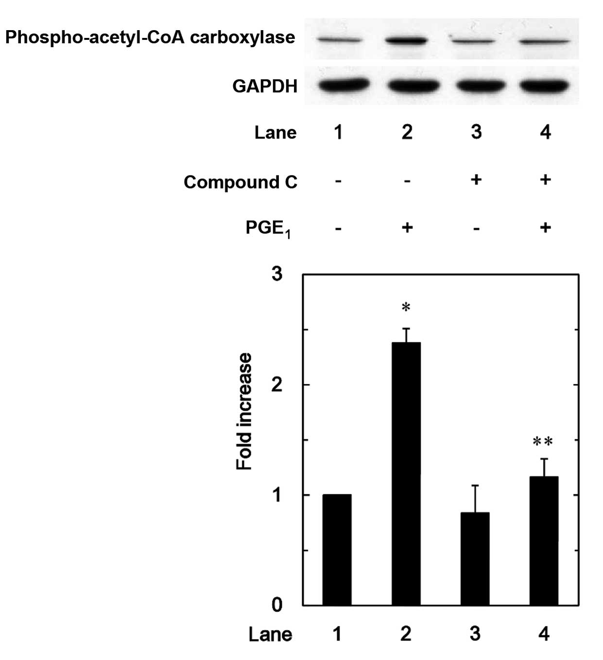
Additionally, the effect of compound C on
PGE1-induced phosphorylation of acetyl-CoA carboxylase
in MC3T3-E1 cells was examined. PGE1-induced
phosphorylation of acetyl-CoA carboxylase was significantly reduced
by compound C compared with PGE1 treatment alone
(P<0.05; Fig. 3).
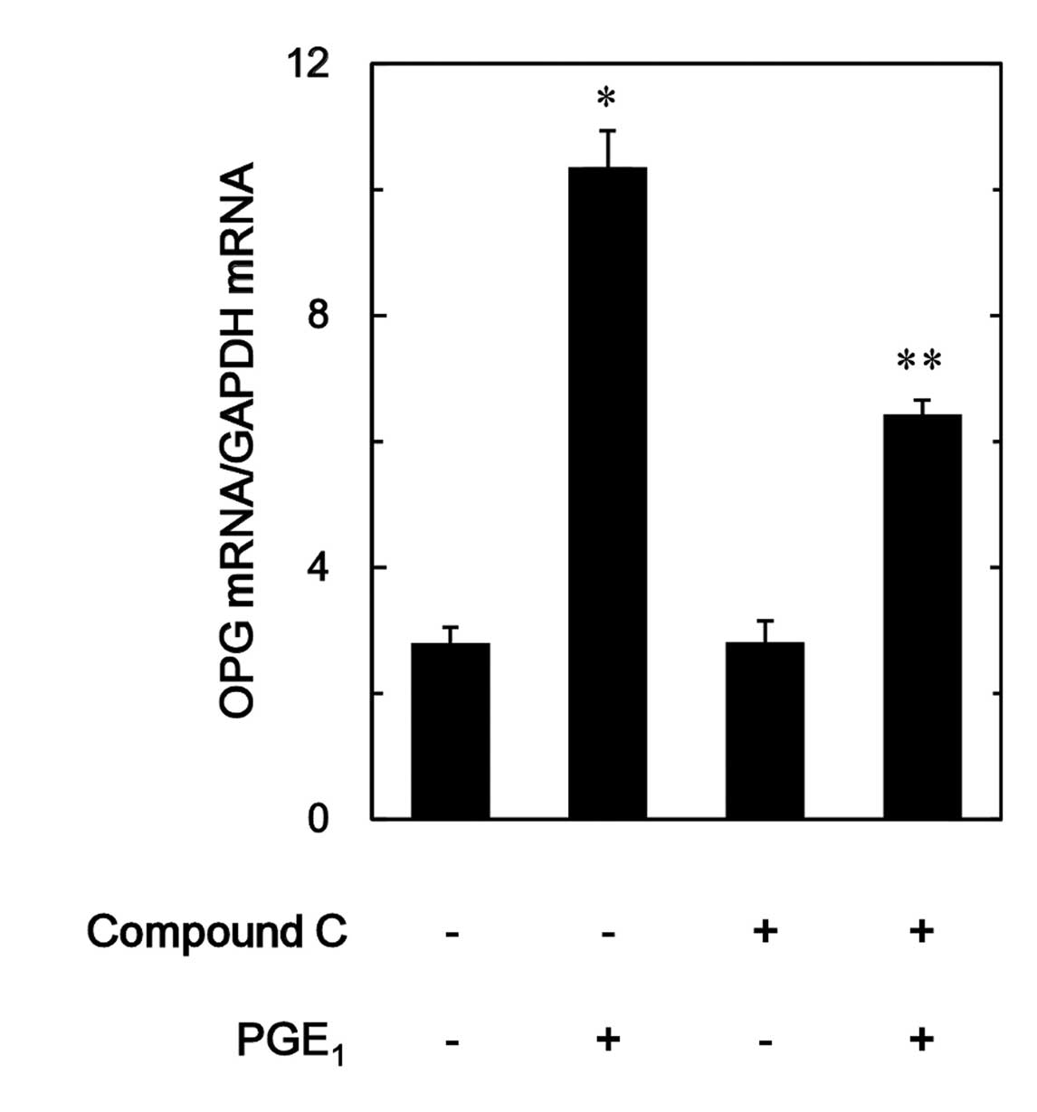
Compound C suppresses
PGE1-induced osteoprotegerin mRNA expression levels in
MC3T3-E1 cells
To determine whether the inhibition of the
PGE1-stimulated osteoprotegerin release by compound C is
mediated through transcriptional events in osteoblast-like MC3T3-E1
cells, the effect of compound C on PGE1-induced
osteoprotegerin mRNA expression levels were examined by RT-qPCR.
The expression levels of osteoprotegerin mRNA induced by
PGE1 were significantly suppressed by compound C
compared with PGE1 treatment alone (P<0.05; Fig. 4).
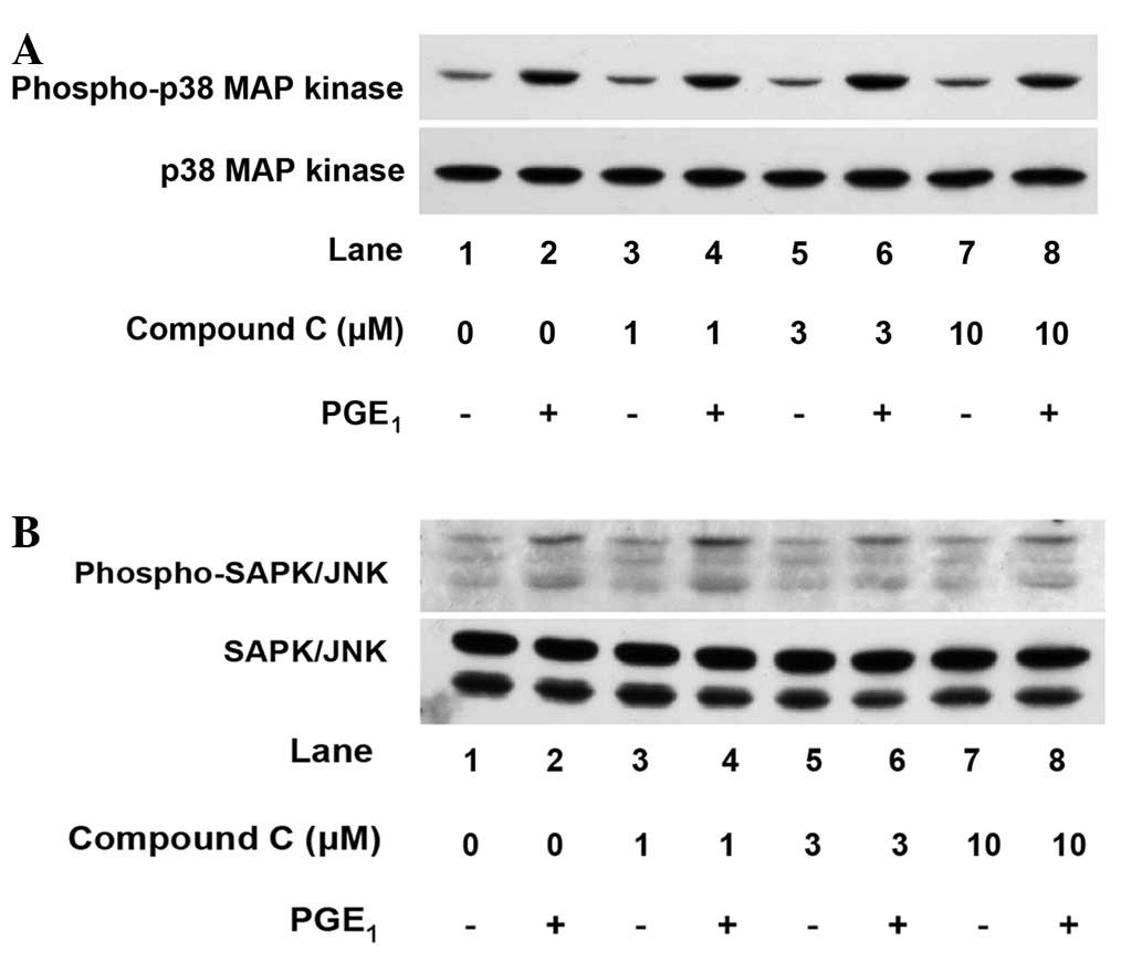
Compound C does not affect
PGE1-stimulated phosphorylation of p38 MAP kinase or
SAPK/JNK in MC3T3-E1 cells
Regarding the intracellular signaling of
PGE1 in osteoblasts, our previous study revealed that
PGE1 induces the activation of p44/p42 MAP kinase, p38
MAP kinase and SAPK/JNK in osteoblast-like MC3T3-E1 cells. In
addition, p38 MAP kinase and SAPK/JNK were implicated in
PGE1-stimulated osteoprotegerin synthesis, whilst
p44/p42 MAP kinase was not (14). To
investigate whether the effect of AMPK on
PGE1-stimulated osteoprotegerin synthesis is associated
with the activation of p38 MAP kinase or SAPK/JNK in MC3T3-E1
cells, the effects of compound C on PGE1-induced
phosphorylation of p38 MAP kinase and SAPK/JNK were examined.
However, compound C did not affect PGE1-induced
phosphorylation of p38 MAP kinase (Fig.
5A) or SAPK/JNK (Fig. 5B)
between 1 and 10 µM.
Discussion
The present study revealed that PGE1
significantly stimulates the phosphorylation of AMPKα (Thr-172) and
AMPKβ (Ser-108) in osteoblast-like MC3T3-E1 cells. The
heterotrimeric AMPK complex is comprised of α, β and γ subunits
(2). Of the aforementioned subunits
of AMPK, the α subunit is a catalytic subunit. Conversely, the β
and γ subunits are regulatory subunits (19). It is currently recognized that the
phosphorylation of Thr-172, located within the α subunit, is
required for AMPK activity, while phosphorylation of the β subunit
is necessary for the activation of AMPK (19). The present study revealed that the
phosphorylation of acetyl-CoA carboxylase was significantly induced
by PGE1 in MC3T3-E1 cells. It is generally accepted that
activated AMPK induces the phosphorylation of acetyl-CoA
carboxylase, a direct substrate of AMPK, resulting in the
stimulation of the oxidation of fatty acid as a result of its
inhibition (3). The maximum effect
of PGE1 on the phosphorylation of acetyl-CoA carboxylase
was observed 3 min after stimulation. Comparatively, the α subunit
demonstrated peak phosphorylation levels within 1 min. Thus, the
time course of AMPKα phosphorylation appeared to be more rapid than
that of acetyl-CoA carboxylase. As a result,
PGE1-induced activation of acetyl-CoA carboxylase occurs
subsequent to AMPKα activation. Considering the findings of the
present study, it is possible that PGE1 induces AMPK
activation in osteoblast-like MC3T3-E1 cells.
Our previous study recently reported that
PGE1 stimulates osteoprotegerin synthesis in
osteoblast-like MC3T3-E1 cells (14). The involvement of AMPK in
osteoprotegerin synthesis has since been investigated, and it was
revealed that the PGE1-stimulated release of
osteoprotegerin was significantly suppressed by compound C in
MC3T3-E1 cells (20). In agreement,
the present study revealed that compound C was able to inhibit
PGE1-induced phosphorylation of acetyl-CoA carboxylase
in these cells. Therefore, it is possible that
PGE1-activated AMPK is involved in osteoprotegerin
release. Additionally, the present study demonstrated that
PGE1-induced mRNA expression levels of osteoprotegerin
were significantly downregulated by compound C, suggesting that the
effect of compound C is exerted through inhibition of the
transcriptional process. Based on the findings of the present
study, it is proposed that PGE1 stimulates AMPK
activation, resulting in the induction of osteoprotegerin synthesis
in osteoblast-like MC3T3-E1 cells.
With regard to the intracellular signaling of
PGE1 in osteoblasts, our previous study demonstrated
that PGE1 induces the activation of p44/p42 MAP kinase,
p38 MAP kinase and SAPK/JNK in osteoblast-like MC3T3-E1 cells
(21,22), and that p38 MAP kinase and SAPK/JNK
(but not p44/p42 MAP kinase) are involved in
PGE1-stimulated osteoprotegerin synthesis (14). The MAP kinase superfamily have a
crucial role in various cellular functions, including
proliferation, differentiation and survival (23). It is generally recognized that p38
MAP kinase, p44/p42 MAP kinase and SAPK/JNK are three major MAP
kinases necessary for the transduction of diverse cellular messages
(24). In order to investigate the
molecular mechanism employed by AMPK in PGE1-stimulated
osteoprotegerin synthesis in MC3T3-E1 cells, the association
between AMPK and the aforementioned MAP kinases were investigated.
However, the present study revealed that compound C failed to
effect the PGE1-induced phosphorylation of p38 MAP
kinase or SAPK/JNK. Therefore, it is likely that AMPK may act
downstream of the aforementioned MAP kinases, or through a
different target located upstream of transcription, resulting in
osteoprotegerin synthesis. Further investigation is required to
identify the exact mechanism by which AMPK is involved in
PGE1-stimulated osteoprotegerin synthesis in
osteoblasts.
It is currently established that AMPK has a central
role in energy homeostasis, particularly in myocytes and adipocytes
(2). Regarding the roles of AMPK in
osteoblasts, it has been revealed that the activation of AMPK
stimulates collagen synthesis and the induction of Runt-related
transcription factor 2 expression, and enhances mineralization
(25,26). Additionally, AMPK has been reported
to have an inhibitory role in palmitate-induced apoptosis (27). Thus, accumulating evidence suggests
that AMPK activation in osteoblasts may stimulate them toward
differentiation. It is accepted that PGE1 acts as an
autocrine/paracrine regulator of osteoblasts and also regulates
bone remodeling (8). Conversely, it
is established that osteoprotegerin acts as a decoy receptor to
prevent RANKL from binding RANK in osteoclast progenitor cells,
resulting in the downregulation of osteoclastogenesis and bone
resorption (11). Based on the
findings of the present study, it is proposed that AMPK activated
by PGE1 in osteoblasts directs the bone remodeling
process to favor formation rather than resorption by stimulating
osteoprotegerin synthesis. Further investigation is necessary to
elucidate the exact roles of AMPK in bone metabolism.
In conclusion, the results of the present study
strongly suggest that AMPK positively regulates the
PGE1-stimulated synthesis of osteoprotegerin in
osteoblasts. The present study may provide novel insights into the
regulatory mechanisms underlying bone metabolism.
Acknowledgements
The present authors are grateful to Mrs. Yumiko
Kurokawa for her skillful technical assistance. The present study
was supported in part by a Grant-in-Aid for Scientific Research
(grant no. 19591042) from the Ministry of Education, Culture,
Sports, Science and Technology of Japan; a Grant-in-Aid for
Scientific Research (grant no. H25-Aging-General-004) from the
Ministry of Health, Labour and Welfare of Japan; and the Research
Funding for Longevity Sciences (grant no. 25-4, 26-12) from the
National Center for Geriatrics and Gerontology (Obu, Japan).
References
|
1
|
Karsenty G and Wagner EF: Reaching a
genetic and molecular understanding of skeletal development. Dev
Cell. 2:389–406. 2002. View Article : Google Scholar : PubMed/NCBI
|
|
2
|
Fogarty S and Hardie DG: Development of
protein kinase activators: AMPK as a target in metabolic disorders
and cancer. Biochim Biophys Acta. 1804:581–591. 2010. View Article : Google Scholar : PubMed/NCBI
|
|
3
|
Mihaylova MM and Shaw RJ: The AMPK
signalling pathway coordinates cell growth, autophagy and
metabolism. Nat Cell Biol. 13:1016–1023. 2011. View Article : Google Scholar : PubMed/NCBI
|
|
4
|
Rutter GA and Leclerc I: The AMP-regulated
kinase family: Enigmatic targets for diabetes therapy. Mol Cell
Endocrinol. 297:41–49. 2009. View Article : Google Scholar : PubMed/NCBI
|
|
5
|
Shah M, Kola B, Bataveljic A, Arnett TR,
Viollet B, Saxon L, Korbonits M and Chenu C: AMP-activated protein
kinase (AMPK) activation regulates in vitro bone formation and bone
mass. Bone. 47:309–319. 2010. View Article : Google Scholar : PubMed/NCBI
|
|
6
|
Kato K, Tokuda H, Adachi S,
Matsushima-Nishiwaki R, Natsume H, Yamakawa K, Gu Y, Otsuka T and
Kozawa O: AMP-activated protein kinase positively regulates
FGF-2-stimulated VEGF synthesis in osteoblasts. Biochem Biophys Res
Commun. 400:123–127. 2010. View Article : Google Scholar : PubMed/NCBI
|
|
7
|
Kato K, Tokuda H, Matsushima-Nishiwaki R,
Natsume H, Kondo A, Ito Y, Kozawa O and Otsuka T: AMPK limits
IL-1-stimulated IL-6 synthesis in osteoblasts: Involvement of
IκB/NF-κB pathway. Cell Signal. 24:1706–1712. 2012. View Article : Google Scholar : PubMed/NCBI
|
|
8
|
Hikiji H, Takato T, Shimizu T and Ishii S:
The roles of prostanoids, leukotrienes, and platelet-activating
factor in bone metabolism and disease. Prog Lipid Res. 47:107–126.
2008. View Article : Google Scholar : PubMed/NCBI
|
|
9
|
Zhu Z, Fu C, Li X, Song Y, Li C, Zou M,
Guan Y and Zhu Y: Prostaglandin E2 promotes endothelial
differentiation from bone marrow-derived cells through AMPK
activation. PLoS One. 6:e235542011. View Article : Google Scholar : PubMed/NCBI
|
|
10
|
Boyce BF and Xing L: Functions of
RANKL/RANK/OPG in bone modeling and remodeling. Arch Biochem
Biophys. 473:139–146. 2008. View Article : Google Scholar : PubMed/NCBI
|
|
11
|
Simonet WS, Lacey DL, Dunstan CR, Kelley
M, Chang MS, Lüthy R, Nguyen HQ, Wooden S, Bennett L, Boone T, et
al: Osteoprotegerin: A novel secreted protein involved in the
regulation of bone density. Cell. 89:309–319. 1997. View Article : Google Scholar : PubMed/NCBI
|
|
12
|
Kong YY, Yoshida H, Sarosi I, Tan HL,
Timms E, Capparelli C, Morony S, Oliveira-dos-Santos AJ, Van G,
Itie A, et al: OPGL is a key regulator of osteoclastogenesis,
lymphocyte development and lymph-node organogenesis. Nature.
397:315–323. 1999. View
Article : Google Scholar : PubMed/NCBI
|
|
13
|
Theoleyre S, Wittrant Y, Tat SK, Fortun Y,
Redini F and Heymann D: The molecular triad OPG/RANK/RANKL:
Involvement in the orchestration of pathophysiological bone
remodeling. Cytokine Growth Factor Rev. 15:457–475. 2004.
View Article : Google Scholar : PubMed/NCBI
|
|
14
|
Yamamoto N, Otsuka T, Kuroyanagi G, Kondo
A, Kainuma S, Nakakami A, Matsushima-Nishiwaki R, Kozawa O and
Tokuda H: Resveratrol reduces prostaglandin E1-stimulated
osteoprotegerin synthesis in osteoblasts: Suppression of
stress-activated protein kinase/c-Jun N-terminal kinase.
Prostaglandins Other Lipid M Mediat. 116(117): 57–63. 2015.
View Article : Google Scholar
|
|
15
|
Sudo H, Kodama HA, Amagai Y, Yamamoto S
and Kasai S: In vitro differentiation and calcification in a new
clonal osteogenic cell line derived from newborn mouse calvaria. J
Cell Biol. 96:191–198. 1983. View Article : Google Scholar : PubMed/NCBI
|
|
16
|
Kozawa O, Tokuda H, Miwa M, Kotoyori J and
Oiso Y: Cross-talk regulation between cyclic AMP production and
phosphoinositide hydrolysis induced by prostaglandin E2 in
osteoblast-like cells. Exp Cell Res. 198:130–134. 1992. View Article : Google Scholar : PubMed/NCBI
|
|
17
|
Laemmli UK: Cleavage of structural
proteins during the assembly of the head of bacteriophage T4.
Nature. 227:680–685. 1970. View
Article : Google Scholar : PubMed/NCBI
|
|
18
|
Kato K, Ito H, Hasegawa K, Inaguma Y,
Kozawa O and Asano T: Modulation of the stress-induced synthesis of
hsp27 and alpha B-crystallin by cyclic AMP in C6 rat glioma cells.
J Neurochem. 66:946–950. 1996. View Article : Google Scholar : PubMed/NCBI
|
|
19
|
Hawley SA, Davison M, Woods A, Davies SP,
Beri RK, Carling D and Hardie DG: Characterization of the
AMP-activated protein kinase kinase from rat liver and
identification of threonine 172 as the major site at which it
phosphorylates AMP-activated protein kinase. J Biol Chem.
271:27879–27887. 1996. View Article : Google Scholar : PubMed/NCBI
|
|
20
|
Zhou G, Myers R, Li Y, Chen Y, Shen X,
Fenyk-Melody J, Wu M, Ventre J, Doebber T, Fujii N, et al: Role of
AMP-activated protein kinase in mechanism of metformin action. J
Clin Invest. 108:1167–1174. 2001. View
Article : Google Scholar : PubMed/NCBI
|
|
21
|
Tokuda H, Kozawa O, Miwa M and Uematsu T:
p38 mitogen-activated protein (MAP) kinase but not p44/p42 MAP
kinase is involved in prostaglandin E1-induced vascular endothelial
growth factor synthesis in osteoblasts. J Endocrinol. 170:629–638.
2001. View Article : Google Scholar : PubMed/NCBI
|
|
22
|
Kanno Y, Tokuda H, Nakajima K, Ishisaki A,
Shibata T, Numata O and Kozawa O: Involvement of SAPK/JNK in
prostaglandin E(1)-induced VEGF synthesis in osteoblast-like cells.
Mol Cell Endocrinol. 220:89–95. 2004. View Article : Google Scholar : PubMed/NCBI
|
|
23
|
Kyriakis JM and Avruch J: Mammalian
mitogen-activated protein kinase signal transduction pathways
activated by stress and inflammation. Physiol Rev. 81:807–869.
2001.PubMed/NCBI
|
|
24
|
Widmann C, Gibson S, Jarpe MB and Johnson
GL: Mitogen-activated protein kinase: Conservation of a
three-kinase module from yeast to human. Physiol Rev. 79:143–180.
1999.PubMed/NCBI
|
|
25
|
Kanazawa I, Yamaguchi T, Yano S, Yamauchi
M and Sugimoto T: Metformin enhances the differentiation and
mineralization of osteoblastic MC3T3-E1 cells via AMP kinase
activation as well as eNOS and BMP-2 expression. Biochem Biophys
Res Commun. 375:414–419. 2008. View Article : Google Scholar : PubMed/NCBI
|
|
26
|
Jang WG, Kim EJ, Lee KN, Son HJ and Koh
JT: AMP-activated protein kinase (AMPK) positively regulates
osteoblast differentiation via induction of Dlx5-dependent Runx2
expression in MC3T3E1 cells. Biochem Biophys Res Commun.
404:1004–1009. 2011. View Article : Google Scholar : PubMed/NCBI
|
|
27
|
Kim JE, Ahn MW, Baek SH, Lee IK, Kim YW,
Kim JY, Dan JM and Park SY: AMPK activator, AICAR, inhibits
palmitate-induced apoptosis in osteoblast. Bone. 43:394–404. 2008.
View Article : Google Scholar : PubMed/NCBI
|