Introduction
Pituitary adenomas are a type of benign intracranial
endocrine tumor that account for ~10% of intracranial tumors
(1,2), and prolactin-secreting adenomas
(prolactinomas) account for 40–60% of pituitary adenomas (3). The treatment of prolactin-secreting
adenomas primarily involves surgery and the administration of drugs
such as bromocriptine (4,5). Some patients choose to undergo
radiation therapy when surgery and drug therapies have been
unsuccessful (6).
The aim of treatment is to eliminate the oppressive
effects of pituitary adenomas on normal tissues, and to reduce or
control hormone levels to their normal ranges. Clinical treatments
include drug therapy, surgery and radiation therapy (7,8). In
2006, the diagnosis and treatment guidelines for pituitary
prolactinoma devised at the 9th International Pituitary Congress
stated that dopamine agonists are the preferred treatment for
patients with prolactinoma (9). In
China, the most commonly used treatment is surgery using a nasal
transsphenoidal approach supplemented with the oral administration
of dopamine agonists, such as bromocriptine. Prolactinomas
typically occur in women of childbearing age (20–40 years old).
Common symptoms are amenorrhea, lactation and infertility in female
patients, and sexual dysfunction in male patients. Prolactinomas
are also associated with vision disorders and hypopituitarism
resulting from compression by the tumor (10–13).
Drug therapy serves a prominent role in the
treatment of prolactinomas; currently, bromocriptine is the most
widely used drug clinically; it is as a semi-synthetic lysergic
alkaloid bromide that acts as a dopamine agonist. Bromocriptine can
reduce tumor size by inhibiting the synthesis and secretion of
prolactin, and inhibiting angiogenesis in the surrounding tissue
(14). Prolactin levels decrease
rapidly following treatment with bromocriptine (7.5–10 mg/day), and
tumors quickly reduce in size or disappear (15,16).
Galactorrhea may improve in female patients after 2 weeks of
treatment with bromocriptine, and menstruation and ovulation can be
recovered after 2 months of treatment (17). In male patients, sexual function can
be recovered following several weeks of treatment and typically
returns to normal within a year (18). However, the use of bromocriptine has
a number of disadvantages; it can cause fibrosis or hardening of
tumors, and capsule thickening, which increases the difficulty of
surgical tumor removal. In addition, a number of adverse reactions
can occur in patients, such as nausea, vomiting and orthostatic
hypotension. Furthermore, bromocriptine is an expensive drug,
requiring long-term administration, and patient compliance is
typically poor (19,20).
In the present study, the data from 102 patients who
had prolactinomas surgically removed between March 2006 and March
2010 were retrospectively analyzed. The effects of bromocriptine
treatment on surgical procedures and postoperative complications
were analyzed these patients.
Materials and methods
Patient information
A total of 102 patients were pathologically
diagnosed with prolactinomas, and were surgically treated at
Xingtai People's Hospital Affiliated to Hebei Medical University
(Xingtai, China) between March 2006 and March 2010. A total of 54
patients (7 males and 47 females; age range, 15–68 years; mean age,
32.7 years), generally those with large adenomas and high prolactin
levels, were treated with bromocriptine. The levels of prolactin in
the blood of these patients ranged between 12.19 and 736.2 ng/ml
(mean, 139.3 ng/ml. Normal levels in females and males are <33
and <17 ng/ml, respectively. The time of treatment with
bromocriptine ranged between 3 weeks and 7 years (mean, 11.8
months), and the treatment doses ranged between 2.5 and 7.5 mg/day.
A total of 48 patients (4 males and 44 females; age range, 17–73
years; mean age, 35.4 years) were not treated with bromocriptine
because of side effects, such as gastrointestinal reactions and
dizziness. The levels of prolactin in the blood ranged between 2.45
and 894.7 ng/ml (mean, 221 ng/ml).
Patients were primarily women of childbearing age,
and disease duration ranged between 2 weeks and 20 years (mean,
13.1 months).
Clinical manifestations
Among the female patients, 69 patients experienced
menstrual disorders, infertility and lactation, 12 patients
experienced a loss of libido, 5 patients experienced acromegaly and
3 patients developed obesity or purple marks on the skin. Among the
male patients, 8 patients experienced impotence and sexual
dysfunction and 1 patient had gynecomastia. Vision loss or
impairment occurred in 43 patients. Elderly patients frequently
experienced headaches and loss of vision.
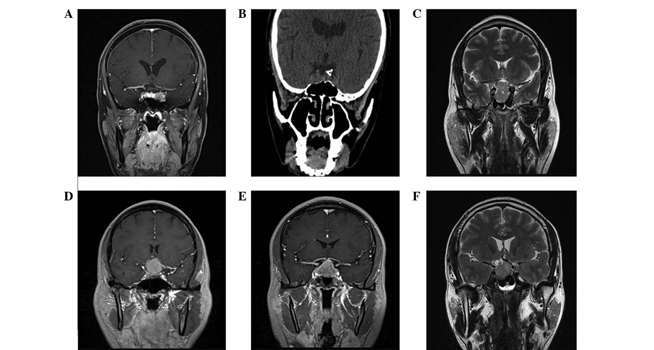
Imaging
All patients were examined by magnetic resonance
imaging (MRI; Achieva 3.0TTX; Philips Healthcare, DA Best, The
Netherlands) of the head and coronal computed tomography (CT)
scanning (SOMATOM Definition AS+ 128; Siemens AG, Munich, Germany).
In addition, 12 patients received a CT angiography (CTA)
examination (tumor diameter, >4 cm surrounding the internal
carotid artery) for intraoperative reference. The tumor diameter
was <1 cm in 56 patients, 1–4 cm in 34 patients and >4 cm in
12 patients. The tumors were invasive in 10 patients, including 9
cases with invasion of the bilateral cavernous sinus, the third
ventricle and the bottom of the frontal lobe; the hypothalamus was
invaded in 2 cases. Coronal CT scanning revealed 13 patients with
sellar floor bone destruction. CTA examination revealed bilateral
anterior cerebral artery pressure resulting in the curved forward
displacement of bilateral internal carotid arteries to one side, so
that they were partly surrounded.
Prolactinoma treatment
All surgical procedures were performed by the same
highly qualified surgeon. The prolactin adenoma was resected using
a nasal transsphenoidal approach in 89 patients, and using a
pterional approach in 13 patients. Prior to surgery, 54 patients
had been treated with bromocriptine (2.5–7.5 mg/day) for between 3
weeks and 7 years (mean, 11.8 months). After complete removal of
the tumor, a small artificial dura mater (Johnson & Johnson,
New Brunswick, NJ, USA) coated with biological glue (Shunkang
Medical Adhesive Co., Ltd., Beijing, China) was placed in the sella
floor to prevent cerebrospinal fluid leakage.
Clinical criteria
Postoperative follow-ups were conducted to analyze
the effectiveness of the surgery in the treatment of the
prolactinomas during the 6 months after surgery. Results for the
patients who were treated with bromocriptine were compared with the
results obtained without bromocriptine treatment with regard to the
efficiency of the surgery, which was classified as cure, remission,
progressive or invalid. The criteria for cure were: Prolactin
levels returned to normal, menstruation patterns were normal,
lactation ceased in females, and the serum testosterone levels
(normal range, 1.75–7.81 ng/ml) and sperm count (normal,
>20×107/ml) were normal in male patients. Remission
criteria were: Prolactinomas returned to normal, menstruation did
not recover in females, and the serum testosterone level and sperm
count were lower than normal in males. Progression was defined as a
prolactinoma reduction of >80%, and invalid was defined as a
prolactinoma reduction of ≤80%.
Statistical analysis
The data were analyzed using SPSS version 17.0
statistical software (SPSS, Inc. Chicago, IL, USA). Data are
represented as the number of patients. The results were analyzed
using the χ2 test. P<0.05 was considered to indicate
a statistically significant difference.
Results
Intraoperative conditions
Total resection surgery was achieved through the
nose and sphenoid sinus in 64 patients, and 25 patients underwent
subtotal resection or substantial removal of the tumor, leaving a
small area of residual tumor or tumor capsule. Craniotomy was
conducted in 13 patients, which were cases where the tumors were
growing invasively with suprasellar extension and it was observed
during surgery that the internal carotid artery was surrounded by
optic chiasm that had unclear boundaries with brain tissue. MRI
results of the head and coronal CT scans are presented in Fig. 1. The surgical observations of
patients with and without bromocriptine treatment are presented in
Table I. Significant differences
(P<0.05) in the tumor texture and the frequency of hard and
tough capsules and pituitary stalk adhesions were detected between
the two groups.
 | Table I.Intraoperative observations. |
Table I.
Intraoperative observations.
|
|
| Texture |
|
|---|
|
|
|
|
|
|---|
| Group | Patients | Soft | Hard | Hard and tough
capsule | Pituitary stalk
adhesions | Rich blood
supply | Tumor apoplexy | Cavernous sinus
bleeding | Cerebrospinal fluid
leakage |
|---|
| Bromocriptine | 54 | 19 | 35 | 41 | 29 | 20 | 13 | 11 | 9 |
| Without
bromocriptine | 48 | 31 | 17 | 22 | 15 | 14 | 8 | 6 | 5 |
| χ2 |
| 8.788 | 9.744 | 5.223 | 0.708 | 0.853 | 1.133 | 0.838 |
| P-value |
| 0.003 | 0.002 | 0.022 | 0.400 | 0.356 | 0.287 | 0.360 |
Postoperative complications
Following the surgery, 69 patients developed
transient diabetes insipidus, and 7 patients developed permanent
diabetes insipidus and required long-term (>6 month) oral 1.25
mg desmopressin acetate tablets (Pfizer, Inc., Zurich, Switzerland)
2–3 times/day to control the quantity of urine produced. A total of
64 patients developed hyponatremia, and 7 patients had varying
degrees of postoperative cerebrospinal fluid rhinorrhea, the
majority of whom were treated within-10 days following conservative
treatment with oral bromocriptine. For 2 of the patients with
cerebrospinal fluid rhinorrhea, the condition was treated by repair
surgery. There were 3 patients who remained unconscious following
the craniotomy and were transferred to the intensive care unit for
treatment. Intracranial infections with gram-negative bacilli
developed in 3 patients and were treated using antibiotics. There
were no mortalities. The major complications observed in the
patients with and without bromocriptine treatment are presented in
Table II.
| Table II.Postoperative complications. |
Table II.
Postoperative complications.
| Group | Patients | Diabetes
insipidus | Electrolyte
imbalance | Hypopituitarism |
|---|
| Bromocriptine | 54 | 45 | 41 | 9 |
| Without
bromocriptine | 48 | 31 | 29 | 6 |
| χ2 |
| 4.704 | 2.839 | 0.352 |
| P-value |
| 0.030 | 0.092 | 0.553 |
Follow-up results
Patients were followed up for between 1 and 9 months
using MRI examination of the head and measurement of serum
prolactin levels. Seven patients were lost to follow-up, and thus
were unable to be assessed. Recurrence of the tumor was observed in
6 patients (5.9%), all of which were a result of residual tissue
from the tumor remaining following the surgery. A total of 14
patients were treated by γ-knife following the surgery, and in 6
patients the residual tumor disappeared completely after 6 months,
as identified by CT. Menstrual disorders and amenorrhea improved in
38 patients, vision disorders improved in 29 patients and loss of
libido improved in 7 patients. The postoperative level of serum
prolactin returned to normal (female, <33 ng/ml, male, <17
ng/ml) in 68 patients; the serum prolactin concentration declined
by >80% in 18 patients, and by ≤80% in 9 patients. In 19
patients, the levels of prolactin in the blood were >60 ng/ml at
1, 3 and 6 months postsurgery, and these patients were treated with
bromocriptine (2.5–5.0 mg/day) for adjuvant therapy. In patients
who developed hypopituitarism, thyroxine and prednisone replacement
therapy was prescribed. The clinical efficacy of bromocriptine in
the treatment of prolactin adenomas is presented in Table III. The cure and remission rate in
patients treated with bromocriptine was 62.7% (32/51); whereas the
cure and remission rate was 81.8% (36/44) in patients without
bromocriptine. A significant difference was detected between the
two groups (χ2=4.224; P=0.04).
| Table III.Comparison of total cure and
remission cases between the two groups. |
Table III.
Comparison of total cure and
remission cases between the two groups.
| Group | Patients | Cure | Remission | Progression | Invalid | Cure and remission,
n (%) |
|---|
| Bromocriptine | 51 | 15 | 17 | 14 | 5 | 32 (62.7) |
| Without
bromocriptine | 44 | 23 | 13 | 4 | 4 | 36 (81.8) |
| χ2 |
| 5.144 | 0.157 |
|
| 4.224 |
| P-value |
| 0.023 | 0.692 |
|
| 0.040 |
Discussion
Transsphenoidal surgery through the nose and
sphenoid sinus is the most common surgical approach for the
treatment of pituitary adenomas (21). It is able to entirely remove the
tumors, significantly reduce the damage to brain tissue, nerves and
blood vessels in during surgery, and results in fewer complications
and low mortality, as compared with the traditional transcranial
approach (22,23). However, a transcranial surgical
approach is required for large pituitary tumors with a hard
texture, rich blood supply, dumbbell-shaped tumor growth and
expansion outside the sella turcica (24). Postoperative complications include
diabetes insipidus, electrolyte imbalance, hypopituitarism and
rhinorrhea of the cerebrospinal fluid (25).
The results in the present study demonstrated that
there was a significant difference between patients with and
without bromocriptine treatment with regards to tumor texture, the
hardness and toughness of adenoma capsules, and pituitary stalk
adhesions (P<0.05). These differences between the groups were
attributable to the preoperative bromocriptine treatment, which
increased the risk of damaging the normal pituitary, pituitary
stalk and sellar diaphragm during the surgery. Bromocriptine,
therefore, increases the difficulty of surgery and promotes
postoperative complications.
Diabetes insipidus is the most common complication
following pituitary adenoma surgery, and is caused by damage to the
pituitary stalk, posterior pituitary, hypothalamus or the blood
supply, which results in an insufficient secretion of antidiuretic
hormones (26–28). The results from the present study
demonstrated that postoperative complications in the bromocriptine
treatment group were more prevalent than those in the group who
were not treated with bromocriptine; however, the only
post-operative complication that was statistically significant
different in prevalence between the two groups was diabetes
insipidus.
Cerebrospinal fluid rhinorrhea results from direct
intraoperative injury to the sellar diaphragm; this can occur when
tumors are large and are excised quickly, causing a rapid reduction
of the sellar diaphragm, or when tumors have eroded the sellar
diaphragm (29–31). It has also been reported that giant
pituitary adenoma, repeat surgery, uneven tumor surface and tough
texture may increase the risk of postoperative cerebrospinal fluid
rhinorrhea (32,33). The results of the present study
demonstrated that 9 patients who were preoperatively prescribed
bromocriptine, and 5 patients who were not prescribed
bromocriptine, experienced intraoperative cerebrospinal fluid
leakage. During the surgery, artificial dura and brain-ear glue
were used to repair the sellar floor, and 7 patients experienced
varying degrees of cerebrospinal fluid rhinorrhea. By conservative
treatment, the majority of patients were cured within 3 to 10 days,
and 2 patients were treated by repair surgery using cerebrospinal
fluid rhinorrhea endoscopy.
In cases where the size of the prolactinoma is not
reduced to its normal size following surgery, the presence of
residual tumors should be considered. Residual tumors can be too
small to be identified using MRI, or they may become confounded
within scars of the sella turcica or the surrounding pituitary
tissue. Therefore, if the prolactinoma does not reduce in size, it
is recommended that patients be closely followed up by observation
of the levels of endocrine indicators and imaging studies (21). If the results from radiological
examinations present a clear residual tumor, surgical exploration
may be considered again with drug or radiation therapy. The purpose
of radiation therapy is to inhibit tumor cell growth and reduce the
secretion of hormones from the tumors (34,35).
Radiation therapy is primarily used as an adjuvant therapy for
patients whose hormone levels have not returned to normal levels,
or who have residual tumors detected following surgical
treatment.
In the present study, removal of prolactinomas was
performed by an experienced neurosurgeon through the nose and
sphenoid sinus; the long-term remission rates for such surgery are
typically 70–80% (36). For
prolactinomas that are confined within the sella turcica, the
long-term remission rates can reach 80–90% (37,38).
Follow-up data from these two clinical trials revealed that there
was a significant difference between patients with and without
bromocriptine treatment with regard to the efficacy following
surgical treatment of prolactinomas. These findings were consistent
with those reported by Landolt et al (39).
In the present study, treatment with bromocriptine
caused difficulties in the removal of the tumors during surgery, as
bromocriptine caused adhesion of the pituitary stalk, pituitary
tissue, or tumor in the rear of pituitary stalk. Thus, the
efficiency of surgery was reduced by bromocriptine; however, the
long-term effects of bromocriptine on prolactin adenomas are
currently unknown.
In conclusion, the oral administration of
bromocriptine is important for the treatment of prolactinoma
tumors. However, large doses or chronic use of bromocriptine can
increase surgical difficulties and result in postoperative
complications due to the hardening of tumor tissues, hardening and
toughening of capsules and aggravated adhesions.
References
|
1
|
Hirohata T, Ishii Y and Matsuno A:
Treatment of pituitary carcinomas and atypical pituitary adenomas:
A review. Neurol Med Chir (Tokyo). 54:966–973. 2014. View Article : Google Scholar : PubMed/NCBI
|
|
2
|
Ciric I, Rosenblatt S, Kerr W Jr, Lamarca
F, Pierce D and Baumgartner C: Perspective in pituitary adenomas:
An end of the century review of tumorigenesis, diagnosis and
treatment. Clin Neurosurg. 47:99–111. 2000.PubMed/NCBI
|
|
3
|
Ozgen T, Oruckaptan HH, Ozcan OE and
Acikgoz B: Prolactin secreting pituitary adenomas: Analysis of 429
surgically treated patients, effect of adjuvant treatment
modalities and review of the literature. Acta Neurochir (Wien).
141:1287–1294. 1999. View Article : Google Scholar : PubMed/NCBI
|
|
4
|
Landolt AM, Minder H, Osterwalder V and
Landolt TA: Bromocriptine reduces the size of cells in
prolactin-secreting pituitary adenomas. Experientia. 39:625–626.
1983. View Article : Google Scholar : PubMed/NCBI
|
|
5
|
Laws ER Jr, Thorner MO and Vance ML:
Bromocriptine therapy for prolactin-secreting pituitary adenomas.
Neurosurg Focus. 1:e4discussion 1p following e6. 1996. View Article : Google Scholar : PubMed/NCBI
|
|
6
|
Bergström M, Muhr C, Lundberg PO,
Bergström K, Gee AD, Fasth KJ and Långström B: Rapid decrease in
amino acid metabolism in prolactin-secreting pituitary adenomas
after bromocriptine treatment: A PET study. J Comput Assist Tomogr.
11:815–819. 1987. View Article : Google Scholar : PubMed/NCBI
|
|
7
|
Kim JO, Ma R, Akagami R, McKenzie M,
Johnson M, Gete E and Nichol A: Long-term outcomes of fractionated
stereotactic radiation therapy for pituitary adenomas at the BC
Cancer Agency. Int J Radiat Oncol Biol Phys. 87:528–533. 2013.
View Article : Google Scholar : PubMed/NCBI
|
|
8
|
Elson A, Bovi J, Kaur K, Maas D, Sinson G
and Schultz C: Effect of treatment modality on the
hypothalamic-pituitary function of patients treated with radiation
therapy for pituitary adenomas: Hypothalamic dose and endocrine
outcomes. Front Oncol. 4:732014. View Article : Google Scholar : PubMed/NCBI
|
|
9
|
Casanueva FF, Molitch ME, Schlechte JA,
Abs R, Bonert V, Bronstein MD, Brue T, Cappabianca P, Colao A,
Fahlbusch R, et al: Guidelines of the Pituitary Society for the
diagnosis and management of prolactinomas. Clin Endocrinol (Oxf).
65:265–273. 2006. View Article : Google Scholar : PubMed/NCBI
|
|
10
|
Sahoo JP, Kamalanathan S, Parida PK and
Pillai V: A giant prolactinoma with nasopharyngeal extension
presenting with nasal blockage and epistaxis. BMJ case reports.
2015:2015. View Article : Google Scholar
|
|
11
|
Nakajima T, Tamura T, Kuroki M, Tanaka R
and Hayashi H: A case of prolactinoma presenting with CSF
rhinorrhea and CSF otorrhea during bromocriptine therapy. No
Shinkei Geka. 20:1091–1095. 1992.(In Japanese). PubMed/NCBI
|
|
12
|
Little AS: Repair of cerebrospinal fluid
fistula from an invasive skull base prolactinoma using a septal
mucosal vascularized flap: Technical case report. J Neurol Surg A
Cent Eur Neurosurg. 74(Suppl 1): e50–e53. 2013.PubMed/NCBI
|
|
13
|
Sunil B, Reddy A, Bryant N, Young DW and
Ashraf AP: Invasive giant prolactinoma presenting as a nasal polyp.
J Pediatr. 162:435. 2013. View Article : Google Scholar : PubMed/NCBI
|
|
14
|
Webster J: Dopamine agonist therapy in
hyperprolactinemia. J Reprod Med. 44(Suppl 12): S1105–S1110.
1999.
|
|
15
|
Acharya SV, Gopal RA, Menon PS, Bandgar TR
and Shah NS: Giant prolactinoma and effectiveness of medical
management. Endocr Pract. 16:42–46. 2010. View Article : Google Scholar : PubMed/NCBI
|
|
16
|
Schettini G, Lombardi G, Merola B, Miletto
P, Fariello C, Cirillo S, Fusco R and Lancranjan I: Effectiveness
of a single injectable dose of bromocriptine long acting in the
treatment of macroprolactinomas. J Endocrinol Invest. 11:47–51.
1988. View Article : Google Scholar : PubMed/NCBI
|
|
17
|
Mroueh AM and Siler-Khodr TM:
Bromocryptine therapy in cases of amenorrhea-galactorrhea. Am J
Obstet Gynecol. 127:291–298. 1977. View Article : Google Scholar : PubMed/NCBI
|
|
18
|
Tyson D, Reggiardo D, Sklar C and David R:
Prolactin-secreting macroadenomas in adolescents. Response to
bromocriptine therapy. Am J Dis Child. 147:1057–1061. 1993.
View Article : Google Scholar : PubMed/NCBI
|
|
19
|
Palmeri CM, Petiti JP, del Sosa LV,
Gutiérrez S, De Paul AL, Mukdsi JH and Torres AI: Bromocriptine
induces parapoptosis as the main type of cell death responsible for
experimental pituitary tumor shrinkage. Toxicol Appl Pharmacol.
240:55–65. 2009. View Article : Google Scholar : PubMed/NCBI
|
|
20
|
Liuzzi A, Chiodini PG, Dallabonzana D,
Oppizzi G and Verde GG: Medical treatment of pituitary adenomas:
Effects on tumor growth. J Endocrinol Invest. 8:273–281. 1985.
View Article : Google Scholar : PubMed/NCBI
|
|
21
|
Sharma M, Ambekar S, Sonig A and Nanda A:
Factors predicting the development of new onset post-operative
Hydrocephalus following trans-sphenoidal surgery for pituitary
adenoma. Clin Neurol Neurosurg. 115:1951–1954. 2013. View Article : Google Scholar : PubMed/NCBI
|
|
22
|
Frank G, Pasquini E and Mazzatenta D:
Extended transsphenoidal approach. J Neurosurg. 95:917–918.
2001.PubMed/NCBI
|
|
23
|
Cappabianca P, Cavallo LM, Esposito F, De
Divitiis O, Messina A and De Divitiis E: Extended endoscopic
endonasal approach to the midline skull base: The evolving role of
transsphenoidal surgery. Adv Tech Stand Neurosurg. 33:151–199.
2008. View Article : Google Scholar : PubMed/NCBI
|
|
24
|
Grabenbauer GG, Fietkau R, Buchfelder M,
Meyer M, Baumann J, Hensen J, Rummelt V, Fahlbusch R and Sauer R:
Hormonally inactive hypophyseal adenomas: The results and late
sequelae after surgery and radiotherapy. Strahlenther Onkol.
172:193–197. 1996.(In German). PubMed/NCBI
|
|
25
|
Goyal N, Borkar SA, Agrawal D and
Mahapatra AK: Pituitary adenoma presenting with cerebrospinal fluid
rhinorrhea as the sole symptom. Neurol India. 60:307–308. 2012.
View Article : Google Scholar : PubMed/NCBI
|
|
26
|
Perrin G, Stevenaert A and Jouanneau E:
Technical aspects and surgical strategy for removal of corticotroph
pituitary adenoma. Neurochirurgie. 48:186–214. 2002.(In French).
PubMed/NCBI
|
|
27
|
Gondim JA, Almeida JP, de Albuquerque LA,
Gomes E, Schops M and Mota JI: Endoscopic endonasal transsphenoidal
surgery in elderly patients with pituitary adenomas. J Neurosurg.
123:31–38. 2015. View Article : Google Scholar : PubMed/NCBI
|
|
28
|
Berkmann S, Schlaffer S, Nimsky C,
Fahlbusch R and Buchfelder M: Intraoperative high-field MRI for
transsphenoidal reoperations of nonfunctioning pituitary adenoma. J
Neurosurg. 121:1166–1175. 2014. View Article : Google Scholar : PubMed/NCBI
|
|
29
|
Mehta GU, Bakhtian KD and Oldfield EH:
Effect of primary empty sella syndrome on pituitary surgery for
Cushing's disease. J Neurosurg. 121:518–526. 2014. View Article : Google Scholar : PubMed/NCBI
|
|
30
|
Berker M, Hazer DB, Yucel T, Gurlek A,
Cila A, Aldur M and Onerci M: Complications of endoscopic surgery
of the pituitary adenomas: Analysis of 570 patients and review of
the literature. Pituitary. 15:288–300. 2012. View Article : Google Scholar : PubMed/NCBI
|
|
31
|
Cho JM, Ahn JY, Chang JH and Kim SH:
Prevention of cerebrospinal fluid rhinorrhea after transsphenoidal
surgery by collagen fleece coated with fibrin sealant without
autologous tissue graft or postoperative lumbar drainage.
Neurosurgery. 68:130–136. 2011.PubMed/NCBI
|
|
32
|
Lehman NL, Horoupian DS and Harsh GR IV:
Synchronous subarachnoid drop metastases from a pituitary adenoma
with multiple recurrences. Case report. J Neurosurg. 98:1120–1123.
2003. View Article : Google Scholar : PubMed/NCBI
|
|
33
|
Kim EH, Ku CR, Lee EJ and Kim SH:
Extracapsular en bloc resection in pituitary adenoma surgery.
Pituitary. 18:397–404. 2015. View Article : Google Scholar : PubMed/NCBI
|
|
34
|
Tanaka S, Link MJ, Brown PD, Stafford SL,
Young WF Jr and Pollock BE: Gamma knife radiosurgery for patients
with prolactin-secreting pituitary adenomas. World Neurosurg.
74:147–152. 2010. View Article : Google Scholar : PubMed/NCBI
|
|
35
|
Wollesen F and Bendsen BB: Effect rates of
different modalities for treatment of prolactin adenomas. Am J Med.
78:114–122. 1985. View Article : Google Scholar : PubMed/NCBI
|
|
36
|
Dai WD, Liu WH and Zhang DL: One
hemodialysis patient with headache, blurred vision and hypotension
induced by pituitary prolactinoma. Chin Med J (Engl).
125:2787–2789. 2012.PubMed/NCBI
|
|
37
|
Domingue ME, Devuyst F, Alexopoulou O,
Corvilain B and Maiter D: Outcome of prolactinoma after pregnancy
and lactation: A study on 73 patients. Clin Endocrinol (Oxf).
80:642–648. 2014. View Article : Google Scholar : PubMed/NCBI
|
|
38
|
Qu X, Wang M, Wang G, Han T, Mou C, Han L,
Jiang M, Qu Y, Zhang M, Pang Q and Xu G: Surgical outcomes and
prognostic factors of transsphenoidal surgery for prolactinoma in
men: A single-center experience with 87 consecutive cases. Eur J
Endocrinol. 164:499–504. 2011. View Article : Google Scholar : PubMed/NCBI
|
|
39
|
Landolt AM, Keller PJ, Froesch ER and
Mueller J: Bromocriptine: Does it jeopardise the result of later
surgery for prolactinomas? Lancet. 2:657–658. 1982. View Article : Google Scholar : PubMed/NCBI
|