Introduction
In the last 20 years, the development of bone tissue
engineering in three major components, namely osteogenic cells,
scaffolds and stimulating factors, has made it a promising
alternative for autologous bone grafting for bone repair and
regeneration (1). Among the
osteogenic cell types, the bone marrow stromal cells (BMSCs) are
considered to be among the most promising cell sources for bone
tissue engineering purposes (2).
However, to direct BMSCs toward osteogenic lineage, essential
regulatory signals, primarily including biochemical and/or
biophysical stimuli, should be applied (3).
To the best of our knowledge, dexamethasone (DEX), a
synthetic glucocorticoid, is the most widely used reagent to
enhance the osteogenic differentiation of BMSCs (4). However, its potential side-effects,
such as inhibition of cell proliferation and induction of
osteoblast apoptosis, impede its further application for engineered
bone constructs (5). Since the early
1990s, increasing numbers of investigators have realized the
effects of mechanical stimulation on the behavior and function of
BMSCs (6–8), and researchers have proposed the
importance of mechanical forces in inducing BMSC differentiation
and full maturation (9). Recently,
it was reported that dynamic compressive loading was as effective
as DEX at inducing matrix production during osteogenic
differentiation of hBMSCs (5).
Furthermore, fluid shear stress was more effective than DEX in the
early osteogenic differentiation of BMSCs (10). In addition to DEX, bone morphogenetic
proteins (BMPs) are also well-known as potent osteogenic
stimulating factors that are able to promote the osteogenic
differentiation of BMSCs and reduce their apoptosis (11). However, to date there have been no
reports focusing on the influence of mechanical forces compared
with that of BMPs on the proliferation and differentiation of
BMSCs. BMP-2 and BMP-7 have been approved for clinical applications
for bone repair (12). However,
compared with BMP-2, BMP-7 is associated with a milder inflammatory
response, particularly when used in high-dose treatments (13), and is capable of attenuating fibrosis
(14), a common complication after
orthopedic surgery. Thus, BMP-7 was employed in the present
investigation.
In addition to various stimuli, a scaffold providing
a three dimensional environment for cell growth and development is
also crucial for bone tissue engineering (15,16).
Previously, the present authors reported that a
1,4-butanediisocyanate-based polyurethane (PU) scaffold could offer
an ideal environment for the attachment and proliferation of human
BMSCs (hBMSCs) in dynamic culture (17). This type of PU scaffold has been
applied in patients for meniscus replacement for several years
(18). Furthermore, a recent study
also revealed promising outcomes of osteochondral repair using this
scaffold (19). There may likewise
be a clinical application potential for constructing
tissue-engineered bone grafts using a patients' own BMSCs for bone
defect repair (20).
In the present study, a PU scaffold consisting of a
hard segment of 1,4-butane diisocyanate and butanediol, and a soft
segment of poly(ε-caprolactone) was employed. Furthermore, a
perfusion bioreactor system was used for optimizing the environment
for rapid differentiation of hBMSCs. The objective of this study
was to identify the effects of perfusion, cyclic compression, DEX
and BMP-7 on the proliferation and differentiation of hBMSCs on the
PU scaffolds in a dynamic culture system and to determine the most
potent factor for enhancing the proliferation and osteogenic
differentiation of hBMSCs.
Materials and methods
Harvest and cultivation of hBMSCs
All procedures were approved by the Institutional
Ethical Committee of Hannover Medical School (Hannover, Germany).
After written informed consent was obtained, bone marrow aspirates
were collected from seven healthy human donors (four males and
three females; mean age, 29±3.5 years) who underwent exposure of
their iliac crests during routine orthopedic procedures. Isolation
and cultivation of hBMSCs was performed as per our previously
described protocol (21). Briefly,
the cells were purified by density gradient centrifugation at 1,200
× g for 20 min at 4°C (Heraeus Labofuge 400R; Thermo Fisher
Scientific, Inc., Waltham, MA, USA). Cells were then cultured in
Dulbecco's modified Eagle's medium (DMEM)/Ham's F12 medium
containing L-glutamine (Biochrom GmbH, Berlin, Germany)
supplemented with 10% fetal calf serum (Gibco; Thermo Fisher
Scientific, Inc.), 5 µg/ml ascorbic acid (Sigma-Aldrich, St. Louis,
MO, USA), 3 ng/ml fibroblast growth factor-2 (FGF-2; PeproTech,
Inc., Offenbach, Germany), 100 U/ml penicillin/streptomycin (Gibco)
and 0.5 µg/ml amphotericin B (Biochrom GmbH) at 37°C and 5%
CO2 in humidified atmosphere. After reaching confluence,
cells were lysed with 0.05% trypsin (Gibco) and combined, and then
the cell pool was subcultured. The hBMSCs from the third passage
were used for the experiments.
PU scaffold preparation, cell seeding
and culture
Biodegradable PU-based scaffolds
(Actifit®; Orteq Ltd., Groningen, The Netherlands) were
fabricated according to the procedure described by van Tienen et
al (22). Cylindrical scaffolds
(diameter, 20 mm; height, 5 mm) with pore sizes ranging between 110
and 455 µm (mean, 301 µm) and porosity of ~80% were used (Fig. 1A and B). Prior to cell seeding, the
scaffolds were immersed in culture medium without FGF-2 for 15 min
with gentle shaking. After removing excess medium from the
scaffolds, 106 hBMSCs from the third passage were
resuspended in 400 µl culture medium without FGF-2 and seeded on
each hydrated scaffold. After incubation at 37°C for 24 h, the
hBMSC-seeded PU scaffolds were cultured under six different
conditions, including: Mechanical stimulation I group, 10% cyclic
compression at 0.5 Hz (perfusion rate, 1 ml/min); mechanical
stimulation II group, 10% cyclic compression at 5 Hz (perfusion
rate, 1 ml/min;); perfusion group, 10 ml/min continuous perfusion;
DEX group, 100 nM DEX (Merck KGaA, Darmstadt, Germany; perfusion
rate, 1 ml/min); BMP-7 group, 100 ng/ml BMP-7 (Stryker Biotech,
Hopkinton, MA, USA; perfusion rate, 1 ml/min); and the control
group, 1 ml/min continuous perfusion in a custom-made perfusion
bioreactor (23) maintained at 37°C
in 5% CO2 in an incubator. The volume of the culture
medium without FGF-2 in each bioreactor was 150 ml, and half of the
medium volume was changed every three days. On days 7 and 14, the
scaffolds were harvested, and each scaffold was split into six
equal parts for the following six analyses.
MTS assay for cell proliferation
On days 7 and 14, the cell-loaded scaffolds were cut
into 1-mm3 pieces for the cell proliferation assay.
Then, 2 ml MTS solution (1:20; Promega Corporation, Madison, WI,
USA) was added to the small pieces. After 2 h of incubation at
37°C, the reaction was stopped using 10% sodium dodecyl sulfate
(Sigma-Aldrich). The solution was removed, and the absorbance was
read at 490 nm using an absorbance reader (BioTek Instruments,
Inc., Winooski, VT, USA). The cell number was determined using the
calibration data (six points, n=3, R2=0.99) obtained
using cells from the same culture (23).
mRNA expression analysis
On days 7 and 14, total cellular RNA was extracted
from the cell-scaffold constructs using the NucleoSpin®
RNA II kit (Macherey-Nagel GmbH & Co. KG, Düren, Germany).
Genomic DNA was removed using DNase supplied with the kit. Total
mRNA was reverse transcribed into cDNA using the High-Capacity cDNA
Reverse Transcriptase Kit (Applied Biosystems; Thermo Fisher
Scientific, Inc., Waltham, MA, USA). Quantitative real-time
polymerase chain reaction (qPCR) was performed to measure
transcript levels of three osteogenic marker genes, namely Runx2
(Hs00231692_m1), COL1A1 (Hs00164004_m1) and osteocalcin
(Hs01587814_g1) and GAPDH (Hs02758991_g1),using a StepOnePlus qPCR
system (4376600; Thermo Fisher Scientific, Inc.). Commercially
available TaqMan primer/probes (4331182) and TaqMan Expression
Master mix (4369016; Applied Biosystems). The reaction mixture
contained 2 µl (100 ng) cDNA, 1 µl primer, 10 µl Master mix and 7
µl nuclease-free water. qPCR cycling conditions were as follows:
50°C for 2 min, 95°C for 10 min, 40 cycles of 95°C for 15 sec and
60°C for 1 min. Negative (nuclease-free water) and reverse
transcriptase controls were used. Three technical replicates were
performed, and experiments were repeated independently three times.
The Cq values of targeted genes were normalized against that of the
housekeeping gene GAPDH (Hs02758991_g1). Data were expressed
relative to control using the 2−ΔΔCq formula (24).
ELISA analysis of osteocalcin
On days 7 and 14, hBMSCs in the scaffolds were lysed
in 1 ml RIPA buffer (Thermo Fisher Scientific, Inc.) overnight at
4°C. After centrifugation at 12,000 × g for 10 min, the supernatant
was gathered and the osteocalcin levels were determined using an
N-MID® Osteocalcin ELISA kit (Immunodiagnostic Systems
Holdings PLC, The Boldons, UK) according to the manufacturer's
instructions. The amount of total protein was quantified using
Coomassie Plus Assay Reagent (Thermo Fisher Scientific, Inc.). The
osteocalcin content was normalized against that of total
protein.
Histological analysis
After collection from the bioreactor on days 7 and
14, the scaffolds were fixed in 4% formaldehyde (Otto Fischar GmbH
& Co. KG, Saarbrücken, Germany) and then embedded in the OCT™
compound (Sakura Finetek Europe B.V., Alphen, The Netherlands).
Next, 30-µm thick sections were processed at −20°C with a freezing
microtome (Microm International Gmbh, Walldorf, Germany) for
histological analysis. Nuclear fast red (Sigma-Aldrich) and
Alizarin Red S (Merck KGaA) staining was performed to observe the
distribution of hBMSCs and calcium deposition in the scaffolds,
respectively. Quantitative analyses of the mineralization from the
Alizarin Red S staining images were performed using Image-Pro Plus
software, version 6.0 (Media Cybernetics, Inc., Rockville, MD, USA)
(25).
Scanning electron microscopy
(SEM)
On days 7 and 14, the scaffolds were collected and
fixed with 2.5% glutaraldehyde (Agar Scientific, Ltd., Stansted,
UK) in 0.1 M cacodylate buffer (pH 7.4; Merck KGaA) for 24 h. After
rinsing in buffer, the scaffolds were dehydrated with graded
ethanol changes (25, 50, 75, 90 and 100%). Then, the specimens were
dried using the liquid carbon dioxide to replace the ethanol within
the scaffolds for 10 cycles (5 min per cycle), coated with Au
(Quorom Technologies, Ltd., Laughton, UK) and observed with SEM. In
addition, at 24 h after seeding, the cell-seeded scaffolds were
observed using the same procedure.
Biomechanical evaluation
Disks of 1-mm thickness and 6-mm diameter were cut
from the central region of the hBMSC-seeded scaffolds from days 7
and 14 and mounted in a cylindrical confining chamber (Zwick/Roell
1445; Zwick GmbH & Co. KG, Ulm, Germany) (17). Each sample was compressed by a
plunger above the chamber using 10 subsequent displacements with 5%
strain, up to a 50% total strain. Based on the force recorded after
each displacement, a stress-strain curve was generated. The
equilibrium modulus of each sample was determined by the slope of
the stress-strain curve (26).
Furthermore, the equilibrium modulus of the empty scaffold was
measured using the same procedure.
Statistical analysis
All experiments were performed in triplicate. Data
are expressed as the mean ± standard deviation. Comparisons among
groups were performed using one-way analysis of variance, with the
Student-Newman-Keuls test. SPSS software, version 15.0 (SPSS, Inc.,
Chicago, IL, USA) was used to conduct all statistical tests.
P<0.05 was considered to indicate a statistically significant
difference.
Results
MTS assay for cell proliferation and
nuclear fast red staining
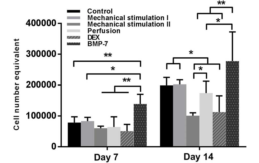
Cell number in all of the groups increased over
time, and no significant difference was observed between controls
and the mechanical stimulation I groups at any time point. On day
7, the BMP-7 group had the highest cell number (P<0.01), and
compared with that of the mechanical stimulation I, the effect of
mechanical stimulation II resulted in lower cell proliferation;
however, there was no significant difference. On day 14, the
mechanical stimulation II and DEX groups had lower cell numbers
(P<0.05), which was in contrast with the control, the mechanical
stimulation I and BMP-7 groups. Furthermore, the maximum cell
number was observed in the BMP-7 group, which was significantly
increased compared with the mechanical stimulation II (P<0.01),
DEX (P<0.01) and perfusion (P<0.05) groups (Fig. 2).
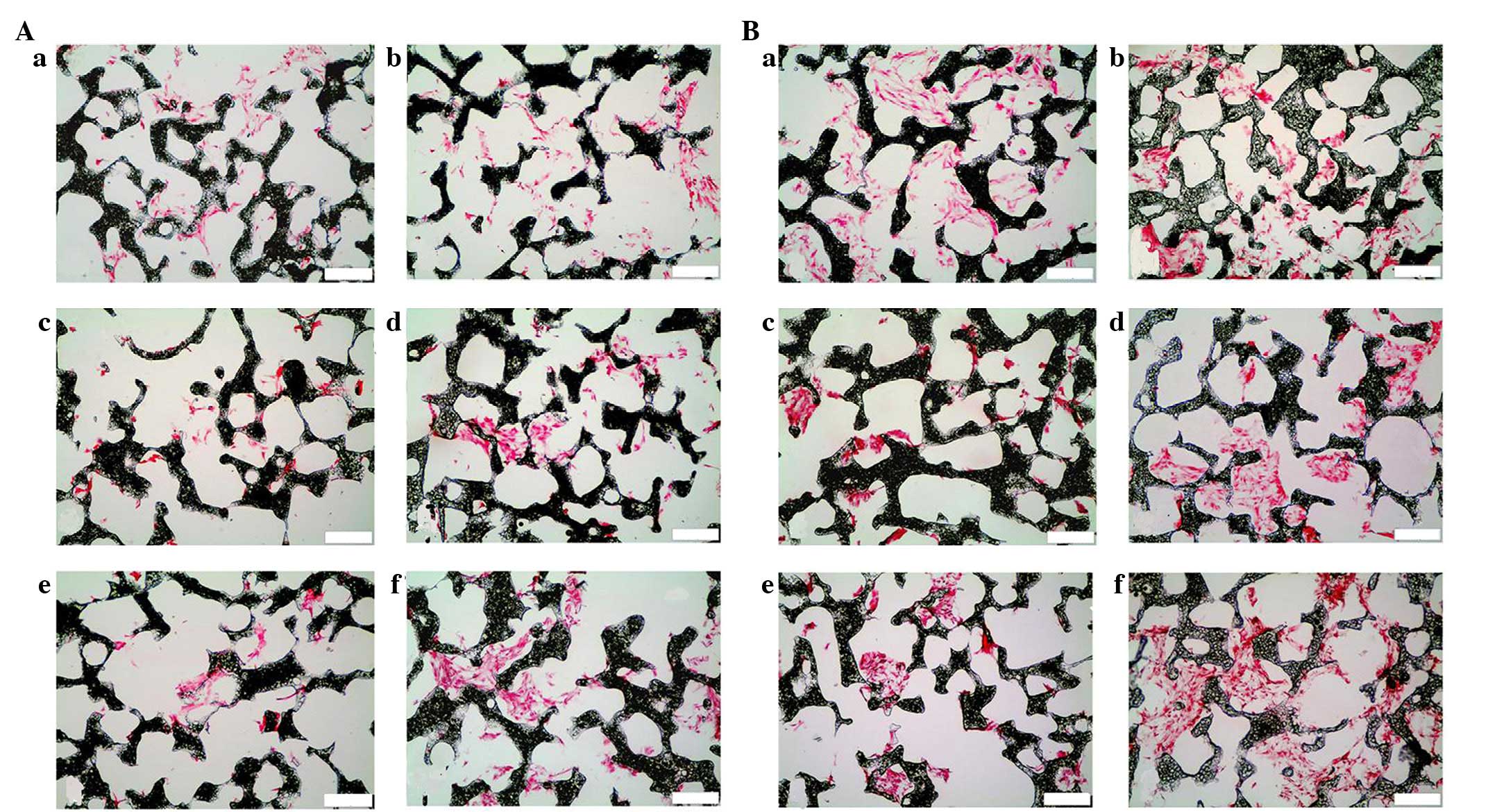
Sections were stained with nuclear fast red to
determine the distribution of the hBMSCs within the scaffolds. The
cell number increased over time in all groups, and the BMP-7 group
always presented the largest number. In contrast, after 14 days,
relatively few cells were observed in the mechanical stimulation II
group. Although the number of BMSCs was low in the scaffolds of the
perfusion group during the first seven days, a high density of
cells was observed in a few parts of the scaffolds after 14 days
(Fig. 3).
SEM analysis
At 24 h after seeding, the attached rounded cells
became polygonal in shape (Fig. 1C).
After seven days, the cells extended pseudopodia and spread
uniformly on the porous walls of the scaffolds in the control,
mechanical stimulation I, perfusion and BMP-7 groups, and there
were noticeably more cells in the BMP-7 groups compared with the
other five groups, which was consistent with the outcome of the MTS
assay. After 14 days, the BMP-7 group continued to exhibit the
highest cell density and quantity of extracellular matrix (ECM),
followed by the mechanical stimulation I group. High cell density
was observed in a few parts of the scaffolds in the perfusion
group. However, the cell density in the mechanical stimulation II
and DEX groups was always lower compared with the other groups
(Fig. 4).
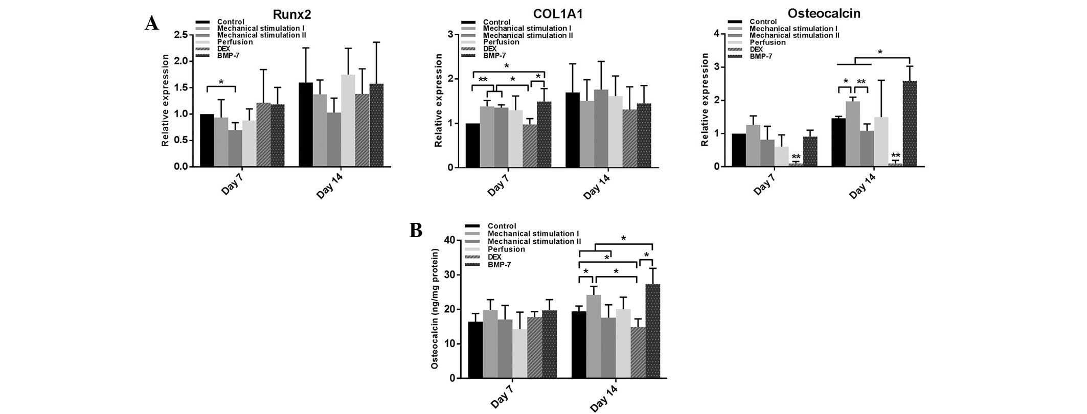
Gene expression analysis
On day 7, compared with the control, the Runx2 mRNA
level in the mechanical stimulation II group was significantly
decreased (P<0.05), and the COL1A1 mRNA levels of the mechanical
stimulation I, mechanical stimulation II and BMP-7 groups were
significantly increased (P<0.05). However, the DEX group
exhibited significantly reduced osteocalcin mRNA levels (P<0.01)
at this time point. On day 14, the maximum and minimum osteocalcin
mRNA levels were observed in the BMP-7 (P<0.05) and DEX groups
(P<0.01), respectively. There was an upregulation of osteocalcin
expression in the mechanical stimulation I group (P<0.05), which
also exhibited markedly increased osteocalcin expression compared
with the mechanical stimulation II group (P<0.01). In addition,
the BMP-7 group had a higher osteocalcin mRNA levels compared with
the mechanical stimulation I group (P<0.05). No significant
difference was detected in the mRNA levels of Runx2 and COL1A1
among the six groups at this time point; however, there was an
overall upward trend during the 14 days of dynamic culture
(Fig. 5A).
Osteocalcin evaluation
During the first seven days, there was no
significant difference among the six groups in osteocalcin levels.
On day 14, the osteocalcin levels in the mechanical stimulation I
and the BMP-7 groups were significantly higher than in the control
(P<0.05). However, although the average level of osteocalcin in
the BMP-7 group was higher compared with the mechanical stimulation
I group, no significant difference was observed. By contrast,
stimulation with DEX resulted in decreased osteocalcin levels
(P<0.05) (Fig. 5B).
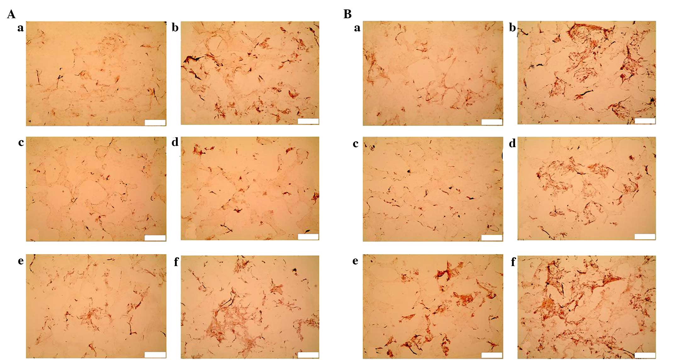
Alizarin Red S staining
Since day 7, calcium deposition was observed in the
mechanical stimulation I and BMP-7 groups. After 14 days, the BMP-7
group displayed the most marked staining, followed by the
mechanical stimulation I and DEX groups. Less intense staining was
exhibited in the perfusion group. No apparent calcium deposition
was detected in the mechanical stimulation II group at either time
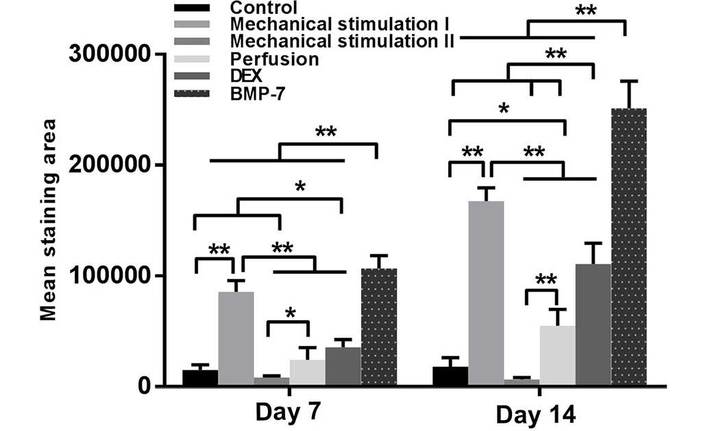
point (Fig. 6). According to the
quantitative analyses, the mechanical stimulation I and BMP-7
groups had significantly higher calcium deposition compared with
the other groups (P<0.01) at each time point. After 14 days,
compared with control, there was significantly increased calcium
deposition in the DEX (P<0.01) and perfusion (P<0.05) groups
(Fig. 7).
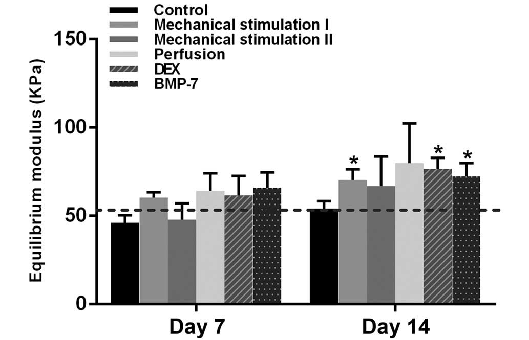
Equilibrium modulus
On day 7, no significant difference in the
equilibrium modulus was identified between the empty scaffold and
each of the cell-seeded scaffolds. After 14 days, a significantly
higher equilibrium modulus was observed in the mechanical
stimulation I, DEX and BMP-7 groups (P<0.05), compared with that
of empty scaffolds (Fig. 8).
Discussion
Osteoprogenitor cells at the fracture sites
experience local physical stimuli, such as fluid shear stress or
cyclic axial compression (27). It
has been reported that mechanical loading plays an important role
in inducing osteoprogenitor cells in the bone marrow stroma to
differentiate towards osteoblasts at the cortical bone surface
in vivo (28). Based on these
results, various studies have been performed to identify the
optimum mechanical stimuli to enhance the osteogenic
differentiation of MSCs, to replace pharmaceutical agents with
potential side effects (5,21,29). As
mentioned above, appropriate mechanical forces may be as effective
or more so than DEX, a main component in the osteogenic medium
(5). In addition to DEX, certain
protein-based growth factors, such as BMPs, are well-known as
potent osteoinductive growth factors. However, their intrinsic
drawbacks, such as short half-life, neoplastic risk, osteoclastic
activation, immunosuppressive properties and high cost (30), necessitate the comparison of the
osteoinductive abilities between mechanical loading and BMPs.
It has been demonstrated that different strain rates
and frequencies may lead to different results (31). Sittichokechaiwut et al
(5) determined the effectiveness of
5% compressive strain at 1 Hz on the enhancement of hBMSC
osteodifferentiation. Michalopoulos et al (32) reported that hBMSCs differentiate to
an osteogenic lineage under 10% cyclic compressive strain and to an
osteochondrogenic lineage under 15% cyclic compressive strain at 1
Hz. The present group previously demonstrated that 10% cyclic
compression at 0.5 Hz was effective for the osteodifferentiation of
hBMSCs (23). To date, little is
known about the influence of strain frequencies on the behavior of
cells. Additionally, fluid flow has been considered to induce or
enhance osteogenesis in MSCs (33).
Therefore, the common mechanical (10% cyclic compression of low and
high frequencies; 10 ml/min continuous perfusion) and biochemical
stimulation (DEX; BMP-7) were included in the present study and
compared their influence on the proliferation and differentiation
of hBMSCs in a perfusion bioreactor.
To date, scaffolds with pore sizes ranging between
20 and 1,500 µm have been utilized in bone tissue engineering;
numerous investigators have suggested that the mean pore size
should be >300 µm for optimal bone regeneration and
vascularization within the constructs (34–37). The
mean pore size of the PU scaffolds in the present investigation
were consistent with the mainstream perspective. In a previous
study by the present authors, 1,4-butane diisocyanate-based
polyurethane scaffolds were found to be non-cytotoxic and could
provide an ideal environment for hBMSC adhesion and proliferation
(17). This result was reconfirmed
in the present study, as the MTS assay demonstrated a continuing
increase in the cell number of all groups without showing any
obvious adverse effects on cell proliferation. Furthermore, as an
elastomeric cancellous bone graft substitute, PU scaffolds are
superior to scaffolds made of rigid polymers, can have intimate
contact with the recipients' bone ends, obviating shear forces at
the bone-implant interface and possess excellent resistance to
plastic deformation and elastic recovery under load (38). Therefore, the PU scaffolds are
speculated to be a promising bone substitute for autologous bone
grafts, particularly in large-scale bone defect reconstruction.
To the best of our knowledge, perfusion is an
effective factor for promoting hBMSC proliferation (23,39).
However, from the MTS assay, no significant difference was observed
between the control and perfusion groups. Unexpectedly, according
to the results of the nuclear fast red staining and the SEM assay,
high cell density was observed in certain areas of the scaffolds in
the perfusion group. It seems that high perfusion rate may detach
the cells from the scaffolds at the beginning (17). Afterwards, the proliferation of the
remaining cells was enhanced under continuous perfusion, which may
improve the transportation of nutrients, such as fetal calf serum
(9). Consistent with the outcomes of
the study by Shea et al (40), BMP-7 promoted cell proliferation
according to the present results. By contrast, DEX markedly
inhibited hBMSC proliferation, a side effect also reported by other
groups (41). Pelaez et al
(42) demonstrated that cyclic
compression maintained the viability of hBMSCs. In the present
study, cyclic compression of low frequency did not affect the
proliferation of hBMSCs, whereas high frequency cyclic compression
had a negative effect, which indicates that even short period high
frequency mechanical stimulation is able to mitigate cell
proliferation. According to the SEM images, the cells spread on the
porous walls and proliferated well, indicating that favorable
circumstance for cell attachment and proliferation can be supplied
by this type of PU scaffold. Consistent with the outcome of the MTS
assay, the BMP-7 group always exhibited the highest cell density in
the scaffolds. Furthermore, substantial quantities of ECM around
the cells was observed in the BMP-7 and mechanical stimulation I
groups. Clustering of cells and synthesis of ECM are considered as
indicators of osteogenic differentiation (43).
It is well known that the expression of genes Runx2,
COL1A1 and osteocalcin are crucially involved in the osteogenic
commitment of MSCs (44). Following
the downregulation of Runx2, high frequency cyclic compression
appeared to inhibit osteogenic differentiation of hBMSCs. There was
no significant difference in Runx2 observed among other groups;
however, there was an overall upward trend. Similar results were
observed in the study of Frank et al (45), which showed that Runx2 mRNA levels in
hBMSCs were slightly elevated during osteogenic differentiation. It
has been proposed that the formation of type I collagen is a
crucial part of the osteogenic differentiation process, as it can
interact with integrins, major cell receptors for collagen, which
is required for the induction of bone-related gene expression
(46). The results of the present
study suggest that the expression of COL1A1 mRNA is sensitive to
mechanical stimulation irrespective of the frequencies of the
stimuli, and mechanical stimulation is as effective as BMP-7 in the
enhancement of COL1A1 expression. As a late-stage marker of
osteoblast maturation, low frequency cyclic compression and BMP-7
resulted in the upregulation of osteocalcin expression at the mRNA
and protein levels, indicating that the hBMSCs differentiated into
preosteoblastic phenotype during the 14 days of dynamic culture,
and that mechanical stimulation is able to promote the osteogenic
differentiation of hBMSCS in the absence of biochemical cues.
However, the addition of DEX resulted in reduced expression of
osteocalcin. Similarly, Fiorentini et al (47) demonstrated that DEX enhanced the ALP
expression and mineralization of hBMSCs, but caused substantially
declined osteocalcin mRNA levels, which reconfirmed the views of
Ito et al (48) that
glucocorticoids failed to induce terminal osteoblast
differentiation.
In the present study, the highest calcium deposition
was observed in the BMP-7-stimulated hBMSCs, and no mineralization
occurred in the high frequency mechanical stimulation group.
Although the stimulation of DEX and ascorbic acid resulted in the
downregulation of osteocalcin, a certain degree of calcium
deposition was observed, which was significantly lower compared
with the low frequency mechanically stimulated hBMSCs. This implies
that the mineralization of hBMSCs can be enhanced without the
presence of biochemical cues. Accumulation of ECM, particularly
calcium, led to the increased equilibrium modulus of the
cell-seeded scaffolds, the importance of which has been well
illustrated (49). Previous studies
reported the positive impact of continuous perfusion on cell
mineralization (50,51). However, in the present study, a small
degree of calcium deposition was observed in the perfusion group,
less than that in the mechanical stimulation I group. According to
our previous results (23),
continuous perfusion is the primary stimulus for hBMSC
proliferation, whereas mechanical stimulation promotes the
osteogenic differentiation of hBMSCs. Duty et al (52) demonstrated that in vivo cyclic
compression loading enhanced the mineralization in the MSC-seeded
constructs, which suggests that the design of engineered constructs
for bone repair should premeditate the interplays with the local
mechanical circumstance.
In conclusion, BMP-7 and perfusion are able to
enhance cell proliferation, and high frequency compression resulted
in decreased proliferation and inhibited osteogenic
differentiation. Low frequency cyclic compression is more effective
than DEX, but less effective than BMP-7 on the osteogenic
differentiation of hBMSCs seeded on a polyurethane scaffold. In the
future, further in vivo studies are required to validate the
safety and functionality of bone substitutes engineered from
different cell lines, following biomechanical or biochemical
stimulation.
Acknowledgements
This study was supported by the German Research
Foundation (Deutsche Forschungsgemeinschaft; DFG Grant JA
1086/3-1). The authors thank Roland Meister for technical
assistance.
References
|
1
|
Murphy CM, O'Brien FJ, Little DG and
Schindeler A: Cell-scaffold interactions in the bone tissue
engineering triad. Eur Cell Mater. 26:120–132. 2013.PubMed/NCBI
|
|
2
|
Colnot C: Cell sources for bone tissue
engineering: Insights from basic science. Tissue Eng Part B Rev.
17:449–457. 2011. View Article : Google Scholar : PubMed/NCBI
|
|
3
|
Wang YK and Chen CS: Cell adhesion and
mechanical stimulation in the regulation of mesenchymal stem cell
differentiation. J Cell Mol Med. 17:823–832. 2013. View Article : Google Scholar : PubMed/NCBI
|
|
4
|
Langenbach F and Handschel J: Effects of
dexamethasone, ascorbic acid and beta-glycerophosphate on the
osteogenic differentiation of stem cells in vitro. Stem Cell Res
Ther. 4:1172013. View
Article : Google Scholar : PubMed/NCBI
|
|
5
|
Sittichokechaiwut A, Edwards JH, Scutt AM
and Reilly GC: Short bouts of mechanical loading are as effective
as dexamethasone at inducing matrix production by human bone marrow
mesenchymal stem cell. Eur Cell Mater. 20:45–57. 2010.PubMed/NCBI
|
|
6
|
Li R, Liang L, Dou Y, Huang Z, Mo H, Wang
Y and Yu B: Mechanical strain regulates osteogenic and adipogenic
differentiation of bone marrow mesenchymal stem cells. Biomed Res
Int. 2015:8732512015.PubMed/NCBI
|
|
7
|
Cardwell RD, Kluge JA, Thayer PS, Guelcher
SA, Dahlgren LA, Kaplan DL and Goldstein AS: Static and cyclic
mechanical loading of mesenchymal stem cells on elastomeric,
electrospun polyurethane meshes. J Biomech Eng. 137:2015.
View Article : Google Scholar : PubMed/NCBI
|
|
8
|
Kim TJ, Joo C, Seong J, Vafabakhsh R,
Botvinick EL, Berns MW, Palmer AE, Wang N, Ha T, Jakobsson E, et
al: Distinct mechanisms regulating mechanical force-induced
Ca2+ signals at the plasma membrane and the ER in human
MSCs. eLife. 4:e048762015. View Article : Google Scholar : PubMed/NCBI
|
|
9
|
Delaine-Smith RM, MacNeil S and Reilly GC:
Matrix production and collagen structure are enhanced in two types
of osteogenic progenitor cells by a simple fluid shear stress
stimulus. Eur Cell Mater. 24:162–174. 2012.PubMed/NCBI
|
|
10
|
Yourek G, McCormick SM, Mao JJ and Reilly
GC: Shear stress induces osteogenic differentiation of human
mesenchymal stem cells. Regen Med. 5:713–724. 2010. View Article : Google Scholar : PubMed/NCBI
|
|
11
|
Liu Y, Wu J, Zhu Y and Han J: Therapeutic
application of mesenchymal stem cells in bone and joint diseases.
Clin Exp Med. 14:13–24. 2014. View Article : Google Scholar : PubMed/NCBI
|
|
12
|
Gautschi OP, Frey SP and Zellweger R: Bone
morphogenetic proteins in clinical applications. ANZ J Surg.
77:626–631. 2007. View Article : Google Scholar : PubMed/NCBI
|
|
13
|
Lee KB, Taghavi CE, Murray SS, Song KJ,
Keorochana G and Wang JC: BMP induced inflammation: A comparison of
rhBMP-7 and rhBMP-2. J Orthop Res. 30:1985–1994. 2012. View Article : Google Scholar : PubMed/NCBI
|
|
14
|
Weiskirchen R and Meurer SK: BMP-7
counteracting TGF-beta1 activities in organ fibrosis. Front Biosci
(Landmark Ed). 18:1407–1434. 2013. View
Article : Google Scholar : PubMed/NCBI
|
|
15
|
Black CR, Goriainov V, Gibbs D, Kanczler
J, Tare RS and Oreffo RO: Bone Tissue Engineering. Curr Mol Biol
Rep. 1:132–140. 2015. View Article : Google Scholar : PubMed/NCBI
|
|
16
|
Gong T, Xie J, Liao J, Zhang T, Lin S and
Lin Y: Nanomaterials and bone regeneration. Bone Res. 3:150292015.
View Article : Google Scholar : PubMed/NCBI
|
|
17
|
Liu C, Abedian R, Meister R, Haasper C,
Hurschler C, Krettek C, von Lewinski G and Jagodzinski M: Influence
of perfusion and compression on the proliferation and
differentiation of bone mesenchymal stromal cells seeded on
polyurethane scaffolds. Biomaterials. 33:1052–1064. 2012.
View Article : Google Scholar : PubMed/NCBI
|
|
18
|
Verdonk P, Beaufils P, Bellemans J, Djian
P, Heinrichs EL, Huysse W, Laprell H, Siebold R and Verdonk R:
Actifit Study Group: Successful treatment of painful irreparable
partial meniscal defects with a polyurethane scaffold: Two-year
safety and clinical outcomes. Am J Sports Med. 40:844–853. 2012.
View Article : Google Scholar : PubMed/NCBI
|
|
19
|
Hannink G, de Mulder EL, van Tienen TG and
Buma P: Effect of load on the repair of osteochondral defects using
a porous polymer scaffold. J Biomed Mater Res B Appl Biomater.
100:2082–2089. 2012. View Article : Google Scholar : PubMed/NCBI
|
|
20
|
Steinert AF, Rackwitz L, Gilbert F, Noth U
and Tuan RS: Concise review: the clinical application of
mesenchymal stem cells for musculoskeletal regeneration: current
status and perspectives. Stem Cells Transl Med. 1:237–247. 2012.
View Article : Google Scholar : PubMed/NCBI
|
|
21
|
Jagodzinski M, Drescher M, Zeichen J,
Hankemeier S, Krettek C, Bosch U and van Griensven M: Effects of
cyclic longitudinal mechanical strain and dexamethasone on
osteogenic differentiation of human bone marrow stromal cells. Eur
Cell Mater. 7:35–41, Discussion 41. 2004.PubMed/NCBI
|
|
22
|
van Tienen TG, Heijkants RG, Buma P, de
Groot JH, Pennings AJ and Veth RP: Tissue ingrowth and degradation
of two biodegradable porous polymers with different porosities and
pore sizes. Biomaterials. 23:1731–1738. 2002. View Article : Google Scholar : PubMed/NCBI
|
|
23
|
Jagodzinski M, Breitbart A, Wehmeier M,
Hesse E, Haasper C, Krettek C, Zeichen J and Hankemeier S:
Influence of perfusion and cyclic compression on proliferation and
differentiation of bone marrow stromal cells in 3-dimensional
culture. J Biomech. 41:1885–1891. 2008. View Article : Google Scholar : PubMed/NCBI
|
|
24
|
Livak KJ and Schmittgen TD: Analysis of
relative gene expression data using real-time quantitative PCR and
the 2(−Delta Delta C(T)) Method. Methods. 25:402–408. 2001.
View Article : Google Scholar : PubMed/NCBI
|
|
25
|
Liu J, Zhang B, Song S, Ma M, Si S, Wang
Y, Xu B, Feng K, Wu J and Guo Y: Bovine collagen peptides compounds
promote the proliferation and differentiation of MC3T3-E1
pre-osteoblasts. PloS One. 9:e999202014. View Article : Google Scholar : PubMed/NCBI
|
|
26
|
Kelly DJ, Crawford A, Dickinson SC, Sims
TJ, Mundy J, Hollander AP, Prendergast PJ and Hatton PV:
Biochemical markers of the mechanical quality of engineered hyaline
cartilage. J Mater Sci Mater Med. 18:273–281. 2007. View Article : Google Scholar : PubMed/NCBI
|
|
27
|
Skerry TM: The response of bone to
mechanical loading and disuse: fundamental principles and
influences on osteoblast/osteocyte homeostasis. Arch Biochem
Biophys. 473:117–123. 2008. View Article : Google Scholar : PubMed/NCBI
|
|
28
|
Turner CH, Owan I, Alvey T, Hulman J and
Hock JM: Recruitment and proliferative responses of osteoblasts
after mechanical loading in vivo determined using sustained-release
bromodeoxyuridine. Bone. 22:463–469. 1998. View Article : Google Scholar : PubMed/NCBI
|
|
29
|
Jones LC, Yeoumans B, Hungerford DS and
Frondoza CG: The response of osteoblast-like cells to dexamethasone
and cyclic loading. Biomed Sci Instrum. 42:273–277. 2006.PubMed/NCBI
|
|
30
|
Makhdom AM and Hamdy RC: The role of
growth factors on acceleration of bone regeneration during
distraction osteogenesis. Tissue Eng Part B Rev. 19:442–453. 2013.
View Article : Google Scholar : PubMed/NCBI
|
|
31
|
Huang C, Dai J and Zhang XA: Environmental
physical cues determine the lineage specification of mesenchymal
stem cells. Biochim Biophys Acta. 1850:1261–1266. 2015. View Article : Google Scholar : PubMed/NCBI
|
|
32
|
Michalopoulos E, Knight RL, Korossis S,
Kearney JN, Fisher J and Ingham E: Development of methods for
studying the differentiation of human mesenchymal stem cells under
cyclic compressive strain. Tissue Eng Part C Methods. 18:252–262.
2012. View Article : Google Scholar : PubMed/NCBI
|
|
33
|
Delaine-Smith RM and Reilly GC:
Mesenchymal stem cell responses to mechanical stimuli. Muscles
Ligaments Tendons J. 2:169–180. 2012.PubMed/NCBI
|
|
34
|
Murphy CM, Haugh MG and O'Brien FJ: The
effect of mean pore size on cell attachment, proliferation and
migration in collagen-glycosaminoglycan scaffolds for bone tissue
engineering. Biomaterials. 31:461–466. 2010. View Article : Google Scholar : PubMed/NCBI
|
|
35
|
Karageorgiou V and Kaplan D: Porosity of
3D biomaterial scaffolds and osteogenesis. Biomaterials.
26:5474–5491. 2005. View Article : Google Scholar : PubMed/NCBI
|
|
36
|
Roosa SM, Kemppainen JM, Moffitt EN,
Krebsbach PH and Hollister SJ: The pore size of polycaprolactone
scaffolds has limited influence on bone regeneration in an in vivo
model. J Biomed Mater Res A. 92:359–368. 2010. View Article : Google Scholar : PubMed/NCBI
|
|
37
|
Kuboki Y, Jin Q and Takita H: Geometry of
carriers controlling phenotypic expression in BMP-induced
osteogenesis and chondrogenesis. J Bone Joint Surg Am. 83-A(Suppl
1): S105–S115. 2001.PubMed/NCBI
|
|
38
|
Gorna K and Gogolewski S: Biodegradable
porous polyurethane scaffolds for tissue repair and regeneration. J
Biomed Mater Res A. 79:128–138. 2006. View Article : Google Scholar : PubMed/NCBI
|
|
39
|
Gaspar DA, Gomide V and Monteiro FJ: The
role of perfusion bioreactors in bone tissue engineering.
Biomatter. 2:167–175. 2012. View Article : Google Scholar : PubMed/NCBI
|
|
40
|
Shea CM, Edgar CM, Einhorn TA and
Gerstenfeld LC: BMP treatment of C3H10T1/2 mesenchymal stem cells
induces both chondrogenesis and osteogenesis. J Cell Biochem.
90:1112–1127. 2003. View Article : Google Scholar : PubMed/NCBI
|
|
41
|
Cheng SL, Yang JW, Rifas L, Zhang SF and
Avioli LV: Differentiation of human bone marrow osteogenic stromal
cells in vitro: Induction of the osteoblast phenotype by
dexamethasone. Endocrinology. 134:277–286. 1994. View Article : Google Scholar : PubMed/NCBI
|
|
42
|
Pelaez D, Huang CY and Cheung HS: Cyclic
compression maintains viability and induces chondrogenesis of human
mesenchymal stem cells in fibrin gel scaffolds. Stem Cells Dev.
18:93–102. 2009. View Article : Google Scholar : PubMed/NCBI
|
|
43
|
Mathews S, Bhonde R, Gupta PK and Totey S:
Novel biomimetic tripolymer scaffolds consisting of chitosan,
collagen type 1 and hyaluronic acid for bone marrow-derived human
mesenchymal stem cells-based bone tissue engineering. J Biomed
Mater Res B Appl Biomater. 102:1825–1834. 2014. View Article : Google Scholar : PubMed/NCBI
|
|
44
|
Kim K, Dean D, Mikos AG and Fisher JP:
Effect of initial cell seeding density on early osteogenic signal
expression of rat bone marrow stromal cells cultured on
cross-linked poly(propylene fumarate) disks. Biomacromolecules.
10:1810–1817. 2009. View Article : Google Scholar : PubMed/NCBI
|
|
45
|
Frank O, Heim M, Jakob M, Barbero A,
Schäfer D, Bendik I, Dick W, Heberer M and Martin I: Real-time
quantitative RT-PCR analysis of human bone marrow stromal cells
during osteogenic differentiation in vitro. J Cell Biochem.
85:737–746. 2002. View Article : Google Scholar : PubMed/NCBI
|
|
46
|
Xiao G, Wang D, Benson MD, Karsenty G and
Franceschi RT: Role of the alpha2-integrin in osteoblast-specific
gene expression and activation of the Osf2 transcription factor. J
Biol Chem. 273:32988–32994. 1998. View Article : Google Scholar : PubMed/NCBI
|
|
47
|
Fiorentini E, Granchi D, Leonardi E,
Baldini N and Ciapetti G: Effects of osteogenic differentiation
inducers on in vitro expanded adult mesenchymal stromal cells. Int
J Artif Organs. 34:998–1011. 2011. View Article : Google Scholar : PubMed/NCBI
|
|
48
|
Ito S, Suzuki N, Kato S, Takahashi T and
Takagi M: Glucocorticoids induce the differentiation of a
mesenchymal progenitor cell line, ROB-C26 into adipocytes and
osteoblasts, but fail to induce terminal osteoblast
differentiation. Bone. 40:84–92. 2007. View Article : Google Scholar : PubMed/NCBI
|
|
49
|
Kim HJ, Kim UJ, Vunjak-Novakovic G, Min BH
and Kaplan DL: Influence of macroporous protein scaffolds on bone
tissue engineering from bone marrow stem cells. Biomaterials.
26:4442–4452. 2005. View Article : Google Scholar : PubMed/NCBI
|
|
50
|
Dahlin RL, Gershovich JG, Kasper FK and
Mikos AG: Flow perfusion co-culture of human mesenchymal stem cells
and endothelial cells on biodegradable polymer scaffolds. Ann
Biomed Eng. 42:1381–1390. 2014. View Article : Google Scholar : PubMed/NCBI
|
|
51
|
Kim J and Ma T: Bioreactor strategy in
bone tissue engineering: Pre-culture and osteogenic differentiation
under two flow configurations. Tissue Eng Part A. 18:2354–2364.
2012. View Article : Google Scholar : PubMed/NCBI
|
|
52
|
Duty AO, Oest ME and Guldberg RE: Cyclic
mechanical compression increases mineralization of cell-seeded
polymer scaffolds in vivo. J Biomech Eng. 129:531–539. 2007.
View Article : Google Scholar : PubMed/NCBI
|