Introduction
Renal cell carcinoma (RCC) is a particularly lethal
genitourinary neoplasm, with an incidence of 5–10 per 100,000 and
accounting for 2–3% of all malignancies (1). Clear-cell renal cell carcinoma (ccRCC)
is the most common histological subtype of human RCC (2), with which ~40,000 patients are
diagnosed annually in the US (1) and
650,000 patients are diagnosed annually in China (3). In ~30% of patients who present with
localized RCC at the time of diagnosis, distant metastases will
develop within 3 years (4). RCC is
resistant to chemotherapy and radiotherapy (5). The 5-year survival rate amongst
patients with early stage RCC is ~55%; however, once RCC develops,
the prognosis of advanced RCC is extremely poor and the 5-year
survival rate for advanced RCC is 10% (6). Although numerous genetic and epigenetic
alterations have been shown to be correlated with RCC (7), the mechanism of carcinogenesis and
progression is poorly understood. Therefore, a reliable biomarker
is required to predict early metastasis and to serve as a novel
therapeutic target for RCC.
Kinesin family member 18A (KIF18A) has important
roles in various cellular processes, including motility, cell
division, microtubule dynamics and organelle transportation
(8–11). During mitosis, KIF18A is concentrated
at the plus ends of microtubules and facilitates microtubule
depolymerization (12). KIF18A
attenuates chromosome oscillation by depolymerizing microtubules
during cellular division, and therefore enhances chromosome
congregation (10). In addition,
loss of KIF18A activity influences chromosome segregation and leads
to chromosome instability (13). It
has been reported that KIF18A suppresses the movement of
kinetochores to regulate mitotic chromosome alignment in the
pre-anaphase state of the cell cycle (14). Furthermore, it has been confirmed
that KIF18A plays critical roles in the modulation of motility and
mitosis, suggesting that KIF18A acts as a critical regulator in
cell transformation and carcinogenesis (15). Notably, several studies have
indicated that KIF18A is involved in breast carcinogenesis and
colorectal cancer progression (16,17).
Moreover, KIF18A has also been identified as a potential biomarker
of lung cancer and cholangiocarcinoma using proteomic analysis
(18,19).
The expression and function of KIF18A in RCC is
currently unclear. To the best of our knowledge, there have been no
previous reports regarding the role of KIF18A in carcinogenesis,
progression and prognostication in patients with RCC. In the
current study, the expression of KIF18A and the effect of KIF18A on
proliferation in human RCC were determined. KIF18A expression
levels were compared in clinical specimens of RCC and normal kidney
tissues, and the effects of transfection with KIF18A cDNA or small
interfering RNA (siRNA) on the proliferation of ccRCC cells were
determined in vitro and in vivo. The potential of
KIF18A as an independent prognostic marker for patients with RCC,
and as a therapeutic target for the treatment of RCC were also
evaluated.
Materials and methods
Patients and specimens
This study included 273 consecutive patients who
underwent radical or partial nephrectomies in the Department of
Urology at The Second Affiliated Hospital of Xi'an Jiaotong
University (Xi'an, China) between May 2005 and May 2012. The
histological cell type of all resections was determined by two
experienced pathologists and all specimens were confirmed to be
ccRCC. The baseline clinical and pathological data and follow-up
outcomes were recorded. TNM stage and Fuhrman grading were
identified according to the TNM system of the 2010 American Joint
Committee on Cancer (AJCC) (20) and
the 2004 World Health Organization (WHO) criteria (21), respectively. RCC tissues and
corresponding normal healthy kidney tissues were collected
immediately after surgical resection, and all tissue specimens were
formalin-fixed and paraffin-embedded; tissues were also maintained
in liquid nitrogen until protein or RNA extraction. The study was
approved by the Ethics Committee of the Second Affiliated Hospital
of Xi'an Jiaotong University.
Cell culture
Four ccRCC cell lines (ACHN, A498, Caki-1 and
Caki-2) were used in this study, all of which were purchased from
the American Type Culture Collection (ATCC; Rockville, MD, USA) and
were cultured in complete medium (RPMI-1640) supplemented with 10%
fetal bovine serum (Gibco; Thermo Fisher Scientific, Inc., Waltham,
MA, USA), streptomycin (100 mg/ml), penicillin (100 U/ml), 25 mM
4-(2-hydroxyethyl)-1-piperazineethanesulphonic acid (HEPES) and 2
mM glutamine. All RCC cell lines were maintained as monolayers in a
10-cm plastic dish and cultured in an incubator with a humidified
atmosphere containing 5% CO2 at 37°C.
Animal xenograft experiment
BALB/c nude mice (n=30; male; 4 weeks old; weight,
40–50 g) were obtained from the Experimental Animal Center of Xi'an
Jiaotong University, and housed in a specific pathogen-free
environment with temperature maintained from 26–28°C and a humidity
of 30–40% with a 12-h dark:light cycle and access to food and
water, supplied by the Experimental Animal Center of Xi'an Jiaotong
University. The mice were randomly divided into ACHN, A498, Caki-1
and Caki-2 groups, each of which was further divided into two
groups with 15 mice in each group: Control and KIF18A vector
groups. The control group received untreated RCC cells, and the
KIF18A group received RCC cells transfected with KIF18A cDNA, as
described later. A total of 3×10⁸ RCC cells were
injected into the back region of each mouse and the mice were fed
continuously for 5 weeks. The volume of each xenograft was recorded
once a week. At the end of the 5-week period, the mice were
sacrificed under deep anesthesia, and the final volume of each
xenograft was measured. Xenograft volume was calculated using the
following formula: v = ab2π/6, where a represents the
longest diameter and b represents the diameter perpendicular to the
longest diameter of the tumor.
Proliferation assay
The proliferative ability of the RCC cells was
evaluated in vitro using the WST-1 assay. Untransfected RCC
cell lines and RCC cells transfected with KIF18A cDNA or siRNA, as
described later, were used. Exponentially growing cells [(2,000 RCC
cells with 100 µl complete medium(Sigma-Aldrich, St. Louis, MO,
USA)] were seeded into 96-well microtiter plates. Following
continuous incubation for 24, 48 and 72 h, 10 µl WST-1 (Roche
Diagnostics, GmbH, Penzberg, Germany) was added to each well, and
the RCC cells were cultured for an additional 2 h. The absorbance,
which represents the RCC cell count in each well, was analyzed with
a microculture plate reader (Immunoreader NJ-2000, Nihon Intermed
Co., Ltd., Tokyo, Japan) at 450 nm.
Reverse transcription-polymerase chain
reaction (RT-PCR) and quantitative PCR (qPCR)
The levels of KIF18A expression in RCC and
corresponding non-cancerous tissues were detected using RT-PCR and
RT-qPCR assays. Total RNA was extracted with TRizol reagent
(Invitrogen; Thermo Fisher Scientific Thermo Fisher Scientific,
Inc.) and reverse transcription was performed using a first-strand
cDNA synthesis kit (Amersham; GE Healthcare, Chalfont, UK)
according to the manufacturer's protocol. The primer sequences of
KIF18A were determined using Primer Premier 5.0 software (Premier
Biosoft, Palo Alto, CA, USA) and glyceraldehyde-3-phosphate
hydrogenase (GAPDH) was used as a control. The primer sequences for
KIF18A were as follows: 5′-AAAAAGTGGTAGTTTGGGCTGA-3′ (sense); and
5′-CTTTCAAGGGAGATGGCATTAG-3′ (antisense). The primer sequences for
GAPDH were as follows: 5′-ATCAAGAAGGTGGTGAAGCAG-3′ (sense); and
5′-TGGAGGAGTGGGTGTCGC-3′ (antisense). Products were amplified by
PCR using a TaqMan Universal PCR Master Mix kit (Applied
Biosystems; Thermo Fisher Scientific, Inc.) and data was obtained
and analyzed with a LightCycler 480 (Roche Diagnostics,
Indianapolis, IN, USA). All RT reactions were performed in
triplicate, and experimental procedures of qPCR were based on MIQE
guidelines (22). The relative
expression levels determined by the 2−ΔΔcq method
(23).
Immunohistochemistry
All sections were dewaxed with xylene and rehydrated
in graded alcohols. Antigen retrieval was conducted in citrate
buffer and the sections were then washed in phosphate-buffered
saline (PBS). Endogenous peroxidase activity was blocked with 3%
hydrogen peroxide for 20 min. Then, sections were incubated with
10% goat serum (Thermo Fisher Scientific, Inc.) at room temperature
for 30 min to block non-specific binding. Sections were incubated
with rabbit polyclonal anti-KIF18A antibody (cat no. 19245;
1:1,1000; Proteintech™ Group, Inc., Chicago, IL, USA) for 16 h at
4°C, washed with PBS, incubated with biotinylated goat anti-rabbit
antibody (cat no. E0432; 1:2,000; Dako, Glostrup, Denmark) for 2 h
at 37°C, and stained with 3,3′-diaminobenzidine
tetrahydrochloride.
Western blot analysis
Total protein was isolated from RCC tissues, normal
kidney tissues or RCC cells using lysis buffer (cat no. ab152163,
Abcam, Cambridge, UK). The protein concentration was measured using
a bicinchoninic acid (BCA) protein assay kit (Pierce Biotechnology,
Inc., Rockford, IL, USA). A 100-µm quantity of total protein was
separated by 10% sodium dodecyl sulfate-polyacrylamide gel
electrophoresis and transferred to a polyvinylidene difluoride
membrane (Amersham; GE Healthcare). The membrane was then probed
for 2 h at 37°C with rabbit polyclonal anti-KIF18A antibody (cat
no. ab72416; 1:1,000; Abcam) with anti-β-actin monoclonal antibody
(cat no. ab8226; 1:5,000; Abcam) as a loading control. Next,
membranes were incubated with goat anti-rabbit IgG H&L (HRP)
(cat no. ab6721; 1:5,000; Abcam) at 37°C for 2 h). Finally, the
immune reaction complexes were visualized using an ECL Plus Western
Blotting Detection Reagents (Amersham; GE Healthcare).
Cell transfection
siRNA oligonucleotide sequences targeting KIF18A
were designed using siDirect software (sidirect2.rnai.jp). The four
ccRCC cell lines were transiently transfected with the
oligonucleotides using Lipofectamine 2000 (Invitrogen; Thermo
Fisher Scientific Inc.) according to the manufacturer's protocol.
Moreover, an expression vector (pcDEF3; Sigma-Aldrich) containing
full-length cDNA for KIF18A was also used to stably transfect the 4
RCC cell lines using Lipofectamine 2000. Monoclonal RCC cells were
collected with G418 (Sigma-Aldrich) and transfection efficiency was
evaluated by western blot analysis.
Statistical analysis
Statistical calculations were carried out using SPSS
software (version 19.0; IBM SPSS, Armonk, NY, USA). Differences
between numerical variables were calculated using the Student's
t-test and the results are presented as the mean ± standard
deviation (SD), while categorical variables were analyzed using the
χ2 test. Survival curves were established using the
Kaplan-Meier method. All experiments were performed in triplicate.
All P-values were two-sided and statistical significance was set at
P<0.05.
Results
Patient clinical characteristics
Data were collected from 273 patients with RCC (149
males and 124 females) and 182 corresponding normal renal tissues.
The ages of the patients ranged from 41 to 87 years (median, 65
years), and the tumor diameter ranged from 0.7 to 23.8 cm (median,
4.2 cm). Among the 273 RCC patients, 147 had stage I, 47 had stage
II, 44 had stage III and 35 had stage IV disease. RCC was localized
in 242 patients, and 31 patients had distant metastases at the time
of diagnosis. Moreover, 21 patients with RCC had previously been
treated with radical nephrectomies on the contralateral side. All
patient clinical characteristics are shown in Table I.
 | Table I.Characteristics of RCC patients and
KIF18A protein and mRNA expression detected by immunohistochemistry
and reverse transcription-quantitative polymerase chain reaction,
respectively. |
Table I.
Characteristics of RCC patients and
KIF18A protein and mRNA expression detected by immunohistochemistry
and reverse transcription-quantitative polymerase chain reaction,
respectively.
|
|
| KIF18A protein |
|
|
|
|---|
|
|
|
|
|
|
|
|---|
| Characteristic | n | − | + | ++ | +++ | P-value | KIF18A mRNA | P-value |
|---|
| Renal cell
carcinoma | 273 | 29 | 126 | 72 | 46 |
| 2.30±0.29 |
|
| Non-cancerous renal
tissue | 182 | 139 | 32 | 11 | 0 | <0.05 | 0.62±0.08 | <0.05 |
| Gender |
|
|
|
|
| >0.05 |
| >0.05 |
| Male | 149 | 17 | 69 | 39 | 24 |
| 2.27±0.28 |
|
|
Female | 124 | 12 | 57 | 33 | 22 |
| 2.33±0.29 |
|
| Age |
|
|
|
|
| >0.05 |
| >0.05 |
| <60
years | 152 | 16 | 69 | 42 | 25 |
| 2.30±0.26 |
|
| ≥60
years | 121 | 13 | 57 | 30 | 21 |
| 2.28±0.28 |
|
| Tumor size |
|
|
|
|
| <0.05 |
| <0.05 |
| ≤7
cm | 147 | 23 | 103 | 14 | 7 |
| 1.53±0.21 |
|
| >7
cm | 126 | 6 | 23 | 58 | 39 |
| 3.18±0.35 |
|
| Histological
grade |
|
|
|
|
| <0.05 |
| <0.05 |
| G1 | 145 | 23 | 97 | 21 | 4 |
| 1.22±0.18 |
|
| G2 | 90 | 6 | 29 | 46 | 9 |
| 2.57±0.29 |
|
| G3 | 38 | 0 | 0 | 5 | 33 |
| 5.88±0.68 |
|
| Tumor stage |
|
|
|
|
| <0.05 |
| <0.05 |
| I | 147 | 23 | 103 | 14 | 7 |
| 1.34±0.15 |
|
| II | 47 | 4 | 17 | 21 | 5 |
| 2.30±0.29 |
|
|
III | 44 | 2 | 5 | 16 | 21 |
| 3.64±0.41 |
|
| IV | 35 | 0 | 1 | 21 | 13 |
| 4.59±0.52 |
|
| Metastasis |
|
|
|
|
| <0.05 |
| <0.05 |
|
Absent | 242 | 29 | 125 | 54 | 34 |
| 1.98±0.22 |
|
|
Present | 31 | 0 | 1 | 18 | 12 |
| 4.76±0.53 |
|
KIF18A protein expression in RCC
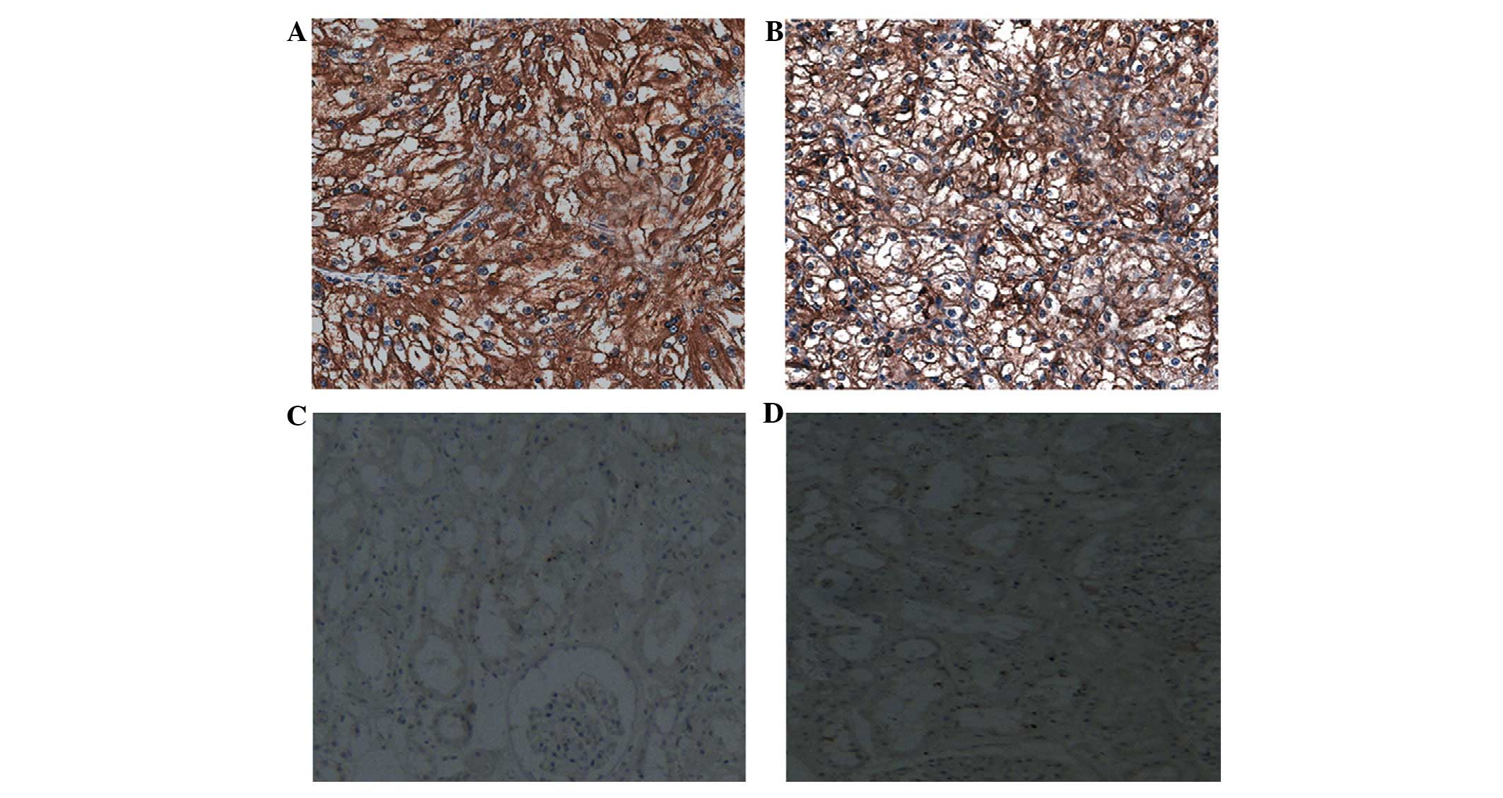
The expression of KIF18A in human RCC and normal
kidney tissues was examined using immunohistochemistry. The
expression of KIF18A protein was observed to be upregulated
significantly in RCC tissues compared with the levels in
corresponding normal kidney tissues (Fig. 1). KIF18A expression was detected in
244/273 RCC tissues (89.4%), but only 43/182 (23.6%) normal kidney
tissues. The association between the expression of KIF18A protein
and various clinicopathologic characteristics was investigated; a
significant association existed between increased KIF18A expression
and high tumor stage, high histological grade, metastasis and large
tumor size (P<0.05). Neither of the other characteristics, age
and gender, showed a significant correlation with the expression of
KIF18A protein (Table I). Together,
these findings indicate that upregulation of KIF18A might be
involved in the carcinogenesis and development of human RCC.
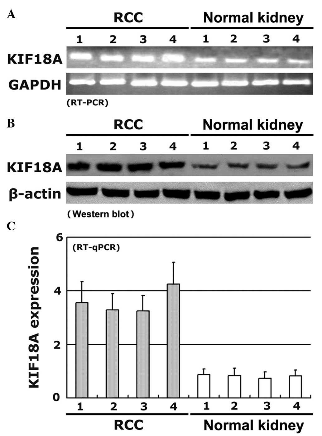
KIF18A expression detected by RT-PCR,
RT-qPCR and western blot analysis
To confirm the expression levels of KIF18A protein
in human RCC as indicated by immunohistochemistry, the expression
of KIF18A in RCC and normal kidney tissues was also evaluated by
RT-PCR, RT-qPCR and western blot analysis. The level of KIF18A
expression was analyzed with reference to an internal control; the
results suggested that KIF18A expression was upregulated
significantly in the RCC tissues compared with the corresponding
normal kidney tissues. Moreover, the expression levels of KIF18A
mRNA were comparable with the levels detected by
immunohistochemistry (Table I).
Representative results for four pairs of RCC and corresponding
normal kidney tissues are shown in Fig.
2.
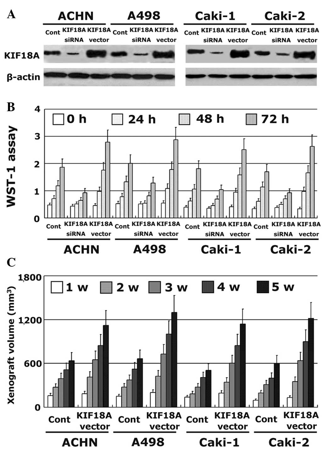
Effect of KIF18A on the proliferation
of RCC cells
KIF18A expression was stably increased by
transfecting a vector containing the full-length cDNA of KIF18A
into ACHN, A498, Caki-1 and Caki-2 cell lines. In addition, KIF18A
expression was also decreased using siRNA technology. The
transfection efficiency was evaluated by western blotting. The
expression of KIF18A protein was significantly increased by the
KIF18A vector insert and decreased by the siRNA (Fig. 3A). The effect of KIF18A on the
proliferative ability of RCC cells in vitro was analyzed by
WST-1 assay. RCC cells with a high level of KIF18A expression had
significantly increased proliferation compared with untreated
control cells. By contrast, RCC cells with a low level of KIF18A
expression had lower proliferative ability (Fig. 3B). These results were also confirmed
in vivo by animal xenograft experiments with BALB/c nude
mice, which showed that KIF18A transfection increased xenograft
volume in the four cell lines (Fig.
3C).
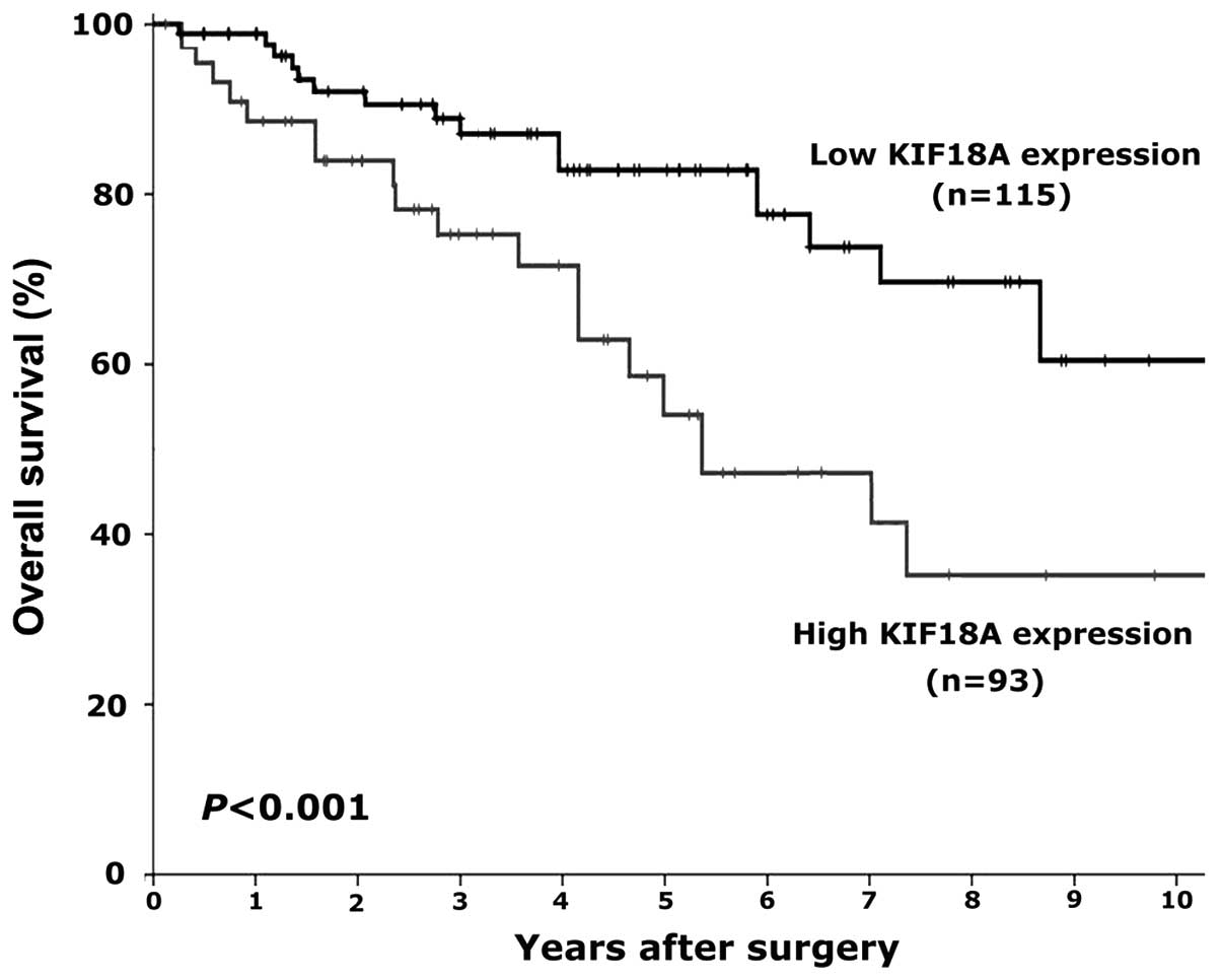
Prognostic significance of KIF18A
expression in RCC
Since significant correlations were demonstrated
between KIF18A expression and clinical stage, pathological grade,
and tumor metastasis in human RCC, it was further evaluated whether
or not KIF18A could be regarded as a novel prognostic marker in
human RCC. Kaplan-Meier analysis was carried out to calculate the
association between KIF18A expression and overall survival in RCC
patients. The overall survival of patients with RCC was found to be
significantly different between the high and low KIF18A expression
groups (P<0.05). After 10 years of follow-up, the overall
survival time of RCC patients who expressed a low level of KIF18A
expression by immunohistochemistry (− or +) was significantly
longer compared with that of the patients who expressed a high
level of KIF18A (++ or +++) (Fig.
4). These findings indicate that high KIF18A expression might
be an independent marker to predict poor prognosis in patients with
RCC.
Discussion
Various motor proteins of the kinesin family have
emerged as targets for chemotherapeutic interventions of
malignancies due to the crucial effects on cell mitosis (24–26).
KIF18A, a member of the kinesin family, plays a key role in the
carcinogenesis and progression of tumors (14,15). A
recent study indicated that the expression of KIF18A is
significantly upregulated and correlated with aggressive
characteristics in patients with hepatocellular carcinoma (27). The same study also suggested the
possibility that KIF18A can serve as a prognostic marker in
patients with hepatocellular carcinoma (27); however, the function of KIF18A in
carcinogenesis is unclear. KIF18A has been shown to be upregulated
in a colorectal cancer model, and KIF18A-deficient mice were found
to be protected from carcinogenesis of colorectal cancer via
inactivation of the phosphoinositide 3-kinase-Akt pathway (28). De Wever et al (29) found that KIF18A directly interacts
with protein phosphatase 1γ, a serine/threonine protein
phosphatase, through a conserved RVxF motif, and KIF18A
participates in phosphatase-induced biological progress. Another
study demonstrated that cells treated with estrogen express higher
levels of KIF18A mRNA and protein, which added novel insight into
the role of estrogen in the regulation of KIF18A expression
(30). In addition, a study
conducted by Zusev and Benayahu (31) indicated that KIF18A is involved in
post-translational modifications and plays a potential role in
regulating protein distribution and the co-association with
cytoskeletal proteins. KIF18A has generated considerable attention
with respect to carcinogenesis, and the expression of KIF18A has
also been detected in tumors. The expression of KIF18A and the
function of KIF18A in human RCC, however, remains elusive to
date.
On the basis of a review of the literature, to the
best of our knowledge, this is the first study involving KIF18A
expression in RCC. In the present study, the level of KIF18A
expression in human RCC was detected, and the expression of KIF18A
mRNA in RCC tissues was detected by RT-PCR and RT-qPCR. The
expression of KIF18A mRNA was similar to the levels of protein
detected by immunohistochemistry and western blotting. KIF18A
expression was significantly upregulated in RCC tissues compared
with normal kidney tissues. Moreover, the expression of KIF18A was
significantly correlated with tumor stage, histological grade,
metastasis and tumor size. These findings demonstrate that KIF18A
may act as an oncogene and plays a crucial role in the
carcinogenesis and progression of human RCC. The effect of KIF18A
on the proliferation of RCC cells was also evaluated. KIF18A
significantly prompted the proliferation of RCC cells; a similar
finding was observed in an animal xenograft experiment with BALB/C
nude mice. The association between the level of KIF18A expression
and overall survival of patients with RCC was further calculated
using Kaplan-Meier analysis. High expression of KIF18A was found to
be associated with poor prognosis of patients with RCC. Thus, the
present study demonstrated that KIF18A can be considered as an
independent marker to predict prognosis in RCC patients. It appears
that the KIF18A gene plays an important role in carcinogenesis of
the adult kidney, and a high level of KIF18A expression may enhance
the progression of human RCC. Moreover, KIF18A is a novel candidate
prognostic gene for RCC patients, thus raising the intriguing
possibility that patients with RCC expressing a high level of
KIF18A expression may be genetically predisposed to RCC. As the
function of KIF18A has not been fully elucidated, it is necessary
to analyze the detailed molecular mechanism of KIF18A in human RCC
in future studies.
In summary, the results of the present study suggest
that KIF18A expression is significantly increased in human RCC and
KIF18A enhances the proliferation of RCC cells in vitro and
in vivo. These findings indicate that KIF18A plays a key
role in the carcinogenesis and progression of RCC, and is a novel
prognostic candidate marker for RCC patients. Silencing KIF18A
expression may serve as a new therapeutic strategy against RCC.
References
|
1
|
Siegel R, Naishadham D and Jemal A: Cancer
statistics, 2013. CA Cancer J Clin. 63:11–30. 2013. View Article : Google Scholar : PubMed/NCBI
|
|
2
|
Deng FM and Melamed J: Histologic variants
of renal cell carcinoma: Does tumor type influence outcome? Urol
Clin North Am. 39:119–132. 2012. View Article : Google Scholar : PubMed/NCBI
|
|
3
|
Yang L, Parkin DM, Ferlay J, Li L and Chen
Y: Estimates of cancer incidence in China for 2000 and projections
for 2005. Cancer Epidemiol Biomark Prev. 14:243–250. 2005.
|
|
4
|
Athar U and Gentile TC: Treatment options
for metastatic renal cell carcinoma: A review. Can J Urol.
15:3954–3966. 2008.PubMed/NCBI
|
|
5
|
Janowitz T, Welsh SJ, Zaki K, Mulders P
and Eisen T: Adjuvant therapy in renal cell carcinoma - past,
present and future. Semin Oncol. 40:482–491. 2013. View Article : Google Scholar : PubMed/NCBI
|
|
6
|
Ljungberg B, Cowan NC, Hanbury DC, Hora M,
Kuczyk MA, Merseburger AS, Patard JJ, Mulders PF and Sinescu IC:
European Association of Urology Guideline Group: EAU guidelines on
renal cell carcinoma: The 2010 update. Eur Urol. 58:398–406. 2010.
View Article : Google Scholar : PubMed/NCBI
|
|
7
|
Bratslavsky G, Sanford T, Srinivasan R,
Aprelikova O, Liu J, Quezado M, Merino M and Linehan WM:
Differential genetic expression in large versus small clear cell
renal cell carcinoma: Results from microarray analysis. J Cancer.
2:271–279. 2011. View Article : Google Scholar : PubMed/NCBI
|
|
8
|
Jordan MA and Wilson L: Microtubules as a
target for anticancer drugs. Nat Rev Cancer. 4:253–265. 2004.
View Article : Google Scholar : PubMed/NCBI
|
|
9
|
Gardner MK, Odde KJ and Bloom K: Kinesin-8
molecular motors: Putting the brakes on chromosome oscillations.
Trends Cell Biol. 18:307–310. 2008. View Article : Google Scholar : PubMed/NCBI
|
|
10
|
Du Y, English CA and Ohi R: The kinesin-8
Kif18A dampens microtubule plus-end dynamics. Curr Biol.
20:374–380. 2010. View Article : Google Scholar : PubMed/NCBI
|
|
11
|
Weaver LN, Ems-McClung SC, Stout JR,
LeBlanc C, Shaw SL, Gardner MK and Walczak CE: Kif18A uses a
microtubule binding site in the tail for plus-end localization and
spindle length regulation. Curr Biol. 21:1500–1506. 2011.
View Article : Google Scholar : PubMed/NCBI
|
|
12
|
Mayr MI, Hümmer S, Bormann J, Grüner T,
Adio S, Woehlke G and Mayer TU: The human kinesin Kif18A is a
motile microtubule depolymerase essential for chromosome
congression. Curr Biol. 17:488–498. 2007. View Article : Google Scholar : PubMed/NCBI
|
|
13
|
Hartwell LH and Kastan MB: Cell cycle
control and cancer. Science. 266:1821–1828. 1994. View Article : Google Scholar : PubMed/NCBI
|
|
14
|
Malumbres M and Barbacid M: Cell cycle,
CDKs and cancer: A changing paradigm. Nat Rev Cancer. 9:153–166.
2009. View
Article : Google Scholar : PubMed/NCBI
|
|
15
|
Mayr MI, Storch M, Howard J and Mayer TU:
A non-motor microtubule binding site is essential for the high
processivity and mitotic function of kinesin-8 Kif18A. PLoS One.
6:e274712011. View Article : Google Scholar : PubMed/NCBI
|
|
16
|
Zhang C, Zhu C, Chen H, Li L, Guo L, Jiang
W and Lu SH: Kif18A is involved in human breast carcinogenesis.
Carcinogenesis. 31:1676–1684. 2010. View Article : Google Scholar : PubMed/NCBI
|
|
17
|
Nagahara M, Nishida N, Iwatsuki M,
Ishimaru S, Mimori K, Tanaka F, Nakagawa T, Sato T, Sugihara K,
Hoon DS and Mori M: Kinesin 18A expression: Clinical relevance to
colorectal cancer progression. Int J Cancer. 129:2543–2552. 2011.
View Article : Google Scholar : PubMed/NCBI
|
|
18
|
Tooker BC, Newman LS, Bowler RP,
Karjalainen A, Oksa P, Vainio H, Pukkala E and Brandt-Rauf PW:
Proteomic detection of cancer in asbestosis patients using
SELDI-TOF discovered serum protein biomarkers. Biomarkers.
16:181–191. 2011. View Article : Google Scholar : PubMed/NCBI
|
|
19
|
Rucksaken R, Khoontawad J, Roytrakul S,
Pinlaor P, Hiraku Y, Wongkham C, Pairojkul C, Boonmars T and
Pinlaor S: Proteomic analysis to identify plasma orosomucoid 2 and
kinesin 18A as potential biomarkers of cholangiocarcinoma. Cancer
Biomark. 12:81–95. 2012.PubMed/NCBI
|
|
20
|
Elmore JM, Kadesky KT, Koeneman KS and
Sagalowsky AI: Reassessment of the 1997 TNM classification system
for renal cell carcinoma. Cancer. 98:2329–2334. 2003. View Article : Google Scholar : PubMed/NCBI
|
|
21
|
Hong SK, Jeong CW, Park JH, Kim HS, Kwak
C, Choe G, Kim HH and Lee SE: Application of simplified Fuhrman
grading system in clear-cell renal cell carcinoma. BJU Int.
107:409–415. 2011. View Article : Google Scholar : PubMed/NCBI
|
|
22
|
Johnson GL, Bibby DF, Wong S, Agrawal SG
and Bustin SA: A MIQE-compliant real-time PCR assay for Aspergillus
detection. PLoS One. 7:e400222012. View Article : Google Scholar : PubMed/NCBI
|
|
23
|
Grimholt RM, Urdal P, Klingenberg O and
Piehler AP: Rapid and reliable detection of α-globin copy number
variations by quantitative real-time PCR. BMC Hematol. 14:42014.
View Article : Google Scholar : PubMed/NCBI
|
|
24
|
Rath O and Kozielski F: Kinesins and
cancer. Nat Rev Cancer. 12:527–539. 2012. View Article : Google Scholar : PubMed/NCBI
|
|
25
|
Wood KW, Cornwell WD and Jackson JR: Past
and future of the mitotic spindle as an oncology target. Curr Opin
Pharmacol. 1:370–377. 2001. View Article : Google Scholar : PubMed/NCBI
|
|
26
|
Huszar D, Theoclitou ME, Skolnik J and
Herbst R: Kinesin motor proteins as targets for cancer therapy.
Cancer Metastasis Rev. 28:197–208. 2009. View Article : Google Scholar : PubMed/NCBI
|
|
27
|
Liao W, Huang G, Liao Y, Yang J, Chen Q,
Xiao S, Jin J, He S and Wang C: High KIF18A expression correlates
with unfavorable prognosis in primary hepatocellular carcinoma.
Oncotarget. 5:10271–10279. 2014. View Article : Google Scholar : PubMed/NCBI
|
|
28
|
Zhu H, Xu W, Zhang H, Liu J, Xu H, Lu S,
Dang S, Kuang Y, Jin X and Wang Z: Targeted deletion of Kif18a
protects from colitis-associated colorectal (CAC) tumors in mice
through impairing Akt phosphorylation. Biochem Biophys Res Commun.
438:97–102. 2013. View Article : Google Scholar : PubMed/NCBI
|
|
29
|
De Wever V, Nasa I, Chamousset D, Lloyd D,
Nimick M, Xu H, Trinkle-Mulcahy L and Moorhead GB: The human
mitotic kinesin KIF18A binds protein phosphatase 1 (PP1) through a
highly conserved docking motif. Biochem Biophys Res Commun.
453:432–437. 2014. View Article : Google Scholar : PubMed/NCBI
|
|
30
|
Zusev M and Benayahu D: The regulation of
MS-KIF18A expression and cross talk with estrogen receptor. PLoS
One. 4:e64072009. View Article : Google Scholar : PubMed/NCBI
|
|
31
|
Zusev M and Benayahu D: New insights on
cellular distribution, microtubule interactions and
post-translational modifications of MS-KIF18A. J Cell Physiol.
217:618–625. 2008. View Article : Google Scholar : PubMed/NCBI
|