Introduction
Acute lung injury (ALI) and its more severe form,
acute respiratory distress syndrome (ARDS) comprise diffuse
heterogeneous lung injury characterized by dyspnea, severe
hypoxemia and non-cardiogenic pulmonary edema, with significant
morbidity and mortality (1). Studies
have suggested that an imbalance between oxidant and antioxidant
systems is involved in the pathogenesis of ALI/ARDS (2,3). In the
context of ALI/ARDS, the generation of reactive oxygen species
(ROS) is increased (2,3). There are numerous potential sources of
ROS, including infiltrated leukocytes (neutrophils), parenchymal
cells (epithelial and endothelial cells), circulating
oxidant-generating enzymes (xanthine oxidase), and inhaled gases
with high oxygen concentrations that are frequently used during
mechanical ventilation (2,3). Concurrently, the expression levels and
activity of ROS scavengers, including superoxide dismutase (SOD),
are decreased significantly in ALI/ARDS (3,4). Other
antioxidant defenses, specifically, urate, glutathione and
ascorbate, are also reduced in the distal airspaces in patients
with ALI (5), resulting the
overproduction of ROS and leading to an imbalance between oxidation
and antioxidation. Increased ROS production has the potential to
induce DNA damage, lipid peroxidation and the activation of nuclear
factor (NF)-κB and activator protein 1 (AP-1) leading to an
expansive inflammatory reaction (2,3).
Therefore, reducing oxidation or restoring the balance of oxidant
and anti-oxidant systems may be a new strategy in the management of
ALI/ARDS.
AMP-activated protein kinase (AMPK) is a
serine/threonine protein kinase composed of a catalytic α subunit
and regulatory β and γ subunits. Pathological changes, including
glucose deprivation, hypoxia, ischemia and heat shock, causing
cellular depletion of ATP or elevation of AMP induce AMPK
activation. Studies have demonstrated that AMPK is also activated
by metformin (6), berberine
(7) and resveratrol (8), independent of energy crisis (9). AMPK has also been shown to negatively
modulate inflammatory reactions. For example, the activation of
AMPK has been found to protect endothelial cells by inhibition of
the NF-κB pathway (10). A further
study has indicated that the activation of AMPK attenuated
neutrophil proinflammatory activity and decreased the severity of
ALI in a lipopolysaccharide (LPS)-induced mouse model of ALI
(11), yet its detailed molecular
mechanisms have not yet been determined.
In the present study, LPS was used to induce a mouse
model of ALI, and the effects of the activation of AMPK by
metformin on oxidant production and the inhibition of ALI were
examined. In addition, further investigations were conducted to
clarify the potential molecular mechanisms.
Materials and methods
Animals and reagents
BALB/c mice were provided by the Experimental Animal
Center of the Medical College of Xi'an Jiaotong University (Xi'an,
China). The animal protocol was approved by the Institutional
Animal Ethics Committee of Xi'an Jiaotong University.
Malondialdehyde (MDA) and SOD assay kits were obtained from
Jiancheng Bioengineering Institute (Nanjing, China). LPS from
Escherichia coli 055:B5 (Sigma-Aldrich, St. Louis, MO, USA)
was used to induce ALI.
Generation of the ALI model
Male BALB/c mice (6–8 weeks old, 20–25 g) were
randomly divided into three groups with 8 mice per group. Control
mice received intratracheal instillation of sterile
phosphate-buffered saline (PBS) alone. Models of ALI were induced
by the intratracheal instillation of 50 µl LPS (100 µg) for 24 h as
previously described (12). Mice of
the third group were pre-treated with metformin (250 mg/kg) by
intraperitoneal injection at 0.5 h prior to stimulation with LPS.
All the mice were maintained in an animal holding room under
special pathogen-free conditions with free access to food and
water.
Harvesting of bronchoalveolar lavage
fluid (BALF)
Animals were sacrificed at 24 h after LPS exposure
by exsanguination under anesthesia with 80 mg/kg ketamine (K2573,
Sigma-Aldrich). Bronchoalveolar lavage (BAL) was performed by
intratracheal injection. The lungs were lavaged three times with
ice-cold sterile PBS at a volume of 0.8 ml/wash and the average
fluid recovery was >85%. The BALF was centrifuged at 800xg for
10 min at 4°C to pelletize cells. The cell pellets were resuspended
in PBS. Total cells were counted under light microscopy using a
hemocytometer and neutrophils were determined as described
previously (12).
Histology
Histopathological evaluation was performed on mice
that were not subjected to BAL. Lungs were inflated and fixed with
10% buffered formalin for 48 h and embedded in paraffin. Tissue
sections (4 µm thick) were cut and stained with hematoxylin and
eosin (H&E) according to the regular staining method for
histological analysis.
MDA content assay
In order to determine the lipid peroxidation level,
MDA contents in the lung tissues were examined using an assay kit.
Briefly, the lung tissue samples were homogenized in cool normal
saline (lung tissue to normal saline ratio, 1:10). The homogenate
was then assessed according to the manufacturer's protocol. The
concentration of MDA was measured by absorbance at 523 nm and was
expressed in units of nmol/mg protein.
SOD activity assay
To examine the oxidation resistance of the lung
tissue samples from each group, the activity of SOD in the lung
tissue was determined using an assay kit following the
manufacturer's protocol. The activity of SOD was quantized by
absorbance at 550 nm and was expressed in units of U/mg
protein.
Western blotting
Lungs were removed and homogenized in ice-cold
radioimmunoprecipitation assay lysis buffer (Sigma-Aldrich) with
phosphatase inhibitor and protease inhibitor. Lysates were
centrifuged at 14,000 × g for 20 min at 4°C, and the supernatant
was collected as the total protein. Protein was separated by 10%
sodium dodecyl sulfate polyacrylamide gel electrophoresis and
transferred to a trans-blot nitrocellulose membrane (Bio-Rad
Laboratories, Inc., Hercules, CA, USA). Polyclonal antibodies
against total-AMPK (2532; 1:500), phospho-AMPK (2535; 1:500; Cell
Signaling Technology, Inc., Danvers, MA, USA), peroxisome
proliferator-activated receptor γ coactivator 1α (PGC-1α;
NBP1-04676; 1:800; Novus Biologicals, LLC, Littleton, CO, USA),
SOD1 (P00441; 1:1,000; Bioworld Technology, Inc., St. Louis Park,
MN, USA) and glyceraldehyde 3-phosphate dehydrogenase (9545;
1:5,000; GAPDH; Sigma-Aldrich) were used following the
manufacturer's protocols. Horseradish peroxidase-conjugated goat
anti-rabbit IgG (1:5,000) was used as the secondary antibody
(A9169; Sigma-Aldrich). Reactions were developed with SuperSignal
West Pico Chemiluminescent Substrate (A9169; Pierce; Thermo Fisher
Scientific, Inc., Waltham, MA, USA) and exposure to
autoradiographic film. Signaling was quantified from scanned films
using Scion Image software version 4.03 (Scion Corporation,
Frederick, MD, USA).
Statistical analysis
Values are presented as the mean ± standard
deviation. Data were analyzed using one way analysis of variance
followed by Tukey's post hoc test. P<0.05 was considered to
indicate a significant difference between groups.
Results
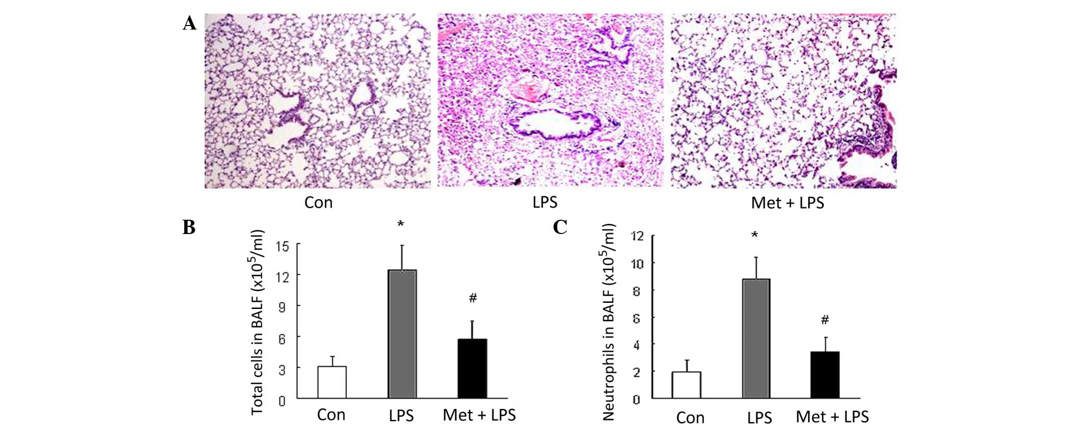
Metformin inhibits LPS-induced
ALI
The results of the present study demonstrated that
LPS induced significant ALI in mice. Histological analysis revealed
substantial hemorrhage, infiltration by inflammatory cells and
damage of epithelial and endothelial cells in mice with LPS-induced
ALI in comparison with the control (Fig.
1A). Furthermore, the number of total inflammatory cells and
neutrophils in the BALF was increased in the mice that received LPS
stimulation (P<0.01 vs. control; Fig.
1B and C). The aforementioned alterations were substantially
ameliorated by prior treatment of the mice with metformin (Fig. 1), suggesting that metformin
effectively inhibits the development of LPS-induced ALI in
mice.
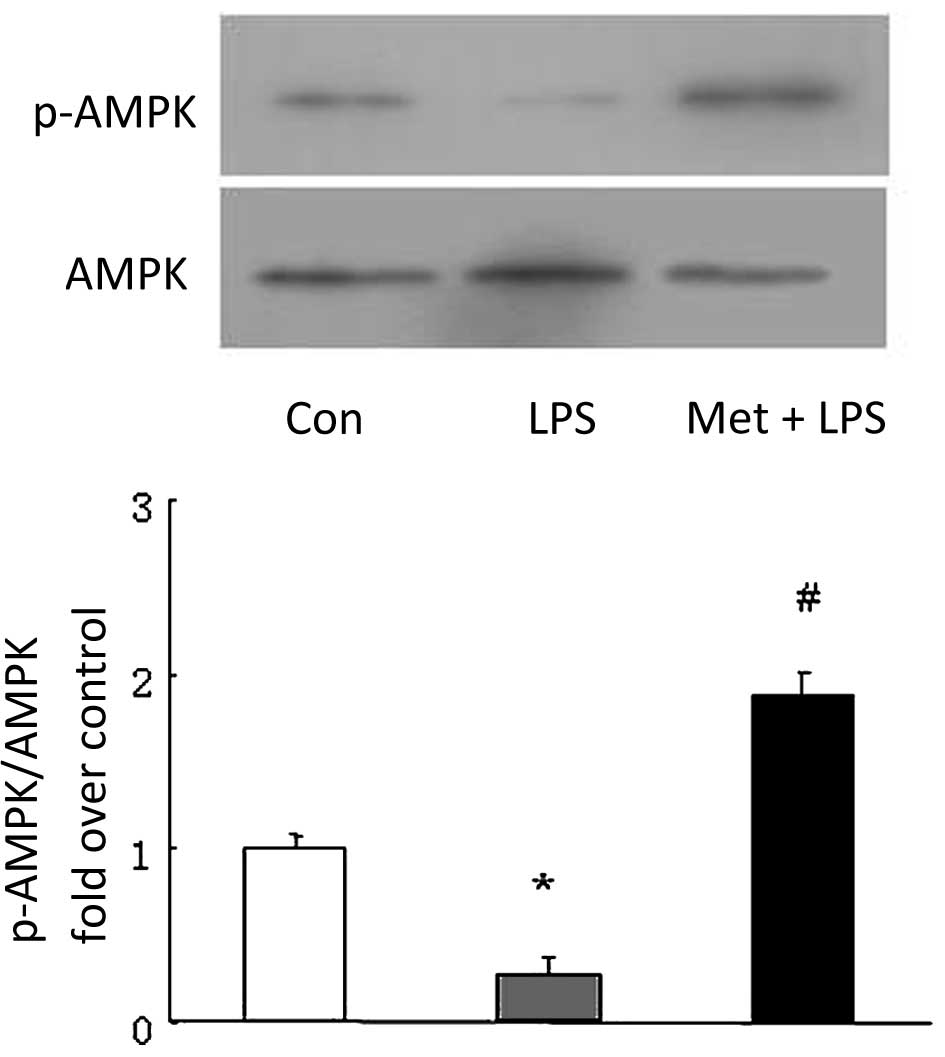
Metformin attenuates the LPS-induced
reduction of AMPK phosphorylation
To investigate the mechanisms by which metformin
inhibits LPS-induced ALI, the phosphorylation of AMPK was examined
using western blotting. As shown in Fig.
2, the phosphorylation level of AMPK declined to 28.3% of the
control value in LPS-treated mice (P<0.05 vs. control). However,
pretreatment of the mice with metformin (250 mg/kg) at 0.5 h prior
to LPS instillation significantly increased AMPK phosphorylation,
which exhibited a 1.86-fold increase compared with that in the LPS
group (P<0.05 vs. LPS). These results suggest that metformin
attenuates the impairment of AMPK activity in LPS-induced ALI,
which might be associated with the development of ALI.
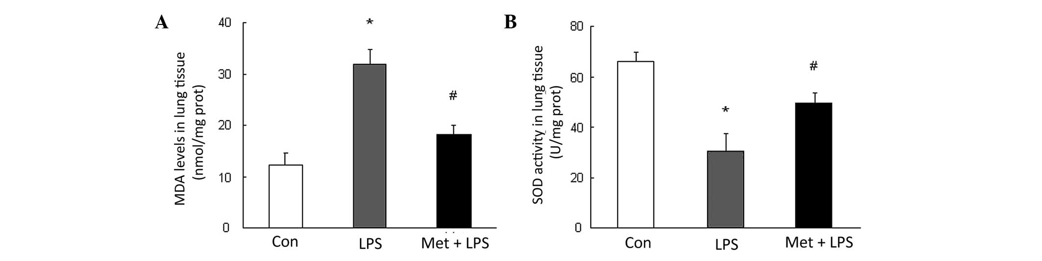
Metformin increases SOD activity and
decreases MDA production
To determine the mechanisms by which the
metformin-stimulated AMPK activation inhibits LPS-induced ALI, the
production of MDA and activity of SOD were determined. The results
shown in Fig. 3 reveal that LPS
instillation caused a 2.58-fold increase over control in MDA
content and a 2.16-fold reduction in SOD activity in lung tissues
compared with those in the control group (P<0.05). However,
pre-treatment with metformin markedly reduced LPS-induced MDA
production and increased SOD activity in mice with LPS-induced ALI
(Fig. 2), suggesting that metformin
restores the balance between oxidation and antioxidation.
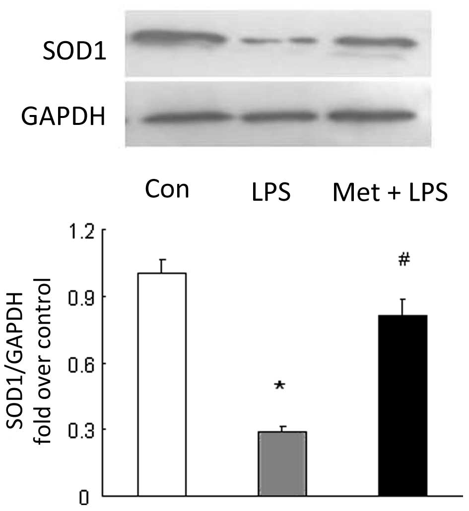
Metformin induces SOD1 expression
SOD1 is a most important enzyme that catalyzes the
elimination of oxygen free radicals produced by infiltrated
leukocytes and parenchymal cells in the antioxidative system
(13). To examine whether metformin
induces the expression of SOD1 and benefits the lungs, the protein
levels of SOD1 were determined in the lung tissues. The results
shown in Fig. 4 indicate that LPS
reduced the SOD1 protein level to 0.28-fold of that in the control
group (P<0.05 vs. control mice). Prior treatment of the mice
with metformin attenuated the LPS-induced reduction in SOD1 level;
the SOD1 protein level was raised to 2.86-fold compared with that
in untreated mice with LPS-induced ALI (P<0.05), suggesting that
metformin induces SOD1 expression in the pulmonary system.
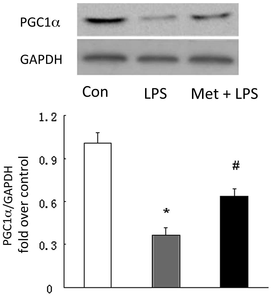
Metformin increases PGC1α
production
To further clarify the mechanisms of induction of
SOD1 by metformin, the level of PGC1α, a transcriptional factor
shown to upregulate SOD1 in non-pulmonary tissues was examined
(14). As shown in Fig. 5, the protein level of PGC1α was
0.36-fold that of the control in the LPS-induced mice model of ALI
(P<0.05 vs. control), while mice treated with metformin prior to
the induction of ALI displayed a 1.74-fold increase in PGC1α
protein level in the lung compared with untreated LPS model mice
(P<0.05 vs. LPS treated mice), indicating that metformin
upregulates the expression of PGC1α in the lung.
Discussion
The present study demonstrates that the activation
of AMPK by metformin suppresses the development of LPS-induced ALI;
this effect is coupled with the upregulation of PGC-1α upregulation
and the subsequent induction of SOD1. The present study also
reveals a novel molecular mechanism by which activation of AMPK
benefits LPS-induced ALI. This study further confirms that the
strategy of activation of AMPK might have potential value in the
treatment of ALI/ARDS.
A disruption of oxidant-antioxidant balance is
likely to be important in the pathogenesis of ALI/ARDS (15). In ALI/ARDS, increased ROS production
induces lipid peroxidation, DNA damage and protein inactivation,
and stimulates the expression of proinflammatory transcriptional
factors, such as NF-κB and AP-1, which in turn upregulate the
expression of adhesion molecules, chemokines and inflammatory
cytokines (for example, tumor necrosis factor-α), leading to cell
injury and death (15). The present
study suggests that the production of MDA, which indicates the
level of lipid peroxidation, was significantly elevated while the
activity of SOD was decreased in the LPS-induced mouse model of
ALI, and this was accompanied by the infiltration of a large number
of neutrophils into the pulmonary tissue. These results further
confirm the involvement of oxidative stress in the pathogenesis of
ALI.
The activation of AMPK using pharmacological ligands
has been demonstrated to induce anti-inflammatory and
immunosuppressive effects in a variety of cell types, and thus is
potentially useful in the treatment of various diseases (7,8,16,17). A
study has shown that activation of the AMPK signaling pathway by
5-aminoimidazole-4-carboxamide-1-β-D-ribofuranoside and berberine
ameliorates the LPS-induced mice model of ALI (11). However, the mechanisms responsible
for AMPK activation exerting an inhibitory effect on ALI remain
largely unknown. In the present study, it was found that the
activation of AMPK by metformin suppressed the development of
LPS-induced ALI, which was accompanied by significantly increased
activity and expression of SOD1 and reduced concentration of MDA in
lung tissues, counteracting the changes induced in the LPS-induced
ALI model. These indicate that the activation of AMPK by metformin
rebuilds the balance between oxidation and antioxidation has a
beneficial effect on LPS-induced ALI.
The induction of PGC-1α has been shown to confer
protective against oxidative stress with AMPK activation in
non-pulmonary systems (16,18). Therefore, it is worthwhile to examine
the expression level of PGC-1α in LPS-induced ALI. The present
study demonstrated that ALI model mice exhibited reduced levels of
PGC-1α, and the application of metformin attenuated the decline of
PGC-1α in the ALI model. This was accompanied by a reduction in the
level of MDA production, suggesting that the induction of PGC-1α
mediates the protective effects of AMPK activation on lung
tissues.
PGC-1α is a transcriptional coactivator that acts to
regulate lipid catabolism, mitochondrial number and function.
PGC-1α has been identified to be a central regulator of energy
metabolism, and is expressed at high levels in tissues with high
metabolic rates (19). A previous
study suggests that PGC-1α is required for the induction of
numerous ROS-detoxifying enzymes, including SOD1, SOD2 and
glutathione peroxidase 1, and cells lacking PGC-1α have an
increased sensitivity to oxidative stress (14). Overexpression of PGC-1α in
vitro and in vivo significantly upregulates the
expression of antioxidant enzymes, including SOD and catalase,
thereby reducing ROS levels, suppressing oxidative damage and
protecting cells and tissues (20,21).
Consistent with the pattern of changes of PGC-1α levels, the
expression of SOD1 was markedly raised in metformin-treated ALI
model mice, whereas the level was reduced in the ALI model compared
with that in the control. Thus, it is proposed that the protective
effect of metformin on LPS-induced ALI is mediated via the
AMPK/PGC-1α/SOD1 axis.
Metformin is an in vitro synthetic AMPK
agonist which has been commonly used clinically to treat type 2
diabetes for many years (22). The
wide clinical experience and safety record of metformin suggest
that it may be an optimal therapeutic approach for human ALI/ARDS.
The present study also provides novel insights into the mechanism
of metformin in the regulation of the balance between oxidation and
antioxidation, and the subsequent amelioration or prevention of the
development of ALI/ARDS.
Acknowledgements
This study was supported by the National Natural
Science Foundation of China (Grant No. 81070045 and No.
81330002).
References
|
1
|
Walkey AJ, Summer R, Ho V and Alkana P:
Acute respiratory distress syndrome: Epidemiology and management
approaches. Clin Epidemiol. 4:159–169. 2012. View Article : Google Scholar : PubMed/NCBI
|
|
2
|
Rosanna DP and Salvatore C: Reactive
oxygen species, inflammation and lung diseases. Curr Pharm Des.
18:3889–3900. 2012. View Article : Google Scholar : PubMed/NCBI
|
|
3
|
Park HS, Kim SR and Lee YC: Impact of
oxidative stress on lung diseases. Respirology. 14:27–38. 2009.
View Article : Google Scholar : PubMed/NCBI
|
|
4
|
Crimi E, Sica V, Williams-Ignarro S, Zhang
H, Slutsky AS, Ignarro LJ and Napoli C: The role of oxidative
stress in adult critical care. Free Radic Biol Med. 40:398–406.
2006. View Article : Google Scholar : PubMed/NCBI
|
|
5
|
Bowler RP, Velsor LW, Duda B, Chan ED,
Abraham E, Ware LB, Matthay MA and Day BJ: Pulmonary edema fluid
antioxidants are depressed in acute lung injury. Crit Care Med.
31:2309–2315. 2003. View Article : Google Scholar : PubMed/NCBI
|
|
6
|
Xu JN, Zeng C, Zhou Y, Peng C, Zhou YF and
Xue Q: Metformin inhibits StAR expression in human endometriotic
stromal cells via AMPK-mediated disruption of CREB-CRTC2 complex
formation. J Clin Endocrinol Metab. 99:2795–2803. 2014. View Article : Google Scholar : PubMed/NCBI
|
|
7
|
Jeong HW, Hsu KC, Lee JW, Ham M, Huh JY,
Shin HJ, Kim WS and Kim JB: Berberine suppresses proinflammatory
responses through AMPK activation in macrophages. Am J Physiol
Endocrinol Metab. 296:E955–E964. 2009. View Article : Google Scholar : PubMed/NCBI
|
|
8
|
Han Y, Jiang C, Tang J, Wang C, Wu P,
Zhang G, Liu W, Jamangulova N, Wu X and Song X: Resveratrol reduces
morphine tolerance by inhibiting microglial activation via AMPK
signalling. Eur J Pain. 18:1458–1470. 2014. View Article : Google Scholar : PubMed/NCBI
|
|
9
|
Yee SW, Chen L and Giacomini KM: The role
of ATM in response to metformin treatment and activation of AMPK.
Nat Genet. 44:359–360. 2012. View
Article : Google Scholar : PubMed/NCBI
|
|
10
|
Su RY, Chao Y, Chen TY, Huang DY and Lin
WW: 5-Aminoimidazole-4-carboxamide riboside sensitizes TRAIL- and
TNF{alpha}-induced cytotoxicity in colon cancer cells through
AMP-activated protein kinase signaling. Mol Cancer Ther.
6:1562–1571. 2007. View Article : Google Scholar : PubMed/NCBI
|
|
11
|
Zhao X, Zmijewski JW, Lorne E, Liu G, Park
YJ, Tsuruta Y and Abraham E: Activation of AMPK attenuates
neutrophil proinflammatory activity and decreases the severity of
acute lung injury. Am J Physiol Lung Cell Mol Physiol.
295:L497–L504. 2008. View Article : Google Scholar : PubMed/NCBI
|
|
12
|
Wang G, Liu L, Zhang Y, Han D, Liu J, Xu
J, Xie X, Wu Y, Zhang D, Ke R, et al: Activation of PPARγ
attenuates LPS-induced acute lung injury by inhibition of
HMGB1-RAGE levels. Eur J Pharmacol. 726:27–32. 2014. View Article : Google Scholar : PubMed/NCBI
|
|
13
|
Fukai T and Ushio-Fukai M: Superoxide
dismutases: Role in redox signaling, vascular function and
diseases. Antioxid Redox Signal. 15:1583–1606. 2011. View Article : Google Scholar : PubMed/NCBI
|
|
14
|
St-Pierre J, Drori S, Uldry M, Silvaggi
JM, Rhee J, Jäger S, Handschin C, Zheng K, Lin J, Yang W, et al:
Suppression of reactive oxygen species and neurodegeneration by the
PGC-1 transcriptional coactivators. Cell. 127:397–408. 2006.
View Article : Google Scholar : PubMed/NCBI
|
|
15
|
Tasaka S, Amaya F, Hashimoto S and
Ishizaka A: Roles of oxidants and redox signaling in the
pathogenesis of acute respiratory distress syndrome. Antioxid Redox
Signal. 10:739–753. 2008. View Article : Google Scholar : PubMed/NCBI
|
|
16
|
Kim MY, Lim JH, Youn HH, Hong YA, Yang KS,
Park HS, Chung S, Ko SH, Shin SJ, Choi BS, et al: Resveratrol
prevents renal lipotoxicity and inhibits mesangial cell
glucotoxicity in a manner dependent on the AMPK-SIRT1-PGC1 α axis
in db/db mice. Diabetologia. 56:204–217. 2013. View Article : Google Scholar : PubMed/NCBI
|
|
17
|
Tamaki N, Cristina Orihuela-Campos R,
Inagaki Y, Fukui M, Nagata T and Ito HO: Resveratrol improves
oxidative stress and prevents the progression of periodontitis via
the activation of the Sirt1/AMPK and the Nrf2/antioxidant defense
pathways in a rat periodontitis model. Free Radic Biol Med.
75:222–229. 2014. View Article : Google Scholar : PubMed/NCBI
|
|
18
|
Marmolino D, Manto M, Acquaviva F, Vergara
P, Ravella A, Monticelli A and Pandolfo M: PGC-1alpha
down-regulation affects the antioxidant response in Friedreich's
ataxia. PLoS One. 5:e100252010. View Article : Google Scholar : PubMed/NCBI
|
|
19
|
Liu C and Lin JD: PGC-1 coactivators in
the control of energy metabolism. Acta Biochim Biophys Sin
(Shanghai). 43:248–257. 2011. View Article : Google Scholar : PubMed/NCBI
|
|
20
|
Valle I, Alvarez-Barrientos A, Arza E,
Lamas S and Monsalve M: PGC-1alpha regulates the mitochondrial
antioxidant defense system in vascular endothelial cells.
Cardiovasc Res. 66:562–573. 2005. View Article : Google Scholar : PubMed/NCBI
|
|
21
|
Tsunemi T, Ashe TD, Morrison BE, Soriano
KR, Au J, Roque RA, Lazarowski ER, Damian VA, Masliah E and La
Spada AR: PGC-1α rescues Huntington's disease proteotoxicity by
preventing oxidative stress and promoting TFEB function. Sci Transl
Med. 4:142ra972012. View Article : Google Scholar : PubMed/NCBI
|
|
22
|
Liu SC, Tu YK, Chien MN and Chien KL:
Effect of antidiabetic agents added to metformin on glycaemic
control, hypoglycaemia and weight change in patients with type 2
diabetes: A network meta-analysis. Diabetes Obes Metab. 14:810–820.
2012. View Article : Google Scholar : PubMed/NCBI
|