Introduction
Complement component 1, q subcomponent binding
protein (C1QBP) is a multifunctional protein. Its sequence is
identical to that of p32, a protein that co-purifies with splicing
factor SF2, and that of hyaluronan-binding protein 1, a member of
the hyaladherin family (1). Its
terminal α-helices are important in protein-protein interactions
and regulate its multifunctional nature. C1QBP is commonly
localized in the mitochondrial matrix, although it exhibits
differential localization among cell lines under various
physiological conditions (1).
Moreover, it serves important roles in various biological
processes, including inflammation, infection, cell signaling and
chemotaxis (2–4).
However, it remains unclear whether C1QBP exerts
anti- or pro-tumorigenic effects during cancer pathogenesis
(5). Upregulated expression of C1QBP
has been reported in various different types of cancer, including
breast, prostate, thyroid, colon, pancreatic, gastric, esophageal
and lung cancer (6–9). This elevated expression has been linked
to the metastasis of ovarian cancer (10), poor prognosis or metastasis of breast
cancer (7,9) and early relapse following surgery in
prostate cancer (11). By contrast,
other studies have reported that C1QBP is required for the
induction of mitochondria-dependent apoptotic cell death (12,13). The
constitutive expression of C1QBP in normal fibroblast cells
perturbs its growth characteristics and induces apoptosis (14). In addition, it has been demonstrated
that C1QBP is upregulated following the induction of apoptosis in
HeLa cells in response to a chemotherapeutic agent (cisplatin),
suggesting that C1QBP may be involved in apoptosis (15,16).
Over one million people worldwide develop colorectal
cancer annually, and the disease has a mortality rate of ~33%
(17). Carcinogenesis and biology of
colorectal cancer have been recognized as multistep processes
involving various molecular alterations (18). Knowledge of the specific molecular
markers or pathways that are responsible for disease progression
and poor prognosis may be beneficial in the development of more
effective treatment.
The present proteomic study revealed that C1QBP was
cleaved in the tumor tissues of patients with colon cancer, and
that apolipoprotein A-I (ApoA-I) interacts with C1QBP. Furthermore,
the possible role of C1QBP in colon cancer via its interaction with
ApoA-I is discussed.
Materials and methods
Tissues from colorectal cancer
patients
Fresh tissues (normal colon mucosa and primary colon
tumor) were obtained from 22 patients with colon cancer (8 female
and 14 male, 61.5±10.5 years old) who were enrolled in the current
study between January 2006 and May 2007 at the National Cancer
Center, Republic of Korea. Following dissection of necrotic
exudates and stromal components, the overall cellularity of the
normal epithelium and tumors was >75%. The present study was
approved and performed in accordance with the guidelines of the
Institutional Review Board of the National Cancer Center (Goyang,
Republic of Korea), and informed consent was obtained from all
patients.
Human cancer cell lines
The human colon cancer cell lines (SNU-81, SNU-407,
SNU-C4, NCI-H498, NCI-H508, CaCo2, DLD-1, HCT-116, LoVo and SW620),
a cervical cancer cell line (HeLa) and an embryonic kidney cell
line (293T) were all obtained from the Korean Culture Line Bank
(Seoul, Korea).
Mass analysis
Mass analysis was performed as described previously
(19). The bands of SDS-PAGE
corresponding to potential proteins of interest were excised,
destained using 50% acetonitrile in 0.1 M ammonium bicarbonate
(Thermo Fisher Scientific, Inc., Waltham, MA USA) and dried using a
SpeedVac evaporator (Savant SVC-100H, Thermo Fisher Scientific,
Inc.). Dried gel pieces were swollen with 30 µl of 25 mM sodium
bicarbonate (pH 8.8) (Thermo Fisher Scientific, Inc.) containing 50
ng trypsin (Promega Corporation, Madison, WI, USA) at 37°C
overnight. Samples were subsequently desalted using Zip-Tips C18
(EMD Millipore, Billerica, MA, USA) and dissolved in 10 µl of 2%
acetonitrile (Thermo Fisher Scientific, Inc.) in 0.1% formic acid
(Thermo Fisher Scientific, Inc.). Analyses were performed using a
linear trap quadrupole (LTQ) XL linear ion trap mass spectrometer
(MS; Thermo Fisher Scientific, Inc.) in the Proteomics Core of the
National Cancer Center (Goyang, Republic of Korea). The mass
spectrometer was set for nanospray ionization (NSI) in the positive
mode. Moreover, a syringe pump was used to introduce the
calibration solution for the automatic tuning and calibration of
the LTQ in an NSI-positive ion mode. The infusion of
trypsin-digested samples into the ionization source of the mass
spectrometer was performed following liquid chromatographic
separation. The spray voltage was set at +1.1 kV, the temperature
of the capillary apparatus was maintained at 200°C, the capillary
voltage was set at +20 V and the tube lens voltage was +100 V.
Moreover, the auxiliary gas was set to zero. Full scan experiments
were performed to linear trap in the m/z range of 150–2,000.
Systematic MS/MS experiments were performed by changing the
relative collision energy and monitoring the intensities of the
fragment ions. All MS/MS samples were analyzed using Sequest
(version v.27, rev.11; Thermo Fisher Scientific) to search the
uniprot_sprot (http://www.uniprot.org) and IPI human
databases (European Bioinformatics Institute, Hinxton, UK;
ftp://ftp.ebi.ac.uk/pub/databases/IPI/last_release/current/)
assuming digestion with trypsin. Sequest was searched with a
fragment ion mass tolerance of 1.00 Da and a parent ion tolerance
of 1.2 Da. Methionine oxidation was specified as a variable
modification.
Western blot analysis
Western blotting was performed as described
previously (20). Briefly, cell
homogenates containing equivalent quantities (20 µg) of protein
were centrifuged at 4,000 × g (4°C) for 5 min and the supernatant
fractions were separated using 4–10% gradient SDS-PAGE. Following
electrophoresis, the proteins were transferred to polyvinylidene
fluoride membranes (EMD Millipore), blocked for 2 h at 4°C in 1%
Tween 20-Tris buffered saline buffer containing 1.5% non-fat dry
milk (Bio-Rad Laboratories, Inc., Hercules, CA, USA) and 1 mM
MgCl2. The blocked membranes were then incubated for 2 h
at room temperature with primary antibodies against C1QBP (dilution
1:1,000, cat no. ab131284, Abcam, Cambridge, MA, USA), ApoA-I
(dilution 1:1,000, cat no. ab52945, Abcam) or β-actin (dilution
1:2,000, cat no. 04-1116, Merck Millipore, Darmstadt, Germany).
They were then washed three times in blocking solution (1% Tween
20-TBS buffer containing 1.5% non-fat dry milk and 1 mM
MgCl2) for 15 min each and incubated with diluted
horseradish-peroxidase conjugated secondary antibody (dilution
1:2,000, cat no. 4010-05, Southern Biotech Associates, Inc.,
Birmingham, AL, USA) for 1 h at room temperature. Membranes were
then washed with blocking solution (3×15 min), incubated with
WEST-ZOL® plus chemiluminescence reagent (iNtRON
Biotechnology, Inc., Gyeonggi, Korea) for 1 min and exposed to film
(Kodak Blue XB-1, Kodak, Rochester, NY, USA).
Overexpression of C1QBP in 293T cells
and immunoprecipitation
In order to generate a pFLAG-CMV2-C1QBP plasmid
containing the full-length coding sequence of human C1QBP,
polymerase chain reaction (PCR) was performed from a human brain
cDNA library (Clontech Laboratories, Inc., Rochester, NY, USA) with
the following oligomers: Sense, 5′-GGAATTCTATGCTGCCTCTGCTGCGCTGC-3′
and antisense, 5′-TAACCCGGGCTACTGGCTCTTGACAAAACTCTTGAGG-3′. The
amplified PCR product was digested using EcoRI-XmaI (Clontech
Laboratories), then inserted into pFLAG-CMV2 (Merck Millipore). The
293T cells were transfected with control or FLAG-C1QBP plasmids. At
48 h after transfection, cells were harvested and aliquots of the
total protein from each sample were analyzed using western
blotting, as indicated.
For immunoprecipitation-coupled MS analyses, cells
were lysed in immunoprecipitation buffer containing 150 mM NaCl, 25
mM HEPES-KOH (pH 7.5), 10% (v/v) glycerol, 1 mM MgCl2, 2
mM sodium orthovanadate, 2 mM glycerophosphate, 1 mM PMSF, 1 mM
DTT, 2 mM EDTA, 0.5% Triton X-100 and 1X protease inhibitor
cocktail (Roche Applied Science, Madison, WI, USA). Following a
brief sonication and incubation on ice, the lysates were
centrifuged at 15,000 × g for 5 min to remove the insoluble
materials. The lysates were then incubated with anti-FLAG M2
affinity agarose beads (Sigma-Aldrich; Merck Millipore) for 2 h at
4°C, and the collected beads were washed four times with washing
buffer (0.05% Triton X-100 immunoprecipitation buffer without a
protease inhibitor cocktail), and boiled in SDS sample buffer for
protein immunoprecipitation-coupled MS analysis.
Results
Differential expression of C1QBP in
tissues from colon cancer patients
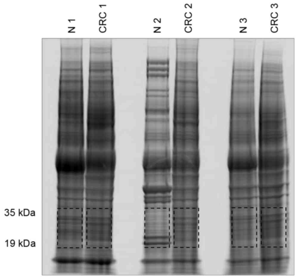
Whole proteins were extracted from both the tumor
and normal tissues of three patients and were separated by SDS-PAGE
(Fig. 1). Sections of gel containing
proteins were then cut according to their molecular weight and
subjected to proteomic analysis. Proteins of 19–35-kDa were
analyzed using the linear ion trap MS system, which identified
C1QBP to be present only in tumor tissues (Fig. 1 and Table
I).
 | Table I.Proteins identified in the range of
molecular weight between 19 to 35 kDa indicated in Fig. 1. Proteins were extracted from the
paired tissues (tumor, CRC; normal, N) obtained from three colon
cancer patients. ‘O’ indicates successful identification of
protein. |
Table I.
Proteins identified in the range of
molecular weight between 19 to 35 kDa indicated in Fig. 1. Proteins were extracted from the
paired tissues (tumor, CRC; normal, N) obtained from three colon
cancer patients. ‘O’ indicates successful identification of
protein.
|
| Total identification
no. | CRC | Normal |
|---|
|
|
|
|
|
|---|
| Protein accession
number and name | CRC | Normal | CRC1 | CRC2 | CRC3 | N1 | N2 | N3 |
|---|
| IPI:IPI00014230.1
C1QBP Complement component 1 Q subcomponent-binding protein,
mitochondrial | 3 | 0 | O | O | O |
|
|
|
| IPI:IPI00010204.1
SRSF3 Serine/arginine-rich splicing factor 3 | 3 | 1 | O | O | O | O |
|
|
| IPI:IPI00021266.1
SNORD4A;RPL23A 60S ribosomal protein L23a | 3 | 1 | O | O | O | O |
|
|
| IPI:IPI00215914.5
ARF1 ADP-ribosylation factor 1 | 3 | 1 | O | O | O |
|
| O |
| IPI:IPI00219622.3
PSMA2 Proteasome subunit alpha type-2 | 3 | 1 | O | O | O | O |
|
|
| IPI:IPI00306332.4
RPL24 60S ribosomal protein L24 | 3 | 1 | O | O | O | O |
|
|
| IPI:IPI00003881.5
HNRNPF Heterogeneous nuclear ribonucleoprotein F | 2 | 0 |
| O | O |
|
|
|
| IPI:IPI00007321.2
LYPLA1 cDNA FLJ60607, highly similar to Acyl-protein thioesterase
1 | 2 | 0 | O |
| O |
|
|
|
| IPI:IPI00010105.1
EIF6 Eukaryotic translation initiation factor 6 | 2 | 0 | O | O |
|
|
|
|
| IPI:IPI00016568.1
AK4 Adenylate kinase isoenzyme 4, mitochondrial | 2 | 0 | O |
| O |
|
|
|
| IPI:IPI00031691.1
RPL9 60S ribosomal protein L9 | 2 | 0 | O | O |
|
|
|
|
| IPI:IPI00179964.5
PTBP1 Isoform 1 of Polypyrimidine tract-binding protein 1 | 2 | 0 | O | O |
|
|
|
|
| IPI:IPI00219097.4
HMGB2 High mobility group protein B2 | 2 | 0 | O | O |
|
|
|
|
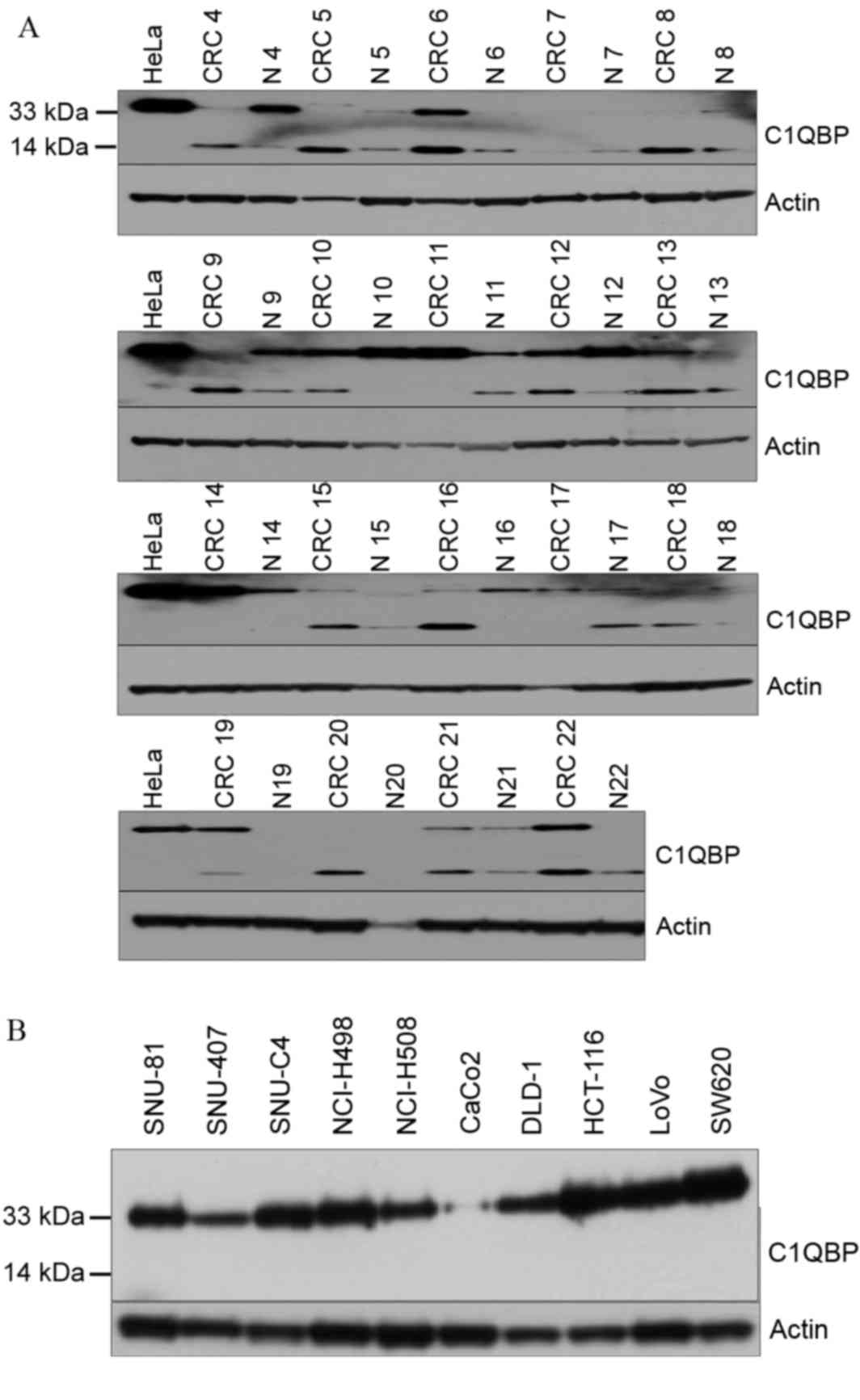
Western blot analysis was then used to confirm the
expression of C1QBP in normal and tumor tissues from patients with
colon cancer (Fig. 2A). Two
immunoreactive signals at 33 and 14 kDa were detected using the
anti-C1QBP antibody (Fig. 2A). In
contrast to the proteomic analysis, 33 kDa C1QBP exhibited no
typical expression pattern in tumor tissues (Fig. 2A). However, among the 19 pairs of
tissues analyzed, 15 showed upregulation of 14 kDa C1QBP in tumor
compared with normal tissues (Fig.
2A). In human colon cancer cell lines, the expression of 33 kDa
C1QBP was variable, whereas the short form was not detected
(Fig. 2B).
Candidate C1QBP-interacting
proteins
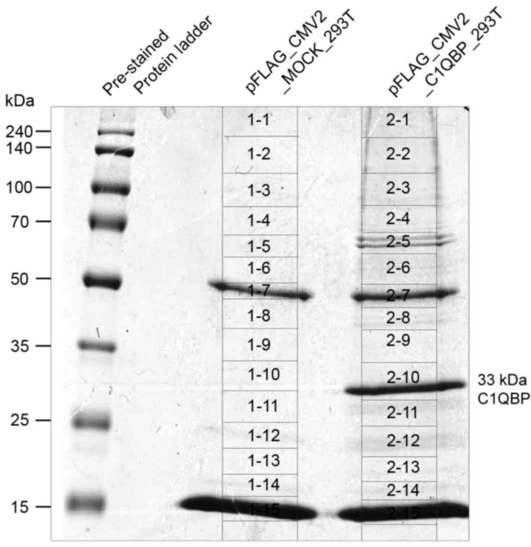
In order to further evaluate the role of C1QBP in
colon cancer at the molecular level, C1QBP-interacting proteins
were screened for in immunoprecipitates from FLAG-tagged
C1QBP-overexpressing 293T cells. Fig.
3 presents the results of immunoprecipitation using FLAG
antibodies in whole cell lysates from C1QBP-overexpressing 293T
cells (pFLAG_CMV2_C1QBP). FLAG-tagged C1QBP was immunoprecipitated
successfully using anti-FLAG antibodies, and was detected as a band
at 33 kDa on the SDS-PAGE gel (Fig.
3). As a control, immunoprecipitation was performed using 293T
cells overexpressing FLAG alone (pFLAG_CMV2_MOCK_293T). The
appropriate bands were excised from the gels as described in
Fig. 3 and the proteins therein were
subjected to in-gel tryptic digestion and MS. The proteins that
co-precipitated with C1QBP are listed in Table II after exclusion of proteins also
identified in the control lane. In total 39 proteins, including
fibrinogen α-, β- and γ-chains, complement C3, complement C4-A,
ApoA-I and ApoA-II, were identified as possible C1QBP-interacting
proteins (Table II).
| Table II.Candidate proteins interacting with
complement component 1, q subcomponent-binding protein. |
Table II.
Candidate proteins interacting with
complement component 1, q subcomponent-binding protein.
| MS/MS sample | Protein name | Accession
number | Molecular weight,
Da | Identification
probability (%) | Total spectra,
% | Sequence coverage,
% |
|---|
| 2–01 | General
transcription factor 3C polypeptide 1 | TF3C1_HUMAN | 238,860.60 | 100.00 | 0.06 |
1.19 |
| 2–01 | Insulin receptor
substrate 4 | IRS4_HUMAN | 133,751.30 |
99.80 | 0.06 |
2.07 |
| 2–02 | Heterogeneous
nuclear ribonucleoprotein U | HNRPU_HUMAN |
90,567.20 | 100.00 | 0.23 |
9.09 |
| 2–02 | General
transcription factor 3C polypeptide 2 | TF3C2_HUMAN | 100,652.80 |
99.80 | 0.07 |
1.98 |
| 2–02 | ATP-dependent RNA
helicase A | DHX9_HUMAN | 140,902.50 | 100.00 | 0.07 |
2.44 |
| 2–02 |
Ubiquitin-associated protein 2-like | UBP2L_HUMAN | 116,780.70 | 100.00 | 0.13 |
4.34 |
| 2–02 | Thyroid hormone
receptor-associated protein 3 | TR150_HUMAN | 108,650.90 |
99.80 | 0.07 |
1.99 |
| 2–03 | General
transcription factor 3C polypeptide 3 | TF3C3_HUMAN | 101,258.20 | 100.00 | 0.27 |
6.77 |
| 2–03 | Nucleolar RNA
helicase 2 | DDX21_HUMAN |
87,328.00 |
99.80 | 0.11 |
2.55 |
| 2–04 |
Alpha-1-antitrypsin | A1AT_HUMAN |
46,719.90 | 100.00 | 0.11 |
8.37 |
| 2–04 | Apolipoprotein
A-I | APOA1_HUMAN |
30,760.50 | 100.00 | 0.11 | 12.40 |
| 2–04 | Protein LAS1
homolog | LAS1L_HUMAN |
83,047.50 | 100.00 | 0.14 |
6.40 |
| 2–06 | Lamin-B
receptor | LBR_HUMAN |
70,688.30 |
99.80 | 0.10 |
3.09 |
| 2–08 | RNA
methyltransferase-like protein 1 | RMTL1_HUMAN |
47,002.50 | 100.00 | 0.24 |
7.62 |
| 2–08 | Keratin, type I
cytoskeletal 14 | K1C14_HUMAN |
51,544.50 | 100.00 | 0.12 |
9.11 |
| 2–09 | Interleukin
enhancer-binding factor 2 | ILF2_HUMAN |
43,044.70 | 100.00 | 0.17 | 11.50 |
| 2–10 | Heterogeneous
nuclear ribonucleoproteins C1/C2 | HNRPC_HUMAN |
33,652.50 | 100.00 | 0.37 | 22.20 |
| 2–10 | Glutaminyl-peptide
cyclotransferase-like protein | QPCTL_HUMAN |
42,907.60 | 100.00 | 0.12 |
8.12 |
| 2–11 | rRNA
2′-O-methyltransferase fibrillarin | FBRL_HUMAN |
33,766.10 |
99.90 | 0.09 |
7.48 |
| 2–11 | Polymerase
delta-interacting protein 2 | PDIP2_HUMAN |
42,015.00 | 100.00 | 0.20 | 20.10 |
| 2–12 | THO complex subunit
4 | THOC4_HUMAN |
26,870.70 | 100.00 | 0.26 | 21.00 |
| 2–13 |
Alpha-1-antichymotrypsin | AACT_HUMAN |
47,634.90 |
99.80 | 0.04 |
4.73 |
| 2–13 |
Alpha-2-macroglobulin | A2MG_HUMAN | 163,271.90 | 100.00 | 0.56 | 18.80 |
| 2–13 | Plasma protease C1
inhibitor | IC1_HUMAN |
55,137.50 |
99.70 | 0.04 |
5.60 |
| 2–13 |
Serotransferrin | TRFE_HUMAN |
77,046.20 | 100.00 | 0.16 | 13.50 |
| 2–13 | Hemopexin | HEMO_HUMAN |
51,658.50 | 100.00 | 0.05 |
8.23 |
| 2–13 | Haptoglobin | HPT_HUMAN |
45,186.90 | 100.00 | 0.16 | 13.10 |
| 2–13 | Fibrinogen alpha
chain | FIBA_HUMAN |
94,955.40 | 100.00 | 0.18 | 14.40 |
| 2–13 | Fibrinogen beta
chain | FIBB_HUMAN |
55,910.60 | 100.00 | 0.15 | 21.60 |
| 2–13 | Fibrinogen gamma
chain | FIBG_HUMAN |
51,495.30 | 100.00 | 0.11 | 14.10 |
| 2–13 | Ig gamma-1 chain C
region | IGHG1_HUMAN |
36,087.00 | 100.00 | 0.33 | 20.00 |
| 2–13 | Ig kappa chain C
region | IGKC_HUMAN |
11,590.50 |
99.80 | 0.18 | 30.20 |
| 2–13 | Ig lambda-2 chain C
regions | LAC2_HUMAN, |
11,275.20 |
99.80 | 0.13 | 32.10 |
| 2–13 | Ig mu chain C
region | IGHM_HUMAN |
49,287.70 | 100.00 | 0.15 | 22.80 |
| 2–13 | Complement C3 | CO3_HUMAN | 187,131.10 | 100.00 | 0.15 |
7.76 |
| 2–13 | Complement
C4-A | CO4A_HUMAN | 192,754.80 | 100.00 | 0.05 |
2.52 |
| 2–13 | Apolipoprotein
A-II | APOA2_HUMAN |
11,202.40 |
99.80 | 0.07 | 21.00 |
| 2–14 | 60S ribosomal
protein L13 | RL13_HUMAN |
24,244.20 |
99.80 | 0.08 | 11.40 |
Interaction between C1QBP and
ApoA-I
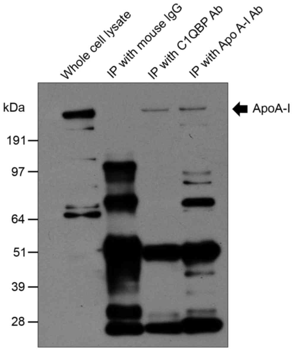
Reverse immunoprecipitation was subsequently used to
confirm the interaction between C1QBP and candidate proteins. Among
the proteins listed in Table II,
ApoA-I was selected for confirmation of its interaction with C1QBP.
Human SW620 colon cancer cells were used because they express high
levels of C1QBP (Fig. 2B, and mouse
IgG or antibodies against C1QBP and ApoA-I were added to SW620
whole-cell lysates. Immunoprecipitation was then performed,
followed by western blotting. Immunoreactive signals produced by
monoclonal antibodies against ApoA-I were detected in
immunoprecipitates prepared using both anti-ApoA-I and -C1QBP
antibodies (Fig. 4). In addition,
SW620 whole cell lysates were immunoreactive for ApoA-I (Fig. 4).
Discussion
The present study revealed that the cleaved form of
C1QBP (14 kDa) is upregulated in colon cancer compared with normal
cells, which is consistent with previous reports of the
pro-tumorigenic properties of C1QBP (5,6,8). Rubinstein et al (8) compared the C1QBP protein levels among
several adenocarcinomas. Immunohistochemical staining of
histological tissue sections revealed pronounced differences in the
expression in colon adenocarcinoma (as well as thyroid, pancreatic,
gastric, esophageal and lung cancer) vs. non-malignant tissues.
Dembitzer et al (6) revealed
strong C1QBP expression in epithelial breast, prostate, liver,
lung, colon and skin tumors. However, increased C1QBP staining was
also detected in inflammatory and proliferative lesions of the same
cell types, as well as in normal and continuously dividing cells.
Moreover, McGee et al (5)
reported that C1QBP expression was upregulated in breast, colon and
lung cancer compared with normal control tissues. Altogether, these
data indicate that C1QBP is important in colon cancer
tumorigenicity.
A novel observation of the present study is that
C1QBP interacts with ApoA-I. Among the many candidate-binding
proteins identified, ApoA-I was selected for confirmation because
it has a relatively well-established role in colon cancer. The
lipid metabolism is closely associated with cancer (21,22). In
particular, it has been speculated that lipids and lipoproteins are
associated with neoplastic processes, including inflammation,
oxidative stress and insulin resistance (23). Moreover, ApoA-I is a major component
of high-density lipoprotein in plasma, which exerts protective
anti-inflammatory, anti-oxidant and anti-microbial functions, and
it is important in innate immunity (21). ApoA-I is synthesized primarily in the
liver (80%) and small intestine (10%) (24), and it is known to be important in
reverse cholesterol transport and for promoting cholesterol efflux
from tissues by acting as a cofactor for lecithin cholesterol
acyltransferase (24). In a study of
a cohort of >520,000 participants from 10 European countries,
high serum concentrations of HDL and ApoA-I were associated with a
decreased risk of colon cancer (23). In addition, HDL mimetics inhibited
tumor development in both induced and spontaneous mouse models of
colon cancer, possibly by inhibiting angiogenesis (21). Zhang et al (25) analyzed the lipid levels of 206
patients with colorectal cancer, 70 patients with benign colorectal
disease, and 300 healthy participants, and revealed that serum
ApoA-I and ApoB levels were significantly lower in colorectal
cancer patients (25). In addition
to colon cancer, ApoA-I inhibited tumor development in a mouse
model of ovarian cancer (26).
Significantly decreased serum levels of ApoA-I were found in
patients with cholangiocarcinoma (27), and increased levels in serum were
associated with a favorable prognosis in patients with metastatic
nasopharyngeal carcinoma (28).
These anti-tumor properties of ApoA-I may be associated with its
binding to and subsequent inhibition of C1QBP. As such, HDL has
received attention as a promising therapeutic strategy for colon
cancer. A novel finding in the present study, that C1QBP binds to
ApoA-I, may assist the future development of such therapeutic
strategies.
Nevertheless, the physiological role of the
interaction between ApoA1 and C1QBP requires further investigation.
Furthermore, one contradictory study indicated that the expression
of ApoA-I was associated with colon adenocarcinoma progression, and
that ApoA-I is a potential marker of tumor aggression (29). However, the novel observations in the
present study may facilitate identification of the molecular
mechanisms underlying the roles of ApoA-I and C1QBP in colon
cancer.
Acknowledgements
The present study was supported by the Soonchunhyang
University Research Fund.
References
|
1
|
Chowdhury AR, Ghosh I and Datta K:
Excessive reactive oxygen species induces apoptosis in fibroblasts:
Role of mitochondrially accumulated hyaluronic acid binding protein
1 (HABP1/p32/gC1qR). Exp Cell Res. 314:651–667. 2008. View Article : Google Scholar : PubMed/NCBI
|
|
2
|
Majumdar M, Meenakshi J, Goswami SK and
Datta K: Hyaluronan binding protein 1 (HABP1)/C1QBP/p32 is an
endogenous substrate for MAP kinase and is translocated to the
nucleus upon mitogenic stimulation. Biochem Biophys Res Commun.
291:829–837. 2002. View Article : Google Scholar : PubMed/NCBI
|
|
3
|
Ghebrehiwet B and Peerschke EI: cC1q-R
(calreticulin) and gC1q-R/p33: Ubiquitously expressed multi-ligand
binding cellular proteins involved in inflammation and infection.
Mol Immunol. 41:173–183. 2004. View Article : Google Scholar : PubMed/NCBI
|
|
4
|
Tahtouh M, Garçon-Bocquet A, Croq F,
Vizioli J, Sautière PE, van Camp C, Salzet M, Nagnan-le Meillour P,
Pestel J and Lefebvre C: Interaction of HmC1q with leech microglial
cells: Involvement of C1qBP-related molecule in the induction of
cell chemotaxis. J Neuroinflammation. 9:372012. View Article : Google Scholar : PubMed/NCBI
|
|
5
|
McGee AM, Douglas DL, Liang Y, Hyder SM
and Baines CP: The mitochondrial protein C1qbp promotes cell
proliferation, migration and resistance to cell death. Cell Cycle.
10:4119–4127. 2011. View Article : Google Scholar : PubMed/NCBI
|
|
6
|
Dembitzer FR, Kinoshita Y, Burstein D,
Phelps RG, Beasley MB, Garcia R, Harpaz N, Jaffer S, Thung SN,
Unger PD, et al: gC1qR expression in normal and pathologic human
tissues: Differential expression in tissues of epithelial and
mesenchymal origin. J Histochem Cytochem. 60:467–474. 2012.
View Article : Google Scholar : PubMed/NCBI
|
|
7
|
Zhang X, Zhang F, Guo L, Wang Y, Zhang P,
Wang R, Zhang N and Chen R: Interactome analysis reveals that C1QBP
(complement component 1, q subcomponent binding protein) is
associated with cancer cell chemotaxis and metastasis. Mol Cell
Proteomics. 12:3199–3209. 2013. View Article : Google Scholar : PubMed/NCBI
|
|
8
|
Rubinstein DB, Stortchevoi A, Boosalis M,
Ashfaq R, Ghebrehiwet B, Peerschke EI, Calvo F and Guillaume T:
Receptor for the globular heads of C1q (gC1q-R, p33,
hyaluronan-binding protein) is preferentially expressed by
adenocarcinoma cells. Int J Cancer. 110:741–750. 2004. View Article : Google Scholar : PubMed/NCBI
|
|
9
|
Chen YB, Jiang CT, Zhang GQ, Wang JS and
Pang D: Increased expression of hyaluronic acid binding protein 1
is correlated with poor prognosis in patients with breast cancer. J
Surg Oncol. 100:382–386. 2009. View Article : Google Scholar : PubMed/NCBI
|
|
10
|
Yu H, Liu Q, Xin T, Xing L, Dong G, Jiang
Q, Lv Y, Song X, Teng C, Huang D, et al: Elevated expression of
hyaluronic acid binding protein 1 (HABP1)/P32/C1QBP is a novel
indicator for lymph node and peritoneal metastasis of epithelial
ovarian cancer patients. Tumour Biol. 34:3981–3987. 2013.
View Article : Google Scholar : PubMed/NCBI
|
|
11
|
Amamoto R, Yagi M, Song Y, Oda Y,
Tsuneyoshi M, Naito S, Yokomizo A, Kuroiwa K, Tokunaga S, Kato S,
et al: Mitochondrial p32/C1QBP is highly expressed in prostate
cancer and is associated with shorter prostate-specific antigen
relapse time after radical prostatectomy. Cancer Sci. 102:639–647.
2011. View Article : Google Scholar : PubMed/NCBI
|
|
12
|
Itahana K and Zhang Y: Mitochondrial p32
is a critical mediator of ARF-induced apoptosis. Cancer Cell.
13:542–553. 2008. View Article : Google Scholar : PubMed/NCBI
|
|
13
|
Sunayama J, Ando Y, Itoh N, Tomiyama A,
Sakurada K, Sugiyama A, Kang D, Tashiro F, Gotoh Y, Kuchino Y and
Kitanaka C: Physical and functional interaction between BH3-only
protein Hrk and mitochondrial pore-forming protein p32. Cell Death
Differ. 11:771–781. 2004. View Article : Google Scholar : PubMed/NCBI
|
|
14
|
Meenakshi J, Goswami SK Anupama and Datta
K: Constitutive expression of hyaluronan binding protein 1
(HABP1/p32/gC1qR) in normal fibroblast cells perturbs its growth
characteristics and induces apoptosis. Biochem Biophys Res Commun.
300:686–693. 2003. View Article : Google Scholar : PubMed/NCBI
|
|
15
|
Kamal A and Datta K: Upregulation of
hyaluronan binding protein 1 (HABP1/p32/gC1qR) is associated with
Cisplatin induced apoptosis. Apoptosis. 11:861–874. 2006.
View Article : Google Scholar : PubMed/NCBI
|
|
16
|
Hosseinimehr SJ, Nobakht R, Ghasemi A and
Pourfallah TA: Radioprotective effect of mefenamic acid against
radiation-induced genotoxicity in human lymphocytes. Radiat Oncol
J. 33:256–260. 2015. View Article : Google Scholar : PubMed/NCBI
|
|
17
|
Han S, Bui NT, Ho MT, Kim YM, Cho M and
Shin DB: Dexamethasone inhibits TGF-β1-induced cell migration by
regulating the ERK and AKT pathways in human colon cancer cells via
CYR61. Cancer Res Treat. 48:1141–1153. 2016. View Article : Google Scholar : PubMed/NCBI
|
|
18
|
Kim ST, Park KH, Kim JS, Shin SW and Kim
YH: Impact of KRAS mutation status on outcomes in metastatic colon
cancer patients without anti-epidermal growth factor receptor
therapy. Cancer Res Treat. 45:55–62. 2013. View Article : Google Scholar : PubMed/NCBI
|
|
19
|
Lee JH, Kim KH, Park JW, Chang HJ, Kim BC,
Kim SY, Kim KG, Lee ES, Kim DY, Oh JH, et al: Low-mass-ion
discriminant equation: A new concept for colorectal cancer
screening. Int J Cancer. 134:1844–1853. 2014. View Article : Google Scholar : PubMed/NCBI
|
|
20
|
Kim KH, Yeo SG, Kim WK, Kim DY, Yeo HY,
Hong JP, Chang HJ, Park JW, Kim SY, Kim BC and Yoo BC: Up-regulated
expression of l-caldesmon associated with malignancy of colorectal
cancer. BMC Cancer. 12:6012012. View Article : Google Scholar : PubMed/NCBI
|
|
21
|
Su F, Grijalva V, Navab K, Ganapathy E,
Meriwether D, Imaizumi S, Navab M, Fogelman AM, Reddy ST and
Farias-Eisner R: HDL mimetics inhibit tumor development in both
induced and spontaneous mouse models of colon cancer. Mol Cancer
Ther. 11:1311–1319. 2012. View Article : Google Scholar : PubMed/NCBI
|
|
22
|
Cho O, Hwang HS, Lee BS, Oh YT, Kim CH and
Chun M: Met inactivation by S-allylcysteine suppresses the
migration and invasion of nasopharyngeal cancer cells induced by
hepatocyte growth factor. Radiat Oncol J. 33:328–336. 2015.
View Article : Google Scholar : PubMed/NCBI
|
|
23
|
van Duijnhoven FJ, Bueno-De-Mesquita HB,
Calligaro M, Jenab M, Pischon T, Jansen EH, Frohlich J, Ayyobi A,
Overvad K, Toft-Petersen AP, et al: Blood lipid and lipoprotein
concentrations and colorectal cancer risk in the European
prospective investigation into cancer and nutrition. Gut.
60:1094–1102. 2011. View Article : Google Scholar : PubMed/NCBI
|
|
24
|
Halley P, Kadakkuzha BM, Faghihi MA,
Magistri M, Zeier Z, Khorkova O, Coito C, Hsiao J, Lawrence M and
Wahlestedt C: Regulation of the apolipoprotein gene cluster by a
long noncoding RNA. Cell Rep. 6:222–230. 2014. View Article : Google Scholar : PubMed/NCBI
|
|
25
|
Zhang X, Zhao XW, Liu DB, Han CZ, Du LL,
Jing JX and Wang Y: Lipid levels in serum and cancerous tissues of
colorectal cancer patients. World J Gastroenterol. 20:8646–8652.
2014. View Article : Google Scholar : PubMed/NCBI
|
|
26
|
Su F, Kozak KR, Imaizumi S, Gao F, Amneus
MW, Grijalva V, Ng C, Wagner A, Hough G, Farias-Eisner G, et al:
Apolipoprotein A-I (apoA-I) and apoA-I mimetic peptides inhibit
tumor development in a mouse model of ovarian cancer. Proc Natl
Acad Sci USA. 107:19997–20002. 2010. View Article : Google Scholar : PubMed/NCBI
|
|
27
|
Wang X, Dai S, Zhang Z, Liu L, Wang J,
Xiao X, He D and Liu B: Characterization of apolipoprotein A-I as a
potential biomarker for cholangiocarcinoma. Eur J Cancer Care
(Engl). 18:625–635. 2009. View Article : Google Scholar : PubMed/NCBI
|
|
28
|
Jiang R, Yang ZH, Luo DH, Guo L, Sun R,
Chen QY, Huang PY, Qiu F, Zou X, Cao KJ, et al: Elevated
apolipoprotein A-I levels are associated with favorable prognosis
in metastatic nasopharyngeal carcinoma. Med Oncol. 31:802014.
View Article : Google Scholar : PubMed/NCBI
|
|
29
|
Tachibana M, Ohkura Y, Kobayashi Y,
Sakamoto H, Tanaka Y, Watanabe J, Amikura K, Nishimura Y and Akagi
K: Expression of apolipoprotein A1 in colonic adenocarcinoma.
Anticancer Res. 23:4161–4167. 2003.PubMed/NCBI
|