Introduction
Squamous cell carcinoma (SCC), sourced from
epithelial keratinocytes, accounts for ~20% of non-melanoma skin
cancer cases (1). It usually
develops from certain skin or precancerous lesions. Currently,
there are ~1,000,000 new cases each year, and the mortality rate is
currently increasing (2,3).
Y-box binding protein 1 (YB-1) has a cold shock
domain and serves multiple functions in cells through binding
directly or indirectly to target gene sequences to regulate gene
transcription and translation (4,5). YB-1 is
expressed in the nucleus and cytoplasm of cells (6,7).
Previous studies have reported that YB-1 upregulation is related to
the growth and resistance of tumour cells to chemotherapy in
multiple tumour cell types. Therefore, it is known as a cancer
prognosis marker (8,9). Its overexpression is an indication of
increased proliferation and invasion of tumour cells (8–10). In
breast cancer tissues, it has previously been demonstrated that
YB-1 upregulation is associated with epidermis growth factor
receptor (EGFR) and human epidermal growth factor receptor-2
(HER-2) upregulation. HER-2 also regulates the nuclear
translocation of YB-1 (10,11). Furthermore, inhibiting the expression
of YB-1 in melanoma cells significantly increased their sensitivity
to chemotherapy, and this process was related to regulation of the
phosphoinositide 3-kinase/Akt signaling pathway (10,11).
These results suggest that YB-1 expression is closely related to
tumour development and progression.
LL-37 is a member of the cathelicidin family and is
a cationic peptide composed of 37 amino acid residues in the human
body. It is a key component of the biological innate immune system
and is mediated by neutrophils (12–14). In
addition, LL-37 serves a key function in angiogenesis and cell
proliferation (13,15). Previous studies have indicated that
LL-37 is upregulated in a variety of solid tumours, and is also
involved in promoting the progression mechanisms of numerous tumour
types, such as the proliferation, migration and invasion of ovarian
cancer, lung cancer, breast cancer and melanoma tumour cells
(16–21). Moreover, previous studies have
reported that these characteristics may be associated with EGFR,
formyl peptide receptor-like 1 (FPRL1) and the insulin-like growth
factor 1 receptor (14,22–24).
However, the mechanisms underlying the effects of LL-37 on the
viability and invasion of SCC cells, and the relationship between
LL-37 and YB-1, are not yet fully understood.
The current study aimed to investigate the effect of
LL-37 on YB-1 expression and the viability and invasion of SCC
cells. The results indicated that LL-37 promoted the viability and
invasion of SCC cells through increasing YB-1 expression, and this
process involved the nuclear factor (NF)-κB signaling pathway.
Materials and methods
Tissue collection
Fresh SCC and adjacent normal tissues were obtained
from 20 patients (12 males and 8 females; age range, 32–58 years)
who underwent skin biopsies between 2013 and 2014 at the Department
of Dermatologic Surgery of The Second Affiliated Hospital of the
Medical College of Xi'an Jiaotong University (Xi'an, China). All
SCC cases were clinically and pathologically verified. Standard
protocols established by the Hospital's Protection of Human
Subjects Committee were followed in this study and informed consent
was provided from all patients in the present study. SCC lesions
were obtained from the center of SCC tissues. The uninvolved
tissues surrounding the SCC were obtained from the normal tissues
surrounding the SCC tissues. Normal skin tissues were obtained from
cosmetic surgeries.
Cell culture
A human squamous cell carcinoma cell line (A431)
(Shanghai Xiang Shi Biotechnology, Co., Ltd., Shanghai, China) was
cultured in F12 medium (Gibco; Thermo Fisher Scientific, Inc.,
Waltham, MA, USA) supplemented with 10% fetal bovine serum (FBS,
Gibco; Thermo Fisher Scientific, Inc.), 100 U/ml penicillin and 100
µg/ml streptomycin (both Sigma-Aldrich; Merck KGaA, Darmstadt,
Germany). Cells were cultured at 37°C with 5% CO2.
Immunohistochemistry
Normal skin tissues, uninvolved tissues surrounding
the SCC and SCC lesions were analyzed by immunohistochemistry.
Paraffin-embedded tissue sections (4-µm thick) were dewaxed in
xylene and re-hydrated in gradient alcohol, then incubated with
rabbit anti-human YB-1 monoclonal antibody (1:50; ab76149; Abcam,
Cambridge, UK) and the slides were placed in a humid environment at
4°C overnight before being incubated with goat anti-rabbit antibody
(1:1,000; ab6721; Abcam) at 37°C for 30 min. Next, the sections
were stained with 3,3′-diaminobenzidine (Sigma-Aldrich; Merck KGaA)
and observed using an optical microscope (magnification, ×200).
Normal skin tissue samples were used as the negative controls.
Inhibiting YB-1 expression using short
interfering RNA (siRNA)
The following siRNA oligonucleotide sequences were
synthesized by Shanghai GenePharma Co., Ltd. (Shanghai, China) in
order to select the most effective one: YB-1 siRNA 1:
5′-GCAGACCGUAACCAUUAUATT-3′ (sense) and 5′-UAUAAUGGUUACGGUCUGCTT-3′
(antisense); YB-1 siRNA 2: 5′-CGGCAAUGAAGAAGAUAAATT-3′ (sense) and
5′-UUUAUCUUCUUCAUUGCCGTT-3′ (antisense); YB-1 siRNA 3:
5′-CUGCCAUAAAGAAGAAUAATT-3′ (sense) and 5′-UUAUUCUUCUUUAUGGCAGTT-3′
(antisense). The negative control duplexes of siRNA were random
sequences and did not target any known mammalian gene according to
GenBank searches (National Center for Biotechnology Information,
Bethesda, MD, USA). A431 cells were transfected with siRNAs
according to the recommended procedures for Lipofectamine 2000
transfection reagent (Invitrogen, Carlsbad, CA). A431 cells were
seeded in 6-well plates at a density of 105 cells/well.
When the fusion reached 70–80%, the cells were treated with
serum-free F12 medium, according to the manufacturer's protocols.
The YB-1 inhibition rate was analyzed using western blot analysis
(described below).
Viability and invasion assay of
siRNA-transfected cells
Transfected cells, which were transfected with
siRNA1, the most effective siRNA of the 3 siRNA sequences, were
seeded at a density of 3×10³ cells/well and cultured in
serum-free F12 medium for 24 h. MTT (5 mg/ml; 10 µl; Sigma-Aldrich;
Merck KGaA) was added to each well and left to incubate at 37°C for
4 h. Subsequently, 150 µl dimethyl sulfoxide (DMSO; Sigma-Aldrich;
Merck KGaA) was added and the absorbance was measured at 490 nm
using a microplate reader (Bio-Rad Laboratories, Inc., Hercules,
CA, USA).
Cell invasion was evaluated using a polycarbonate
membrane with a pore size of 8 µm that was pre-coated with Matrigel
(BD Biosciences, Franklin Lakes, NJ, USA) and Transwell chambers
(Costar; Corning, Inc., Corning NY, USA). The transfected cells
were starved without serum for 12 h, then re-suspended in
serum-free medium at a density of 2.5×10⁵ cells/ml. A
200-µl cell suspension was added to each Transwell chamber and 500
µl culture medium with 10% FBS was added to the lower chamber.
After culturing for 24 h at 37°C, the cells that invaded the lower
chamber were stained with a staining solution (0.1% crystal violet
ethanol). Three representative fields were randomly selected and
the average number of invaded cells was calculated.
Total RNA extraction and quantitative
polymerase chain reaction (qPCR)
TRIzol reagent (Sigma-Aldrich; Merck KGaA) was used
to extract total RNA from cells. Total RNA (3 µg) was then reverse
transcribed to cDNA in a total volume of 20 µl using a reverse
transcription reaction kit (Promega Corporation, Madison, WI, USA)
according to the manufacturer's instructions. Then, qPCR was
performed using an Mx 3000P qPCR system (Applied Biosystems; Thermo
Fisher Scientific, Inc.), according to the manufacturer's
instructions. SYBR Premix Ex Taq II (Takara Biotechnology Co.,
Ltd., Dalian, China) was used as a DNA-specific fluorescent dye.
PCR was performed for 50 cycles of 95°C for 10 sec and 60°C for 30
sec. Primer sequences for detection of mRNA expression were
synthesized as follows: YB-1 specific primers,
5-CAGAATAGTGAGAGTGGGG-3 (forward) and 5-ATGTAGTAAGGTGGGAACC-3
(reverse); human β-actin primers, 5-TTCCATATCGTCCCAGTTGGT-3
(forward) and 5-CCAGGGCGTTATGGTAGGCA-3 (reverse). The YB-1
transcriptional level was normalized against the transcriptional
level of β-actin. All values are from the results of at least three
independent experiments. Relative mRNA expression levels were
calculated using the 2−∆∆Cq method (25).
Immunofluorescence staining
After placing a monolayer of cells on the climbing
film, LL-37 (Sigma-Aldrich; Merck KGaA) of the appropriate
concentration (0, 0.05, 0.5 or 5 µg/ml) was added for 48 h. Then,
the cells were fixed with 4% paraformaldehyde at room temperature
for 10 min, followed by treatment with 5% Triton X-100 for 15 min
at room temperature. They were then sealed with 1% goat serum (200
µl; Thermo Fisher Scientific, Inc.) at 37°C for 30 min. Cells were
incubated with a 1:50 diluted rabbit anti-human YB-1 monoclonal
antibody overnight at 4°C. Then, they were incubated with a
fluorescein isothiocyanate-labeled goat anti-rabbit antibody
(1:2,000) at 37°C for 1 h. Finally, the cells were stained with
DAPI for 1 min and the staining intensity was observed using a
fluorescence inverted microscope (magnification, ×400; Zeiss LSM
700; Zeiss AG, Oberkochen, Germany).
Protein extraction and western blot
analysis
LL-37-stimulated cells were treated with 100–400 µl
of a mixture comprising PBS, 5 mM EDTA, 0.5% Triton X-100, 20 mM
NaF, 1 mM orthovanadate, 1 mM pyrophosphate and protease inhibitors
(0.1 mM phenylmethylsulfonyl fluoride, 10 µM pepstatin A, 10 µM
leupeptin, 25 µg/ml aprotinin) for 30 min at 4°C, then the cell
lysates were separated via centrifugation (13,523 × g at 4°C for 20
min). Approximately 10 µg of the protein isolates were separated
with 10% SDS-PAGE and transferred to a polyvinylidene difluoride
membrane. The membrane was blocked with 5% non-fat milk and 0.1%
PBS with Tween-20 at room temperature for 2 h and then treated with
rabbit anti-human monoclonal antibody YB-1 (1:500) at 4°C
overnight. Subsequently, the membrane was hybridized with goat
anti-rabbit secondary antibody (1:5,000) for 1 h. The results were
detected using an enhanced chemiluminescence kit (EMD Millipore,
Billerica, MA, USA). The sample protein expression was presented
relative to β-actin expression.
Analysis of the signal transduction
pathways of the YB-1 induction by LL-37
SCC cells were seeded at 105 cells/well
and treated with the mitogen-activated protein kinase kinase (MEK)
inhibitor, PD98059 (10 µM; Abcam), the p38/mitogen-activated
protein kinase (MAPK) inhibitor, SB203580 (10 µM; Abcam) or the
NF-κB inhibitor, ammonium pyrrolidinedithiocarbamate (PDTC; 1 µM;
Abcam) for 30 min. Then, 0.5 µM LL-37 was added and incubated at
37°C for 24 h. The protein was then extracted for western blot
analysis (described above).
Cell viability assay after LL-37
stimulation
The cells were seeded at a density of
3×10³ cells/well with serum-free medium for 24 h. In
order to evaluate the effect of dosage, LL-37 at concentrations of
0, 0.05, 0.5 or 5 µg/ml was used to stimulate the cells for 24 h.
In order to evaluate the effect of treatment duration, 0.5 µg/ml
LL-37 was used to stimulate the cells for 0, 24, 48 or 72 h. Then,
MTT (5 mg/ml; 10 µl) was added and the cells were cultured for 4 h
at 37°C. DMSO (150 µl) was added for 10 min. The absorbance was
measured at 490 nm using a microplate reader.
Cell migration and invasion assay
after LL-37 stimulation
The migration assay was conducted using a
polycarbonate membrane with a pore size of 8 µm and Transwell
chambers (EMD Millipore, Billerica, MA, USA). The cells
(2.5×10⁵ cells/ml) were suspended in serum-free F12
culture medium. A 200 µl cell suspension was added to each
Transwell chamber and 500 µl culture medium with 10% FBS was added
to the lower chamber. In the upper chamber, a specific
concentration of 0, 0.05, 0.05 or 5 µg/ml LL-37 was added. After 24
h, the residual cells on the surface were gently wiped out using a
cotton swab. The cells that invaded the lower chamber were stained
with staining solution (0.1% crystal violet ethanol) and observed
under a light microscope (magnification, ×200). Three
representative fields were randomly selected and the average number
of invaded cells was calculated. With the pre-coated Matrigel (BD
Biosciences) Transwell chambers, the invasion experiment was
performed similar to the migration assay.
Statistical analysis
Statistical analysis was conducted using SPSS 17.0
software (SPSS, Inc., Chicago, IL, USA). All data are presented as
the mean ± standard deviation. Comparisons between two groups were
analysed using a two-tailed t-test. Comparisons among three or more
groups were evaluated using analysis of variance and Fisher's least
significant difference test. P<0.05 was considered to indicate a
statistically significant difference.
Results
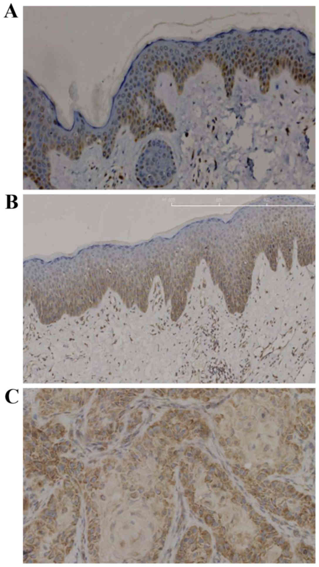
YB-1 is upregulated in SCC
In normal skin tissues and uninvolved skin tissues
adjacent to SCC, YB-1 protein was strongly expressed in the basal
layer and weakly expressed in the lower layers of the stratum
spinosum (Fig. 1A and B). However,
in SCC tissues, YB-1 was strongly expressed in nearly all tumour
cells (Fig. 1C).
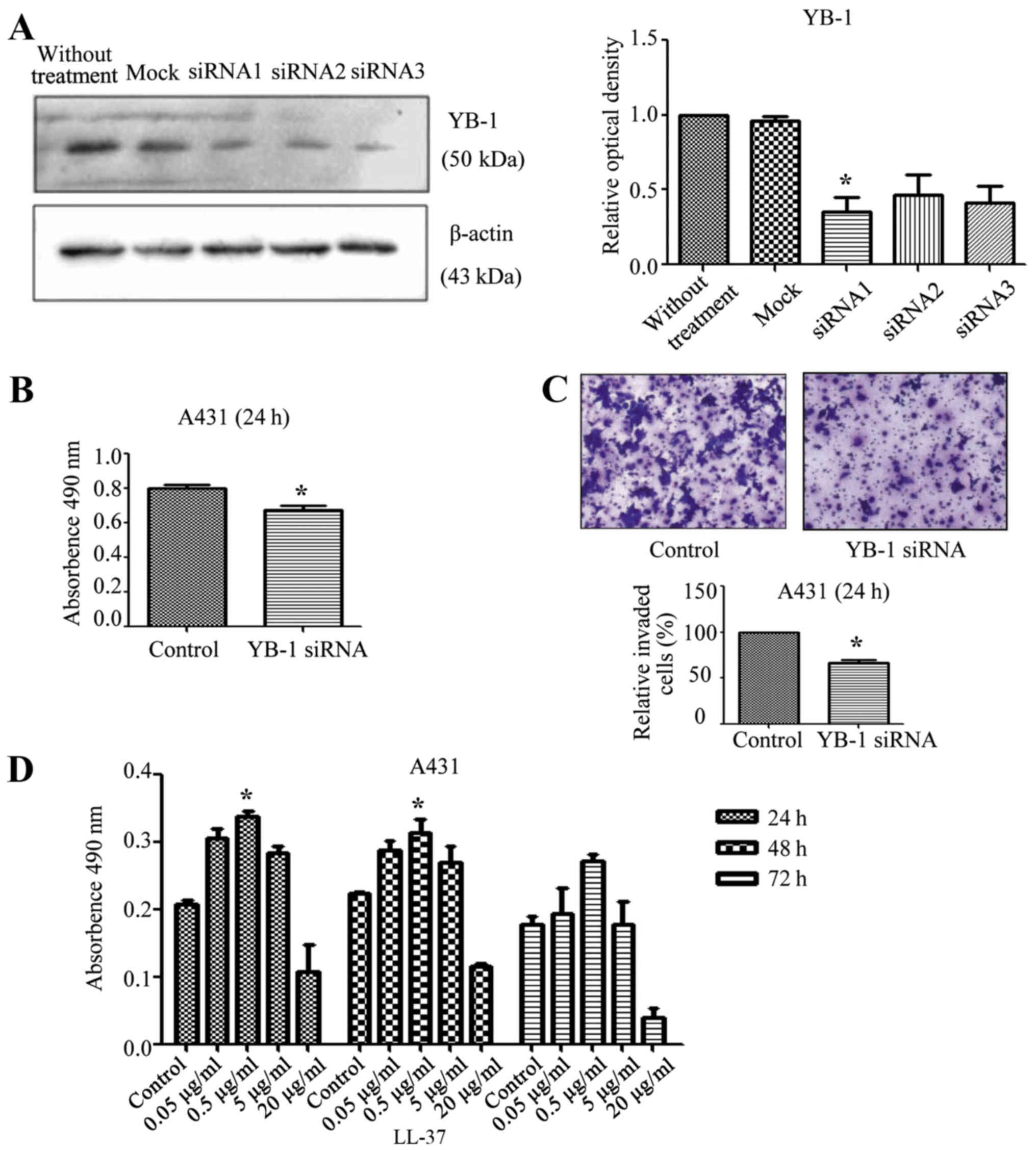
YB-1 siRNA inhibits the protein levels
of YB-1 and reduces the viability and invasion of SCC cells
Total protein was extracted from siRNA-transfected
cells and analysed by western blot analysis. Compared with the
control group, the YB-1 siRNA 1 significantly inhibited the
expression of YB-1 (P<0.05; Fig.
2A). Based on this result, the expression of YB-1 in A431 cells
was then inhibited by transfection with YB-1 siRNA 1. Compared with
the control group, the viability of the cells significantly
decreased (P<0.05; Fig. 2B),
which indicated that the viability of SCC cells was at least partly
dependent on YB-1. The invasion ability of the A431 cells was also
evaluated. Compared with the control group, the proportion of
invaded cells significantly decreased (P<0.05; Fig. 2C), which suggested that the
invasiveness of SCC cells was at least partly dependent on YB-1.
Thus, the inhibition of YB-1 may reduce the viability and
invasiveness of A431 cells.
Effect of LL-37 on the viability of
SCC cells
A431 cells were stimulated with LL-37 (0, 0.05, 0.5,
5 or 20 µg/ml) and the effect on cell viability was observed at
different time intervals (24, 48 or 72 h). Treatment with 0.5 µg/ml
LL-37 significantly increased cell viability at both 24 and 48 h
compared with the respective control groups (P<0.05 for both;
Fig. 2D).
LL-37 promotes the migration and
invasion of SCC cells
The effect of different LL-37 concentrations (0,
0.05, 0.5 or 5 µg/ml) on the migration of A431 cells was
investigated. Compared with the control group, 0.05 µg/ml LL-37
significantly increased the migration of A431 cells (P<0.05;
Fig. 3A). Furthermore, the effect of
different LL-37 concentrations (0, 0.05, 0.5 or 5 µg/ml) on the
invasiveness of A431 cells was investigated. Compared with the
control group, 0.05 µg/ml LL-37 significantly enhanced the
invasiveness of A431 cells (P<0.05; Fig. 3B).
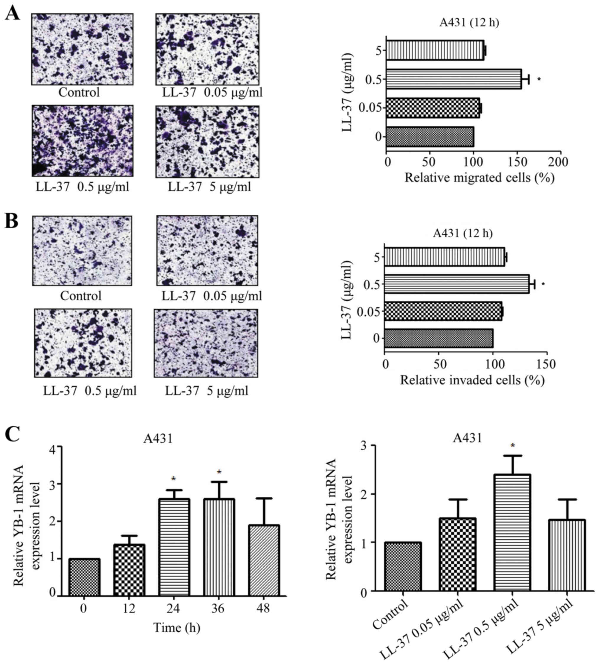 | Figure 3.Effect of LL-37 treatment on
migration, invasion and YB-1 mRNA expression in squamous cell
carcinoma cells. (A) Effect of LL-37 treatment on migration in A431
cells. Serum-starved A431 cells were treated with LL-37 at 0.05,
0.5 or 5 µg/ml for 12 h, and cell migration was analysed using a
Transwell assay. (B) Effect of LL-37 treatment on invasion by A431
cells. Serum-starved A431 cells were treated with LL-37 at 0.05,
0.5 or 5 µg/ml for 24 h, and the cell invasion was analysed using a
Transwell assay. Magnification, ×200. (C) Effect of LL-37 treatment
on YB-1 mRNA expression in A431 cells. Time effect: A431 cells were
stimulated with 0.5 µg/ml LL-37 for 0, 12, 24, 36 or 48 h. Dose
effect: A431 cells were stimulated with 0, 0.05, 0.5 or 5 µg/ml
LL-37 for 24 h. YB-1 mRNA levels were determined by quantitative
polymerase chain reaction. YB-1 mRNA expression is presented
relative to β-actin mRNA expression. *P<0.05 vs. control. YB-1,
Y-box binding protein 1. |
Effect of LL-37 on YB-1 mRNA
expression in SCC cells
qPCR was used to evaluate the effect of LL-37
treatment at different concentrations (0, 0.05, 0.5 or 5 µg/ml) and
durations (0, 12, 24, 36 or 48 h) on the expression of YB-1 in SCC
cells. Compared with the control group, stimulation of A431 cells
with LL-37 for 24 or 36 h significantly increased the mRNA
expression of YB-1 (P<0.05; Fig.
3C). When different concentrations of LL-37 treatment were
evaluated, 0.05 µg/ml LL-37 resulted in a significant increase in
YB-1 mRNA expression (P<0.05; Fig.
3C).
Effect of LL-37 on YB-1 protein
expression in SCC cells
Western blot analysis indicated that treatment with
LL-37 for 24 h and at a concentration of 0.5 µg/ml significantly
increased the protein expression of YB-1 compared with the controls
(P<0.05; Fig. 4A and B).
Furthermore, stimulating A431 cells with various concentrations of
LL-37 (0.05, 0.5 or 5 µg/ml) increased the fluorescence intensity
of YB-1 immunostaining, and 0.5 µg/ml LL-37 was observed to cause
the greatest increase (Fig. 4C).
These results suggested that LL-37 promoted the expression of YB-1
protein in A431 cells.
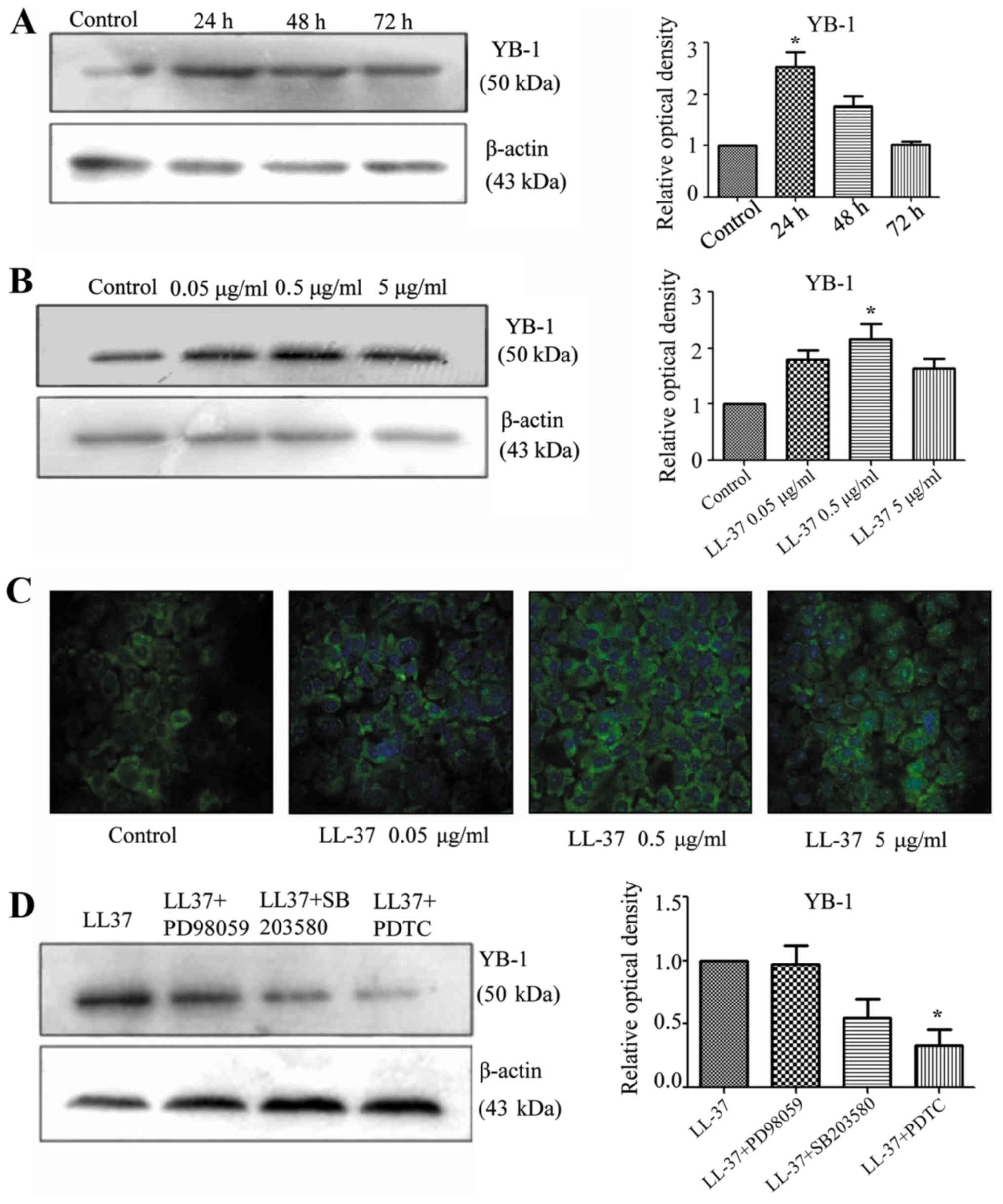 | Figure 4.Effect of LL-37 treatment and
signaling pathway inhibitors on YB-1 protein expression. (A) Effect
of 0.05 µg/ml LL-37 treatment for 0, 24, 48 or 72 h on YB-1 protein
expression in A431 cells. YB-1 protein levels were determined by
western blot analysis. YB-1 protein was measured relative to
β-actin protein. (B) Effect of LL-37 treatment (0, 0.05, 0.5 or 5
µg/ml) for 48 h on YB-1 protein expression in A431 cells. YB-1
protein levels were determined by western blot analysis. YB-1
protein was measured relative to β-actin protein. (C)
Immunofluorescence assay to evaluate the effect of LL-37 on YB-1
protein levels in A431 cells. A431 cells were stimulated with LL-37
(0, 0.05, 0.5 or 5 µg/ml) for 48 h. Magnification, ×400. (D) Effect
of signaling pathway inhibitors on LL-37-induced YB-1 protein
expression. A431 cells were pre-treated for 30 min with
mitogen-activated protein kinase kinase inhibitor (PD98059, 10 µM),
mitogen-activated protein kinase inhibitor (SB203580, 10 µM) or
nuclear factor-κB inhibitor (PDTC, 1 µM), followed by treatment
with 0.5 µg/ml of LL-37 for 48 h. YB-1 protein levels were
determined by western blot analysis. YB-1 protein was measured
relative to β-actin protein. *P<0.05 vs. control. YB-1, Y-box
binding protein 1; PDTC, ammonium pyrrolidinedithiocarbamate. |
Analysis of LL-37 signaling in SCC
cells
In order to investigate the signaling pathway of
LL-37 that induced the expression of YB-1, inhibition experiments
were performed. A431 cells were pre-treated with the MEK inhibitor,
PD98059, the p38/MAPK inhibitor, SB203580, and the NF-κB inhibitor,
PDTC. Treatment with PDTC significantly inhibited the LL-37-induced
expression of YB-1 in the A431 cells (P<0.05; Fig. 4D). This indicated that the increased
expression of YB-1 by treatment with LL-37 involved the NF-κB
signaling pathway.
Discussion
The results of the current study indicated that YB-1
was weakly expressed in the basal layer of normal skin tissue and
strongly expressed in SCC tissue. Treatment with 0.5 µg/ml LL-37
promoted A431 cell viability and invasion, and increased YB-1 mRNA
and protein expression. Inhibiting the NF-κB signaling pathway led
to a decrease in LL-37-induced YB-1 protein expression. This
suggested that LL-37 could regulate YB-1 expression, and that this
process occurred via the NF-κB signaling pathway in A431 cells.
LL-37 is a member of the antibacterial peptide
family and is correlated with the proliferation of epidermal cells
(13,14). Previous studies have indicated that
LL-37 promoted the growth of malignant tumours, primarily via
upregulation of EGFR and the epidermal growth factor receptor 2
(ErbB2), in numerous cancer types, such as lung cancer, melanoma,
prostate cancer, ovarian cancer and oral SCC (23). Through the induction of
membrane-associated protein kinase, EGFR receptors split the metal
protein kinase, and LL-37 activates the EGFR, to depend on
G-protein coupled receptors (GPCRs) in different tumour cell types,
such as lung, prostate and ovarian tumour cells (14,22–24).
LL-37 promotes the proliferation and invasion of tumour cells in
lung SCC and activates the mitogenic effect of EGFR phosphorylation
and the subsequent rat sarcoma (Ras)/MAPK cascade (21). EGFR signaling serves a key function
in proliferation, angiogenesis, anti-apoptosis, invasion and
metastasis in lung cancer cells (14,21).
EGFR ligands, such as transforming growth factor-α and heparin
binding epidermal growth factor, are released from the cells after
splitting by a protease. These precursors can diffuse freely and
activate the EGFR (26,27). Thus, the oncogenic effect of LL-37 is
via an EGFR-mediated transcriptional mechanism in some tumour
tissues, including lung, prostate and ovarian tumour tissues
(23,26,27).
LL-37 has been reported to increase tumour
progression via the ErbB-mediated pathway way in breast cancer, and
upregulate the expression of ErbB2 or EGFR to promote growth and
metastasis (19). FPR2 may also be
involved in this process. The activation of MAPK and Janus
kinase/signal transducer is the result of a biochemical cascade
that involves transcription factor signaling. Previous results
indicate that numerous transcription factors lead to significant
MAPK and Janus kinase/signal transducer activation and this is
dependent on FPR2 (22,24,28). In
ovarian cancer, LL-37 has previously been reported to stimulate
cell proliferation independent of GPCRs (20). However, LL-37 has also been
demonstrated to enhance the invasiveness of ovarian cancer cells
via upregulation of tissue remodeling enzymes, such as matrix
metalloproteinase-2 (MMP-2), a process which is mediated by GPCRs
(23). In ovarian cancer cells, FPR2
has previously been reported to increase the expression of MMP-2,
thus blocking the GPCRs and promoting the invasiveness of tumour
cells (20,23,26,27).
Previous studies have indicated that YB-1 could
regulate the proliferation of epithelial cells and is abnormally
expressed in prostate cancer, lung cancer and other tumor types
(29–32). In transgenic mice, the addition of
FPR2 has been reported to increase YB-1 mRNA levels (11,33).
Previous results have also suggested that Y-box proteins are
involved in the regulation of EGFR and ErbB2 expression, via
binding to the enhancer sequences of EGFR and the promoter region
of ErbB2 (6,8). This suggests that EGFR and ErbB2 are
related to the proliferation and invasion of epidermal tumours.
Furthermore, a previous study demonstrated that the
upregulation of YB-1 during cell proliferation is due to
upregulation of extracellular signal-regulated kinase 2 (ERK2) and
glycogen synthase kinase-3 (GSK-3β) activity. ERK2 and GSK-3β serve
key functions in cell proliferation and apoptosis. Therefore, YB-1
is a downstream target of ERK2 and GSK-3β that promotes cell
proliferation and transformation (10,34).
The results of the current study suggested that the
expression of YB-1 is increased in SCC. A431 cells were transfected
with YB-1 siRNA in order to inhibit the expression of YB-1, and
in vitro MTT and Transwell invasion assays confirmed that a
reduction of YB-1 reduced their viability and invasion rate.
Treatment with 0.5 µg/ml LL-37 increased YB-1 mRNA expression in
A431 cells. Immunofluorescence and western blot analyses confirmed
that LL-37 treatment also increased YB-1 protein expression changes
in A431 cells. Previous studies have reported that LL-37 caused an
increase in the degree of malignancy of tumour cells and this
process was related to the NF-κB pathway and the Ras/MAPK signaling
cascade (12,14). In the current study, LL-37-treated
cells were also treated with MEK, MAPK and NF-κB signaling pathway
inhibitors. It was found that YB-1 protein expression, which was
increased by LL-37, could be reduced by the NF-κB inhibitor. This
result indicated that LL-37 upregulated YB-1 expression via the
NF-κB signaling pathway. NF-κB is a transcription factor that is
known to regulate the expression of multiple genes and is involved
in a range of cellular responses. LL-37 has previously been
reported to increase the levels of NF-κB p65 in some tumour cells,
such as lung, ovarian and breast cancer cells, which can regulate
the expression of genes to promote cell growth. The activation of
NF-κB serves a key function in promoting metastasis and preventing
the apoptosis mechanism of tumour cells (35–38). The
current study demonstrated that the upregulation of YB-1, induced
by LL37, involves the NF-κB signaling pathway in A431 cells.
In conclusion, the current study confirms that YB-1
expression is increased in SCC. LL-37 upregulates YB-1 expression
and promotes the viability and invasion rate of A431 cells. It is
suggested that the activity of LL-37 involves the NF-κB signaling
pathway. These results introduce a novel mechanism of LL-37 in
tumour occurrence and growth, and may be relevant in developing a
new strategy for the clinical treatment of SCC.
Acknowledgements
The present study was supported by the National
Natural Science Foundation of China (grant nos. 81071299, 81371732
and 81573055) and was partially supported by the Fundamental
Research Funds for the Central Universities and for Changjiang
Scholars and Innovative Research Team in University (grant no.
PCSIRT:1171).
References
|
1
|
Knackstedt TJ, Brennick JB, Perry AE, Li
Z, Quatrano NA and Samie FH: Frequency of squamous cell carcinoma
(SCC) invasion in transected SCC in situ referred for Mohs surgery:
The dartmouth-hitchcock experience. Int J Dermatol. 54:830–833.
2015. View Article : Google Scholar : PubMed/NCBI
|
|
2
|
Rogers HW, Weinstock MA, Feldman SR and
Coldiron BM: Incidence estimate of nonmelanoma skin cancer
(keratinocyte carcinomas) in the US population, 2012. JAMA
Dermatol. 151:1081–1086. 2015. View Article : Google Scholar : PubMed/NCBI
|
|
3
|
Sapijaszko M, Zloty D, Bourcier M, Poulin
Y, Janiszewski P and Ashkenas J; Canadian Non-melanoma Skin Cancer
Guidelines Committee, : Non-melanoma skin cancer in canada chapter
5: Management of squamous cell carcinoma. J Cutan Med Surg.
19:249–259. 2015. View Article : Google Scholar : PubMed/NCBI
|
|
4
|
Sakura H, Maekawa T, Imamoto F, Yasuda K
and Ishii S: Two human genes isolated by a novel method encode
DNA-binding proteins containing a common region of homology. Gene.
73:499–507. 1988. View Article : Google Scholar : PubMed/NCBI
|
|
5
|
Eliseeva IA, Kim ER, Guryanov SG,
Ovchinnikov LP and Lyabin DN: Y-box-binding protein 1 (YB-1) and
its functions. Biochemistry (Mosc). 76:1402–1433. 2011. View Article : Google Scholar : PubMed/NCBI
|
|
6
|
Shiota M, Izumi H, Onitsuka T, Miyamoto N,
Kashiwagi E, Kidani A, Yokomizo A, Naito S and Kohno K: Twist
promotes tumor cell growth through YB-1 expression. Cancer Res.
68:98–105. 2008. View Article : Google Scholar : PubMed/NCBI
|
|
7
|
Lasham A, Samuel W, Cao H, Patel R, Mehta
R, Stern JL, Reid G, Woolley AG, Miller LD, Black MA, et al: YB-1,
the E2F pathway, and regulation of tumor cell growth. J Natl Cancer
Inst. 104:133–146. 2012. View Article : Google Scholar : PubMed/NCBI
|
|
8
|
Lasham A, Print CG, Woolley AG, Dunn SE
and Braithwaite AW: YB-1: Oncoprotein, prognostic marker and
therapeutic target? Biochem J. 449:11–23. 2013. View Article : Google Scholar : PubMed/NCBI
|
|
9
|
Takahashi M, Shimajiri S, Izumi H, Hirano
G, Kashiwagi E, Yasuniwa Y, Wu Y, Han B, Akiyama M, Nishizawa S, et
al: Y-box binding protein-1 is a novel molecular target for tumor
vessels. Cancer Sci. 101:1367–1373. 2010. View Article : Google Scholar : PubMed/NCBI
|
|
10
|
Stratford AL, Habibi G, Astanehe A, Jiang
H, Hu K, Park E, Shadeo A, Buys TP, Lam W, Pugh T, et al: Epidermal
growth factor receptor (EGFR) is transcriptionally induced by the
Y-box binding protein-1 (YB-1) and can be inhibited with Iressa in
basal-like breast cancer, providing a potential target for therapy.
Breast Cancer Res. 9:R612007. View
Article : Google Scholar : PubMed/NCBI
|
|
11
|
Schittek B, Psenner K, Sauer B, Meier F,
Iftner T and Garbe C: The increased expression of Y box-binding
protein 1 in melanoma stimulates proliferation and tumor invasion,
antagonizes apoptosis and enhances chemoresistance. Int J Cancer.
120:2110–2118. 2007. View Article : Google Scholar : PubMed/NCBI
|
|
12
|
Durr UH, Sudheendra US and Ramamoorthy A:
LL-37, the only human member of the cathelicidin family of
antimicrobial peptides. Biochim Biophys Acta. 1758:1408–1425. 2006.
View Article : Google Scholar : PubMed/NCBI
|
|
13
|
Bucki R, Leszczyńska K, Namiot A and
Sokolowski W: Cathelicidin LL-37: A multitask antimicrobial
peptide. Arch Immunol Ther Exp (Warsz). 58:15–25. 2010. View Article : Google Scholar : PubMed/NCBI
|
|
14
|
Wu WK, Wang G, Coffelt SB, Betancourt AM,
Lee CW, Fan D, Wu K, Yu J, Sung JJ and Cho CH: Emerging roles of
the host defense peptide LL-37 in human cancer and its potential
therapeutic applications. Int J Cancer. 127:1741–1747. 2010.
View Article : Google Scholar : PubMed/NCBI
|
|
15
|
Coffelt SB and Scandurro AB: Tumors sound
the alarmin(s). Cancer Res. 68:6482–6485. 2008. View Article : Google Scholar : PubMed/NCBI
|
|
16
|
Hensel JA, Chanda D, Kumar S, Sawant A,
Grizzle WE, Siegal GP and Ponnazhagan S: LL-37 as a therapeutic
target for late stage prostate cancer. Prostate. 71:659–670. 2011.
View Article : Google Scholar : PubMed/NCBI
|
|
17
|
Gill K, Mohanti BK, Singh AK, Mishra B and
Dey S: The over expression of cathelicidin peptide LL37 in head and
neck squamous cell carcinoma: The peptide marker for the prognosis
of cancer. Cancer Biomark. 10:125–134. 2011. View Article : Google Scholar : PubMed/NCBI
|
|
18
|
Kim JE, Kim HJ, Choi JM, Lee KH, Kim TY,
Cho BK, Jung JY, Chung KY, Cho D and Park HJ: The antimicrobial
peptide human cationic antimicrobial protein-18/cathelicidin LL-37
as a putative growth factor for malignant melanoma. Br J Dermatol.
163:959–967. 2010. View Article : Google Scholar : PubMed/NCBI
|
|
19
|
Heilborn JD, Nilsson MF, Jimenez CI,
Sandstedt B, Borregaard N, Tham E, Sørensen OE, Weber G and Ståhle
M: Antimicrobial protein hCAP18/LL-37 is highly expressed in breast
cancer and is a putative growth factor for epithelial cells. Int J
Cancer. 114:713–719. 2005. View Article : Google Scholar : PubMed/NCBI
|
|
20
|
Coffelt SB, Waterman RS, Florez L, Höner
zu Bentrup K, Zwezdaryk KJ, Tomchuck SL, LaMarca HL, Danka ES,
Morris CA and Scandurro AB: Ovarian cancers overexpress the
antimicrobial protein hCAP-18 and its derivative LL-37 increases
ovarian cancer cell proliferation and invasion. Int J Cancer.
122:1030–1039. 2008. View Article : Google Scholar : PubMed/NCBI
|
|
21
|
von Haussen J, Koczulla R, Shaykhiev R,
Herr C, Pinkenburg O, Reimer D, Wiewrodt R, Biesterfeld S, Aigner
A, Czubayko F and Bals R: The host defence peptide LL-37/hCAP-18 is
a growth factor for lung cancer cells. Lung Cancer. 59:12–23. 2008.
View Article : Google Scholar : PubMed/NCBI
|
|
22
|
Coffelt SB, Tomchuck SL, Zwezdaryk KJ,
Danka ES and Scandurro AB: Leucine leucine-37 uses formyl peptide
receptor-like 1 to activate signal transduction pathways, stimulate
oncogenic gene expression, and enhance the invasiveness of ovarian
cancer cells. Mol Cancer Res. 7:907–915. 2009. View Article : Google Scholar : PubMed/NCBI
|
|
23
|
Coffelt SB, Marini FC, Watson K, Zwezdaryk
KJ, Dembinski JL, LaMarca HL, Tomchuck SL, zu Bentrup K Honer,
Danka ES, Henkle SL and Scandurro AB: The pro-inflammatory peptide
LL-37 promotes ovarian tumor progression through recruitment of
multipotent mesenchymal stromal cells. Proc Natl Acad Sci USA.
106:pp. 3806–3811. 2009; View Article : Google Scholar : PubMed/NCBI
|
|
24
|
Girnita A, Zheng H, Grönberg A, Girnita L
and Ståhle M: Identification of the cathelicidin peptide LL-37 as
agonist for the type I insulin-like growth factor receptor.
Oncogene. 31:352–365. 2012. View Article : Google Scholar : PubMed/NCBI
|
|
25
|
Livak KJ and Schmittgen TD: Analysis of
relative gene expression data using real-time quantitative PCR and
the 2(−Delta Delta C(T)) Method. Methods. 25:402–408. 2001.
View Article : Google Scholar : PubMed/NCBI
|
|
26
|
Chuang CM, Monie A, Wu A, Mao CP and Hung
CF: Treatment with LL-37 peptide enhances antitumor effects induced
by CpG oligodeoxynucleotides against ovarian cancer. Hum Gene Ther.
20:303–313. 2009. View Article : Google Scholar : PubMed/NCBI
|
|
27
|
Li D, Wang X, Wu JL, Quan WQ, Ma L, Yang
F, Wu KY and Wan HY: Tumor-produced versican V1 enhances
hCAP18/LL-37 expression in macrophages through activation of TLR2
and vitamin D3 signaling to promote ovarian cancer progression in
vitro. PLoS One. 8:e566162013. View Article : Google Scholar : PubMed/NCBI
|
|
28
|
Kittaka M, Shiba H, Kajiya M, Ouhara K,
Takeda K, Kanbara K, Fujita T, Kawaguchi H, Komatsuzawa H and
Kurihara H: Antimicrobial peptide LL37 promotes vascular
endothelial growth factor-A expression in human periodontal
ligament cells. J Periodontal Res. 48:228–234. 2013. View Article : Google Scholar : PubMed/NCBI
|
|
29
|
Yasen M, Kajino K, Kano S, Tobita H,
Yamamoto J, Uchiumi T, Kon S, Maeda M, Obulhasim G, Arii S and Hino
O: The up-regulation of Y-box binding proteins (DNA binding protein
A and Y-box binding protein-1) as prognostic markers of
hepatocellular carcinoma. Clin Cancer Res. 11:7354–7361. 2005.
View Article : Google Scholar : PubMed/NCBI
|
|
30
|
Zhang LL, He DL, Li X, Li L, Zhu GD, Zhang
D and Wang XY: Overexpression of coxsackie and adenovirus receptor
inhibit growth of human bladder cancer cell in vitro and in vivo.
Acta Pharmacol Sin. 28:895–900. 2007. View Article : Google Scholar : PubMed/NCBI
|
|
31
|
Guay D, Garand C, Reddy S, Schmutte C and
Lebel M: The human endonuclease III enzyme is a relevant target to
potentiate cisplatin cytotoxicity in Y-box-binding protein-1
overexpressing tumor cells. Cancer Sci. 99:762–769. 2008.
View Article : Google Scholar : PubMed/NCBI
|
|
32
|
Shiota M, Yokomizo A, Tada Y, Uchiumi T,
Inokuchi J, Tatsugami K, Kuroiwa K, Yamamoto K, Seki N and Naito S:
P300/CBP-associated factor regulates Y-box binding protein-1
expression and promotes cancer cell growth, cancer invasion and
drug resistance. Cancer Sci. 101:1797–1806. 2010. View Article : Google Scholar : PubMed/NCBI
|
|
33
|
Koike K, Uchiumi T, Ohga T, Toh S, Wada M,
Kohno K and Kuwano M: Nuclear translocation of the Y-box binding
protein by ultraviolet irradiation. FEBS Lett. 417:390–394. 1997.
View Article : Google Scholar : PubMed/NCBI
|
|
34
|
Oda Y, Ohishi Y, Basaki Y, Kobayashi H,
Hirakawa T, Wake N, Ono M, Nishio K, Kuwano M and Tsuneyoshi M:
Prognostic implications of the nuclear localization of
Y-box-binding protein-1 and CXCR4 expression in ovarian cancer:
Their correlation with activated Akt, LRP/MVP and P-glycoprotein
expression. Cancer Sci. 98:1020–1026. 2007. View Article : Google Scholar : PubMed/NCBI
|
|
35
|
Sasaki Y and Iwai K: Roles of the NF-κB
pathway in B-lymphocyte biology. Curr Top Microbiol Immunol.
393:177–209. 2016.PubMed/NCBI
|
|
36
|
Huang L, Liu Q, Zhang L, Zhang Q, Hu L, Li
C, Wang S, Li J, Zhang Y, Yu H, et al: Encephalomyocarditis virus
3C protease relieves TRAF family member-associated NF-κB activator
(TANK) inhibitory effect on TRAF6-mediated NF-κB signaling through
cleavage of TANK. J Biol Chem. 290:27618–27632. 2015.PubMed/NCBI
|
|
37
|
Quaglio AE, Castilho AC and Di Stasi LC:
Experimental evidence of heparanase, Hsp70 and NF-κB gene
expression on the response of anti-inflammatory drugs in
TNBS-induced colonic inflammation. Life Sci. 141:179–187. 2015.
View Article : Google Scholar : PubMed/NCBI
|
|
38
|
Tomás A, Lery L, Regueiro V,
Pérez-Gutiérrez C, Martínez V, Moranta D, Llobet E,
González-Nicolau M, Insua JL, Tomas JM, et al: Functional genomic
screen identifies klebsiella pneumoniae factors implicated in
blocking nuclear factor κB (NF-κB) signaling. J Biol Chem.
290:16678–16697. 2015. View Article : Google Scholar : PubMed/NCBI
|