Introduction
Parkinson's disease (PD) is a common chronic
degenerative disease of the nervous system, and the incidence in
people is mainly in middle-age. The motor neuron disorders are the
main lesion of PD, manifested as muscle tremor, mobility and
coordination capacity decrease (1,2). PD not
only affects the health of patients, but also seriously affects the
quality of patient's life (3).
Currently the pathogenesis of PD is not clear, but the main point
now is that it is related to the degeneration of dopaminergic
neurons. With the progress of the disease, neuronal lesion results
in the gradual reduction of dopamine synthesis, leading to abnormal
discharge in cerebral cortex. Tyrosine hydroxylase (TH) is a
rate-limiting enzyme for dopamine synthesis, which plays an
important role in the synthesis of dopamine and may be related to
the development of PD (4,5). Some studies shown that intervening in
the expression and synthesis of TH could effectively improve the
neurological symptoms of PD rats (5,6). In this
study, we investigated the expression of TH in brain tissue, in
order to explore the expression level and significance of TH in
PD.
Materials and methods
Animals
Experiments were performed using healthy adult
Wistar rats weighing 180–240 g, provided by our College Animal
Center. Rats were kept in the animal house under constant
temperature (18–25°C) and humidity (60–70%) on natural light with
the C060 sterilized commercial feed and free water intake, with 12
h before fasting. The study was approved by the Ethics Committee of
Zhengzhou University.
Experimental design
Before modeling, rats were examined repeatedly to
observe whether they had rotational behavior and to ensure that all
rats were healthy.
Modeling method
Rats were fixed and anesthetized with injection of
2% pentobarbital sodium (0.2 ml/100 g). Then the hair was removed
of the rat head and fixed in the stereotaxic device, with the ear
canal and the incisors fixed, it is important to note that the door
hooks plane was 2.4 mm lower than the external auditory canal
connection. The skin around the incision was disinfected, and the
middle incision was used to separate the rat subcutaneous tissue
layer and layer to expose the rat skull. After removal of the
skull, the meninges was removed followed by wiping with 30%
hydrogen peroxide, using the front flotation points as benchmark
and the injection point positioning and methods refer to the
stereotactic parameters for modeling PD introduced by He (7). After the successful injection, the
wound was sutured and disinfected regularly, with intraperitoneal
injection of penicillin for one week continuous infection.
Evaluation of success criteria for rat
modeling
After subcutaneous injection of apomorphine (APO) in
the rat neck with the dose of 0.05 mg/kg, induced rat rotation to
the rotational behavior, the rotations were counted over a period
of 30 min in the rat PD model. All rats were randomly divided
equally into four groups (A-D) according to the random number table
method. Rats were sacrificed before and after the model
establishment for 3, 6 or 8 weeks. The number of revolutions in
rats was counted and the relative expression of TH mRNA in brain
tissue was measured by qPCR.
Observation and evaluation of
indicators
After modeling operation 3, 6 and 8 weeks, the
revolutions was counted and recorded to evaluate the success of
modeling. Modeling success indicators: Number of revolutions >7
r/min can be determined as successful modeling. The relative
expression level of TH mRNA in both sides of brain tissue in each
group was detected.
Statistical analysis
The statistical analysis was performed with SPSS
21.0 (SPSS, Inc., Chicago, IL, USA) and the obtained data are in
normal distribution. Measurement data are expressed as mean ± SD
and the t-test was performed. The non-parametric data were analyzed
by rank sum test. The data were calculated by Chi-square test and
repeated variance analysis. P<0.05 was considered to indicate a
statistically significant difference.
Results
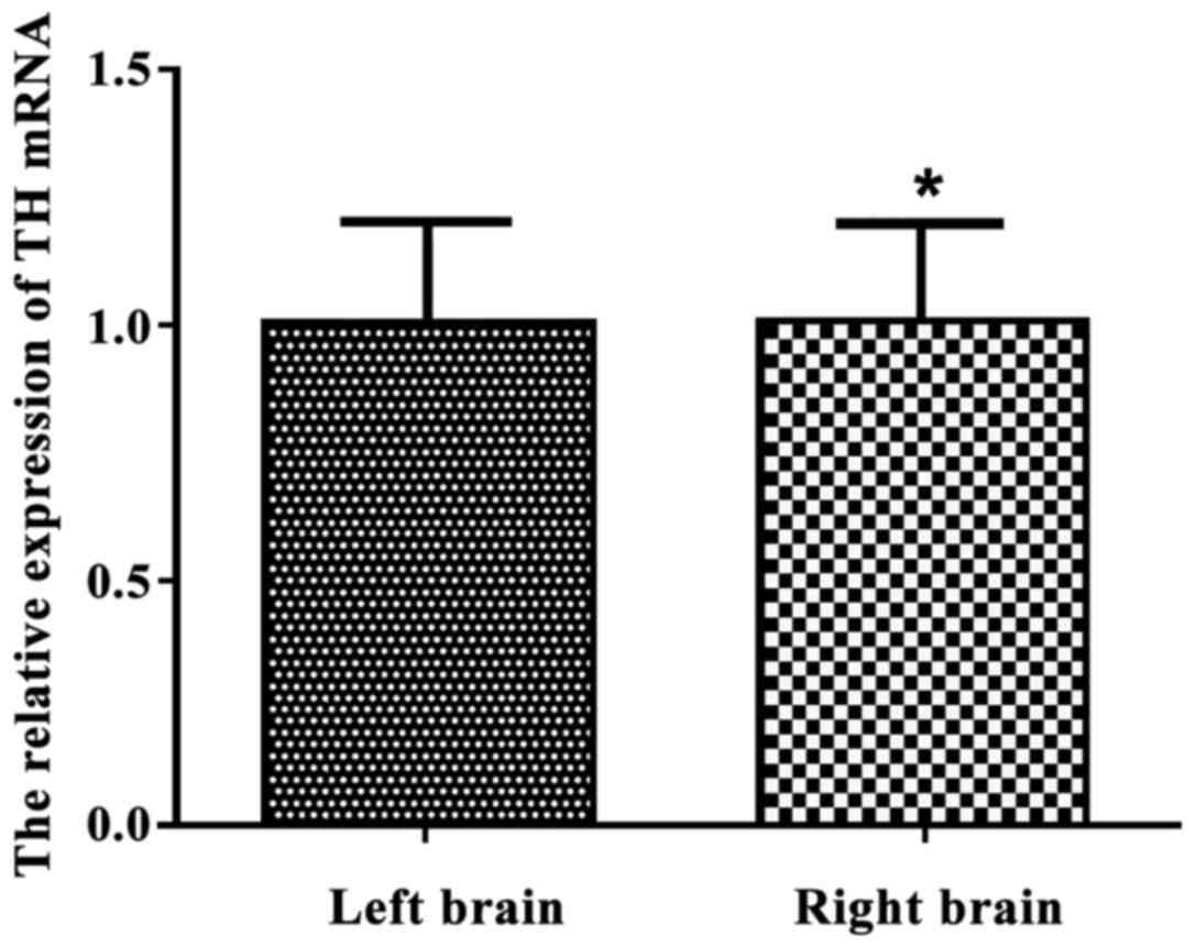
The relative expression level of TH
mRNA in both sides of brain tissues in normal rats
As shown in Fig. 1,
the relative expression of TH mRNA was respectively 1.039±0.112 and
0.956±0.120 in left and right brain tissues of normal rats. There
was no significant difference (p>0.05) between the two sides of
brain tissues in normal rats.
The success rates in each group of
modeling
With the extension of modeling time, the success
rate of modeling was significantly increased, and the success rate
of induction was close to the peak value at 6 weeks after
operation. The success rate was not significantly increased after
(p>0.05), as shown in Table
I.
 | Table I.Induction time of rats with different
induction times. |
Table I.
Induction time of rats with different
induction times.
|
|
| Modeling situation
(n, %) |
|---|
|
|
|
|
|---|
| Group | N | 2 weeks | 3 weeks | 6 weeks | 8 weeks |
|---|
| No. of >7 r/min
rats | 120 | 78 | 68 | 51 | 26 |
| Induction success
rate |
| 78/120 (65.0%) | 68/90
(75.5%)a | 51/60
(85%)a | 26/30
(86.7%)a |
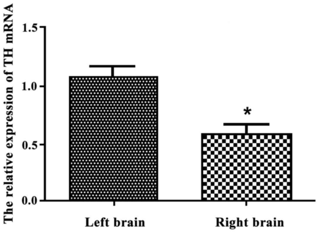
The relative expression of TH mRNA in
the left and right brain tissue of PD rats after operation for 3
weeks
For 3 weeks after operation, the relative expression
of TH mRNA in the left brain tissue of PD rats was more than the
right brain tissue, with 1.056±0.094 and 0.053±0.082, respectively.
There was statistically significant deference between the sides of
brain tissues (p<0.05), as shown in Fig. 2.
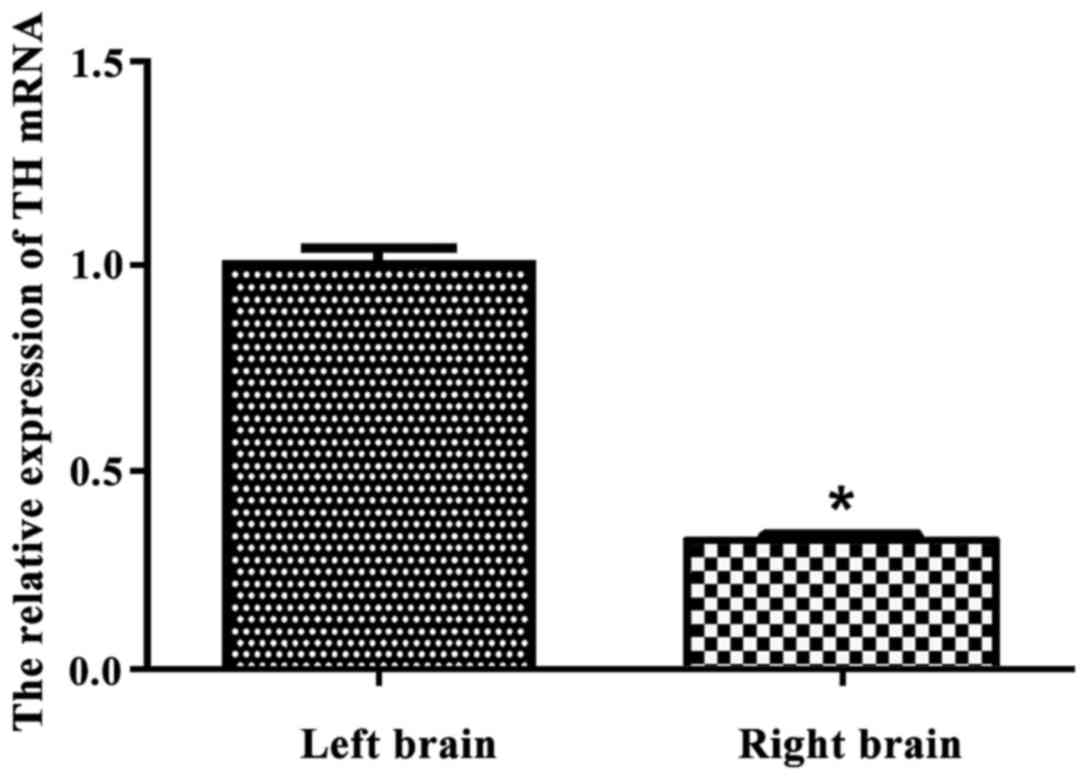
The relative expression of TH mRNA in
the left and right brain tissue of PD rats after operation for 6
weeks
As shown in Fig. 3,
the relative expression of TH mRNA in the left brain tissue of PD
rats was more than that in the right brain tissue for 6 weeks after
operation (p<0.05). The relative expression amount was
1.004±0.034 in left brain tissue and 0.316±0.012 in right brain
tissue.
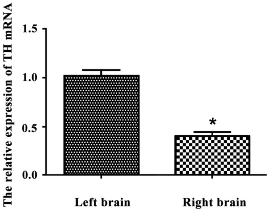
The relative expression of TH mRNA in
the left and right brain tissue of PD rats after operation for 8
weeks
As shown in Fig. 4,
the relative expression of TH mRNA was 1.021±0.0578 in the left
brain tissue and 0.395±0.041 in the right brain tissue in the PD
rats. The expression of TH in left brain tissue was significantly
more than that in the right brain tissue (p<0.05).
Comparison of relative expression of
TH mRNA in both sides of brain tissue
The preoperative expression of TH mRNA had no
significant difference in the sides of the brain tissue (p>0.05)
by the analysis of variance. Comparatively, the expression of TH
mRNA was significantly different between the sides of brain tissue
after model establishment (F=11.64, p=0.001, F=7.48, p=0.021,
F=9.59, p=0.016), with the expression decreased relatively. After 8
weeks of operation, the TH mRNA expression in damaged side of the
brain tissue was somewhat increased, but it was still lower than
the normal side (p<0.05) (Table
II).
| Table II.Comparison of the relative expression
of TH mRNA (mean ± SD). |
Table II.
Comparison of the relative expression
of TH mRNA (mean ± SD).
|
| Relative expression
of TH mRNA |
|---|
|
|
|
|---|
| Group | Preoperative
(n=30) | 3 weeks (n=30) | 6 weeks (n=30) | 8 weeks (n=30) |
|---|
| Left brain
(normal) | 1.039±0.112 | 1.056±0.094 | 1.004±0.034 | 1.021±0.057 |
| Right brain
(damage) | 0.956±0.120 |
0.530±0.082a,b |
0.316±0.012a,b |
0.395±0.041a,b |
| Between groups | F=11.32, p=0.001 |
| Different
time-points | F=7.12, p=0.025 |
| Between groups
different time-points | F=9.53, p=0.014 |
Discussion
PD is a common neurological degeneration in the
elderly, with limb tremor and activity disorders as the main
performance (8,9). PD is hidden, with slow progress, and
has no obvious movement disorders at early stage. As the disease
progress the patient may have symptoms such as static tremor,
muscle rigidity, and slowness of movement (10,11).
Although PD rarely endangers the lives of patients, it seriously
reduces the quality of life and also increased the burden on
patients on the family. So it is of great significance to improve
the therapeutic effect of patients. The most important pathological
changes in PD is the depletion of DA in striatum, and the
degeneration of dopaminergic neurons in substantia nigra is an
important reason for the reduction of striatum content. Studies
suggested that oxidative stress, environmental factors, aging and
genetic factors were all likely to be involved in the process of
denatured death of PD dopaminergic neurons (12). TH, as a key pathway for the catalytic
synthesis of catecholamines neurotransmitters, has an important
role in regulating the rate of dopamine synthesis and is a
rate-limiting enzyme for dopamine synthesis. The role of TH was
played through the mutual coordination between the catalytic
subregion and regulatory subregions, and then catecholamine
neurotransmitters were synthesized. The study (13,14)
showed that the expression of TH mRNA in substantia nigra of the
brain of PD was decreased, with the relationship to its gene
expression restriction. Therefore, if the expression change of TH
was well understood in the PD patients, it could provide a
theoretical basis for clinical treatment of PD.
In this study, fluorogenic quantitative PCR was used
to determine the relative expression of TH mRNA in the brain of
rats. It was found that the relative expression of TH mRNA in the
left and right brain tissue had no significant difference in normal
rats (p>0.05), with 1.039±0.112 and 0.956±0.120, respectively.
With the extension of modeling time, the success rate of model
establishment was significantly improved (p<0.05). The success
rate of modeling and the relative expression of TH mRNA reached the
peak value in the 6 weeks after operation. As the modeling time
increased, the success rate of modeling and the relative expression
of TH mRNA has not changed much compared to the rats at 6 weeks,
with a P-value <0.05. That was to say, after 6 weeks model
establishment, the model of PD rats was basically completed and met
the standard of PD model in rats. The relative expression of TH
mRNA in both sides of the brain tissue was significantly different
(p<0.05) and decreased. The expression of TH mRNA in the injured
brain tissue was significantly lower than that in normal rats. The
results showed that the relative expression of TH mRNA in brain was
of great significance in the pathogenesis of PD rats. With the
impaired brain tissue in PD rats, the relative expression of TH
mRNA also decreased. And the increase of the expression level after
8 weeks may be an experimental error, as the sample size is not big
enough. Similarly, Zhao et al also found that the expression
of TH in PD rats decreased compared to normal rats (15,16),
which may be the important reason for dopaminergic neurons
degeneration.
In conclusion, the decreased expression of TH mRNA
in injured brain tissue was lower than that in normal brain tissue,
which may be related to the occurrence and development of PD.
References
|
1
|
Yang QQ, Sun FL, Al HX, Zhang L and Wang
W: Assessment of 6-hydroxydopamine-lesion induced behavioral
alteration as a rat model of Parkinson's disease. Acta Neuropharm.
1:17–22. 2013.
|
|
2
|
Li J, Wang LN and Xiao HL: Effect of
electroacupuncture on oxidative stress injury of dopaminergic
neurons in rats with Parkinson's disease. Acupunct Res. 39:185–191.
2014.
|
|
3
|
Leem J: Acupuncture for motor symptom
improvement in Parkinson's disease and the potential identification
of respondersto acupuncture treatment. Integr Med Res. 5:332–335.
2006. View Article : Google Scholar
|
|
4
|
Shavali S, Combs CK and Ebadi M: Reactive
macrophages increase oxidative stress and alpha-synuclein nitration
during death of dopaminergic neuronal cells in co-culture:
Relevance to Parkinson's disease. Neurochem Res. 31:85–94. 2006.
View Article : Google Scholar : PubMed/NCBI
|
|
5
|
Feve AP: Current status of tyrosine
hydroxylase in management of Parkinson's disease. CNS Neurol Disord
Drug Targets. 11:450–455. 2012. View Article : Google Scholar : PubMed/NCBI
|
|
6
|
Meng XJ, Pang Q, Ding F, Xin T and Yang
HA: Establishment of PD rat model by injecting Taclo sterically and
directionally in to striatum. J ShandongUniv. 52:16–18. 2014.(In
Chinese).
|
|
7
|
Kashkin VA, Shekunova EV, Makarova MN and
Makarov VG: A study of combination treatment with nacom (levodopa +
carbodope) and citicoline in the model of Parkinson disease inrats.
Zh Nevrol Psikhiatr Im S S Korsakova. 117:59–63. 2017.(In Russian).
View Article : Google Scholar : PubMed/NCBI
|
|
8
|
Roche M, Frison G, Brion JD, Provot O,
Hamze A and Alami M: Csp2-N bond formation via
ligand-free Pd-catalyzed oxidative coupling reaction of
N-tosylhydrazones and indole derivatives. J Org Chem.
78:8485–8495. 2013. View Article : Google Scholar : PubMed/NCBI
|
|
9
|
Nguyen Anh TN, Benatmane N, Fallahi V,
Fang Y, Mohseni SM, Dumas RK and Åkerman J:
[Co/Pd]4-Co-Pd-NiFe spring magnets with highly tunable
and uniform magnetization tilt angles. J Magn Magn Mater.
324:3929–3932. 2012. View Article : Google Scholar
|
|
10
|
McCabe K, Concannon RM, McKernan DP and
Dowd E: Time-course of striatal Toll-like receptor expression
inneurotoxic, environmental and inflammatory rat models of
Parkinson's disease. J Neuroimmunol. 310:103–106. 2017. View Article : Google Scholar : PubMed/NCBI
|
|
11
|
Zheng LF, Zhang Y, Chen CL, Song J, Fan
RF, Cai QQ, Wang ZY and Zhu JX: Alterations in TH- and
ChAT-immunoreactive neurons in the DMV and gastric dysmotility in
an LPS-induced PD rat model. Auton Neurosci. 177:194–198. 2013.
View Article : Google Scholar : PubMed/NCBI
|
|
12
|
Michell AW, Tofaris GK, Gossage H, Tyers
P, Spillantini MG and Barker RA: The effect of truncated human
α-synuclein (1–120) on dopaminergic cells in a transgenic mouse
model of Parkinson's Disease. Cell Transplant. 16:461–474. 2007.
View Article : Google Scholar : PubMed/NCBI
|
|
13
|
Huang Y, Chang C, Zhang JW and Gao XQ:
Alternation of proteins in brain of Parkinson's disease model rats
after the transplantation of TH-NTN gene modified bone marrow
mesenchymal stem cells. Zhonghua Yi Xue Za Zhi. 92:2353–2356.
2012.(In Chinese). PubMed/NCBI
|
|
14
|
Ali F, Taresh S, Al-Nuzaily M, Mok PL,
Ismail A and Ahmad S: Stem cells differentiation and probing their
therapeutic applications in hematological disorders: A critical
review. Eur Rev Med Pharmacol Sci. 20:4390–4400. 2016.PubMed/NCBI
|
|
15
|
Thenral ST and Vanisree AJ: Peripheral
assessment of the genes AQP4, PBP and TH in patients with
Parkinson's disease. Neurochem Res. 37:512–515. 2012. View Article : Google Scholar : PubMed/NCBI
|
|
16
|
Zhao J: Effect of 3-n-butylphthalide on
the expression of Bax caspase-8 and TH in the substantia nigra of
PD rats. Chinese J Clin. 15:1288–1292. 2014.(In Chinese).
|