Introduction
Ovarian cancer represents one of the leading causes
of cancer-related mortality, with an increasing prevalence in Japan
(1). The pathogenesis of ovarian
cancer is complex, and is affected by numerous epigenetic and
genetic factors (2). Endometriosis
is usually a benign disorder, but is associated with an increased
risk for developing ovarian (3) and
endometrial cancers (4).
Endometrioid and clear cell carcinoma of the ovary
(endometriosis-associated ovarian cancer, EAOC) originates from
endometriosis.
Magnetic resonance (MR) imaging may be used as an
adjunctive method to distinguish EAOC from benign ovarian
endometrioma (OE). Specifically, we have recently demonstrated that
the MR transverse relaxation rate provides a noninvasive predictive
tool to discriminate between EAOC and OE (5). However, the implementation of MR
imaging in the outpatient clinic is often difficult.
Recent progress in research on the pathogenesis of
EAOC is based on key developments in two areas: i) New mechanistic
concepts regarding the pathogenesis of EAOC revealed a key role of
hemoglobin (Hb), heme and iron-induced oxidative stress in the OE
cystic fluid, wherein an imbalance between the overproduction of
iron-induced oxidative stress and defense mechanisms could trigger
DNA damage and carcinogenesis; and ii) the establishment of novel
approaches to identify stress biomarkers, such as Hb and iron, for
the prediction of malignant transformation (2,6,7). A recent ex vivo study revealed
that electronic absorption spectroscopy using visible light at 580
and 620 nm provides a measure of Hb species in endometriotic cystic
fluid by monitoring the relative concentrations of oxyhemoglobin
and methemoglobin, respectively (8).
The 620/580 nm peak ratio of cystic fluid in EAOC patients was
significantly lower compared with that measured in women with
benign OE (8). Therefore, the cystic
fluid Hb species may be used as biomarkers in the differential
diagnosis between EAOC and OE. However, one limitation of this
absorption-based method is that the camera must be placed opposite
a halogen white light source, which is disadvantageous as light in
the visible spectrum does not penetrate tissue.
Recent advances in optical technology have led to
innovative quantitative monitoring tools, which include
spatially-resolved reflectance, diffuse optical spectroscopy,
diffuse optical tomography and diffuse correlation spectroscopy
(9,10). Optical spectroscopy and tomography
utilize the near-infrared spectral region to provide quantitative
determination of several important biological chromophores, such as
Hb (11) or cytochrome c
oxidase (12). Furthermore, light in
the near-infrared spectrum efficiently penetrates tissue, including
bone and muscle (11). When
near-infrared light is shone through cystic fluid, the influence of
photon attenuation and scattering varies depending on Hb
concentration. A backscattered photon or an on-axis luminance
measurement can be detected by means of appropriate optical
apparatus. We hypothesized that the changes in luminance across a
fluid can typically be used to determine the Hb concentration of
the cystic fluid using a near-infrared sensor camera. Thus, we
developed a rapid and sensitive ex vivo assay based on the
changes of dynamic light scattering or changes in luminance across
the cystic fluid. Furthermore, we investigated the potential of the
luminance measurement as an objective optical method to
discriminate EAOC from benign OE.
Materials and methods
Study population
The research protocol was approved by the Nara
Medical University Review Boards, and written informed consent was
obtained from all subjects. A total of 46 patients with OE (n=34)
or EAOC (n=12) were recruited between February 2013 and January
2015 at the Department of Gynecology, Nara Medical University
Hospital (Kashihara, Japan). Histopathological examination
confirmed the diagnoses of benign OE and EAOC. All cystic fluid
samples were collected from the patients during surgery, and an
aliquot of each sample was stored at −80°C until testing.
Instrumentation and system design
Narrow-band optical filtering is required to achieve
the highest signal-to-noise ratio (SNR) as an optical enhancement
technology. During our preliminary study, the SNR was determined
analytically using a band-path filter with varying wavelengths
(750–1,000 nm), and the results were validated experimentally. We
found a single optimum for the optical path length of the filter at
800 nm (data not shown). This was applied to in the current
system.
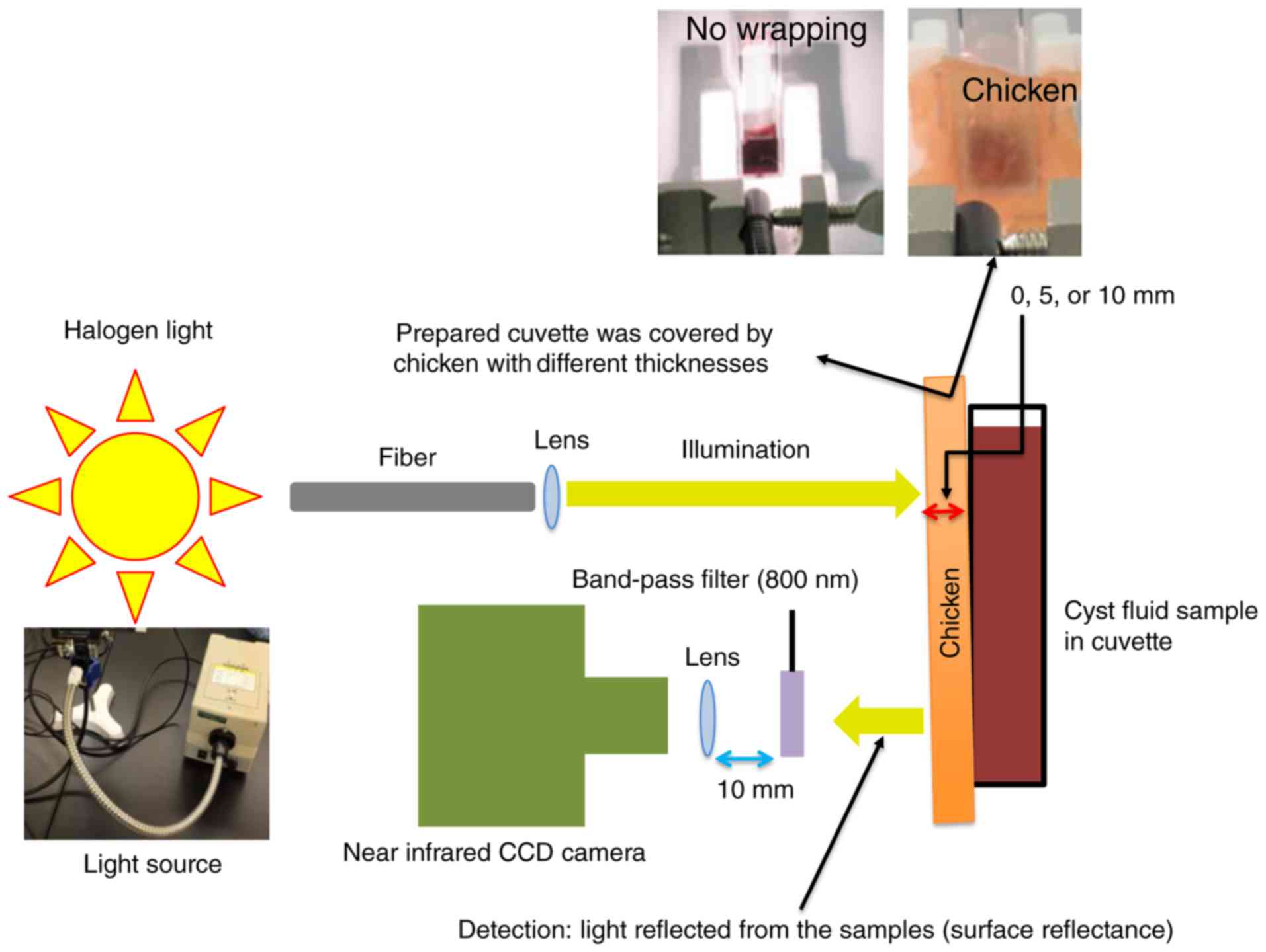
The instrument was designed and set up. A diagram of
the light path is illustrated in Fig.
1. This figure indicates the luminance (light reflected from
the sample) measurement of the cystic fluid sample. Measurements
were obtained by performing ex vivo phantom experiments. An
aliquot of the cystic fluid sample (1 ml) was transferred to a
disposable cuvette (10-mm wide × 10-mm thick). The light source was
a halogen lamp with a 300 W quartz-tungsten-halogen bulb (EXR 82v;
Eiko, Co., Ltd., Hitachinaka, Japan). The distance between the
halogen light and the cystic fluid was 50 mm. Halogen light
illuminated the sample and a near-infrared CCD camera with
band-path filter (800 nm) recorded the light signal reflected from
the sample. The change in luminance [Δl value (cd/m2)]
was calculated by subtracting a sample blank for each specimen (Δl
= background luminance - cystic fluid luminance at 800 nm).
Hb assay
Cystic fluid total Hb concentrations were measured
as described previously (6,8,13). From
the correlation data, a formula was calculated to convert heme
levels (mg/l) to hemoglobin (g/dl). We then investigated the
correlation between Δl and Hb in cystic fluid.
Effects of an anatomical barrier on
surface reflectance
Transvaginal ultrasound imaging is replacing
radiological methods in the investigation of ovarian tumors. Due to
the presence of an anatomical barrier (which may include an ovarian
cyst wall or vaginal connective/muscle tissue) in this type of
imaging, ex vivo experiments using appropriate modeling are
important to establish a clinically relevant model. To investigate
the effect of such an anatomical barrier, the surface of a
disposable cuvette was covered with pieces of commercial Japanese
chicken of different thicknesses (5 and 10 mm). The Δl value was
measured by recording the light scattered (surface luminance) from
the cystic fluid that was covered by these barriers. We generated
three sets of experiments: Experiment 1 (0 mm; the surface of the
cuvette was not covered); experiment 2 (5 mm; the surface of the
cuvette was covered by a 5-mm-thick piece of chicken); and
experiment 3 (10 mm; the surface of the cuvette was covered by a
10-mm-thick piece of chicken).
Statistical analysis
Statistical analysis was conducted using the SPSS
22.0 software package (IBM Corp., Armonk, NY, USA). Comparisons of
non-parametric data (Δl and Hb levels) between the OE and EAOC
groups were performed using the Mann-Whitney U test. Correlation
analysis was performed using Pearson's correlation coefficient. The
optimal cutoff value was defined according to analysis of the
receiver operating characteristic (ROC) curve. The sensitivity and
specificity of detection were calculated on the basis of cutoff
value to differentiate EAOC from benign OE. The area under the ROC
curve (AUC) was also calculated for each marker. P<0.05 was
considered to indicate a statistically significant difference.
Results
Δl and Hb of cystic fluid samples
The clinical characteristics, cystic fluid Δl levels
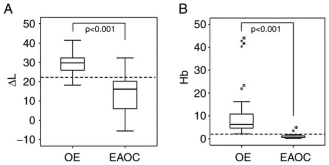
and Hb concentrations of patients are summarized in Table I. Subjects in the EAOC group were
older compared with the OE group (P<0.001). Fig. 2 shows box and whisker plots
representing the median level and interquartile range (box) of Δl
and Hb for each studied group. The EAOC patients showed
significantly lower Δl values compared with the OE group
(P<0.001) (Table I; Fig. 2A). The cystic fluid levels of Hb were
also significantly lower in EAOC patients compared with OE patients
(Table I; Fig. 2B). These results indicated that the
OE and EAOC groups were clearly separated.
 | Table I.Patient demographics and tumor
characteristics of two groups. |
Table I.
Patient demographics and tumor
characteristics of two groups.
| Patient and clinical
characteristics | OE | EAOC | P-value |
|---|
| Number | 34 | 12 |
|
| Age (years) |
|
| <0.001 |
| Median
(range) | 39.0 (26–51) | 49.5 (36–69) |
|
| Mean ±
SD | 38±7 | 49±11 |
|
| Cyst size
(cm)a |
|
| 0.022 |
| Median
(range) | 7.0 (2.7–19.3) | 11.0 (4.2–22.5) |
|
| Mean ±
SD | 7.7±3.2 | 12.1±5.7 |
|
| FIGO
stage | – | Ia (n=5), Ib (n=1),
Ic (n=6) |
|
|
Pathology | Endometriosis | Clear cell carcinoma
(n=6) |
|
|
|
| Endometrioid
carcinoma (n=3) |
|
|
|
| Mucinous carcinoma
(n=1) |
|
|
|
| Serous carcinoma
(n=1) |
|
|
|
| Seromucinous
carcinoma (n=1) |
|
| Δl
(cd/m2) |
|
| <0.001 |
| Median
(range) | 29.0 (18.2–41.4) | 16.2 (−5.5–32.3) |
|
| Mean ±
SD | 29.6±5.0 | 14.4±10.8 |
|
| Hb (g/dl) |
|
| <0.001 |
| Median
(range) | 6.1 (2.2–42.8) | 0.77 (0.2–3.5) |
|
| Mean ±
SD | 9.5±9.3 | 1.1±0.9 |
|
ROC curve in EAOC group vs. benign OE
group
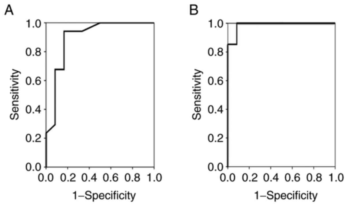
The sensitivity and specificity of cystic fluid Δl
level for the diagnosis of malignant transformation were 83.3 and
94.1%, respectively, using a cutoff value of 21. The AUC was 0.897
(Fig. 3A; Table II, experiment 1). A Hb level of 1.99
g/dl was identified to detect EAOC with a sensitivity of 100% and a
specificity of 91.7%, and an AUC of 0.988 (Fig. 3B). Since the patients with EAOC were
significantly older than those with OE, correlations between age
and each parameter were evaluated using Pearson's correlation
coefficient. The age distribution of the subjects is shown in
Fig. 4. There were no significant
correlations between age and cystic fluid Δl (Fig. 4A; r=−0.128, P=0.470) or Hb level
(Fig. 4B; r=−0.159, P=0.370) in the
OE group. There were also no correlations between age and cystic
fluid Δl (Fig. 4A; r=0.518, P=0.084)
or Hb level (Fig. 4B; r=0.532,
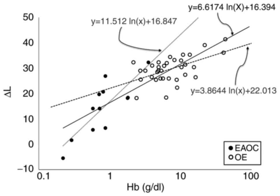
P=0.075) in the EAOC group. Fig. 5
shows a scatter plot of correlation between Δl and cystic fluid Hb
level for the OE and the EAOC groups. When the interaction between
Δl and Hb was analyzed using a linear model, the best model fitted
an exponential function. Therefore, data were linearized by log
transformation. The association of Δl with Hb became steeper with
lower Hb levels (<2 g/dl). Δl was strongly correlated with the
cystic fluid Hb concentration (r=0.558, P<0.001).
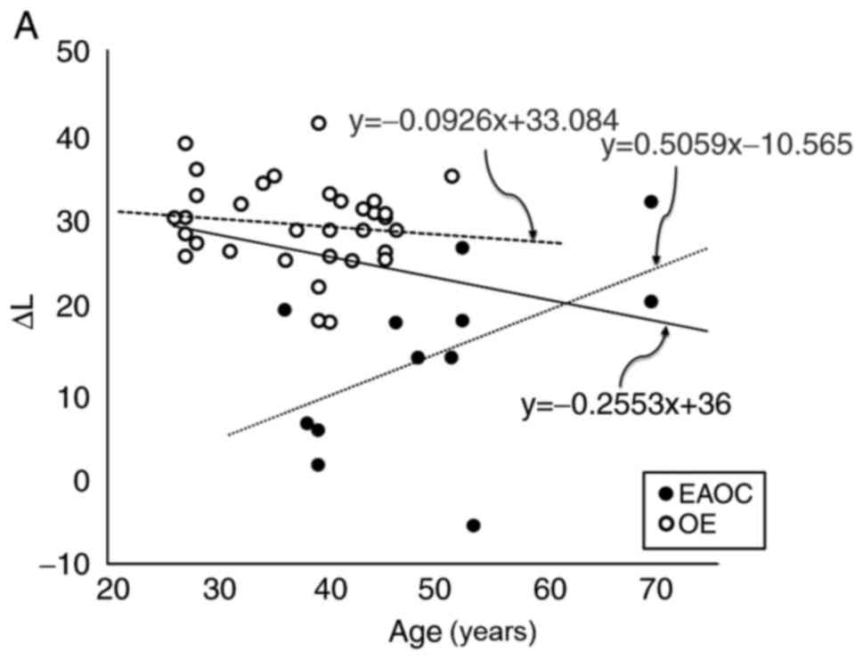 | Figure 4.Cystic fluid (A) Δl and (B) Hb levels
per age for subjects in the OE and EAOC samples. Open circle
represents an individual value of OE subject. Closed circle
represents an individual value of EAOC subject. (A) Correlation
between cystic fluid Δl levels and age at surgery in women with OE
(y = −0.0926× + 33.084, r=−0.128, P=0.470) and in patient with EAOC
(y = 0.5059× - 10.565, r=0.518, P=0.084,). x, age at surgery; y,
cystic fluid Δl level (cd/m2). (B) Correlation between
cystic fluid Hb levels and age at surgery in women with OE (y =
−0.2087× + 17.351, r=−0.159, P=0.370) and in patient with EAOC (y =
0.0443× - 1.1211, r=0.532, P=0.075,). x, age at surgery; y, cystic
fluid Hb level (mg/l). |
| Table II.Effects of an anatomical barrier
against surface reflectance. |
Table II.
Effects of an anatomical barrier
against surface reflectance.
| Patients | OE (n=34) | EAOC (n=12) | AUC | 95% CI | P-value | Cut-off | Sensitivity (%) | Specificity (%) | PPV (%) | NPV (%) |
|---|
| Experiment 1 (Samples
not-covered by a chicken) Δl (cd/m2) | 29.6±5.0 | 14.4±10.8 | 0.897 | 0.772–1.000 | <0.001 | 21.5 | 83.3 | 94.1 | 83.3 | 94.1 |
|
| (18.2–41.4) | (−5.5–32.3) |
|
|
|
|
|
|
|
|
| Experiment 2 (Samples
covered by a 5 mm-thick chicken) Δl (cd/m2) | 2.1±10.2 | −9.7±7.6 | 0.859 | 0.733–0.985 | <0.001 | −5.0 | 75.0 | 91.2 | 75.0 | 91.2 |
|
| (−16.6–49.1) | (−19.8–2.7) |
|
|
|
|
|
|
|
|
| Experiment 3 (Samples
covered by a 10 mm-thick chicken) Δl (cd/m2) | −9.9±5.1 | −17.6±7.6 | 0.778 | 0.609–0.948 | 0.005 | −15.5 | 66.7 | 85.3 | 61.5 | 87.9 |
|
| (−20.1–2.0) | (−26.3–6.3) |
|
|
|
|
|
|
|
|
Effects of an anatomical barrier on
surface reflectance
The results of the ex vivo approach are
summarized in Table II, which shows
the comparison of measurements obtained from each experiment. The
AUC for diagnosing EAOC from OE was 0.897 (experiment 1:
Sensitivity, 83.3%; specificity, 94.1%), 0.859 (experiment 2:
Sensitivity, 75.0%; specificity, 91.2%) and 0.778 (experiment 3:
Sensitivity, 66.7%; specificity, 85.3%).
Discussion
To the best of our knowledge, this is the first
ex vivo study of cystic fluid measurements via optical
properties at 800 nm, which can discriminate malignant
transformation from benign OE. The Δl of cystic fluid from the EAOC
group was significantly lower compared with that of the OE group
(P<0.001). Δl level could serve as a simple, rapid and accurate
method to discriminate EAOC from benign OE, with high sensitivity
(83.3%) and specificity (94.1%). In our ex vivo experiments,
the samples were covered by 5- or 10 mm-thick pieces of chicken.
Our measurements showed that the 10 mm-thick sample attenuated the
power of ∆L to discriminate between benign and malignant specimens,
with relatively lower sensitivity (66.7%) and specificity
(85.3%).
Furthermore, the cystic fluid Hb concentrations were
reduced in patients with EAOC (6–8). The Δl
values and total Hb concentrations in 46 samples exhibited an
exponential correlation (r=0.558), suggesting that the Δl value may
reflect the Hb concentration. The present results were in agreement
with those of Yoshimoto et al (6), who reported the cystic fluid
concentration of total iron, heme iron, free iron and Hb species
(6,7). Previous studies of Hb species have
reported differences between OE and EAOC samples (8). Transvaginal ultrasound-guided luminance
measurements using near-infrared approaches may advance medical
imaging technology as a tool for discriminating malignant
transformation in endometriosis.
Despite the advantages discussed above, there are
several limitations in the present study. Firstly, an exponential
curve of the Hb levels was a better-fitting model compared with the
linear model. However, whether and how the Δl level reflects
absolute Hb concentration has not yet been studied. We could not
exclude the possibility of cross-contamination of other factors,
such as heme iron and free iron, in these data acquired at an
800-nm wavelength. Secondly, a major limitation is the lack of
large-scale evaluation. Finally, the complexity of reproductive
organ anatomy poses several challenges for in vivo luminance
imaging. Non-invasive imaging in deep tissue requires a
near-infrared CCD camera with strong sensitivity and high spatial
resolution. By adding detectors at multiple distances from the
emitted light source, specific algorithms can subtract superficial
light absorption from deep absorption to provide qualitative
information of the cystic fluid Hb level (11). Despite these limitations, there is a
great need to develop a clinically useful, noninvasive and reliable
tool that accurately predicts the malignant transformation of
endometriosis.
In conclusion, the luminance value obtained from
ex vivo cystic fluid samples at an 800-nm wavelength may
discriminate EAOC from benign OE patients. Transvaginal
near-infrared approaches may provide a non-invasive assessment of
malignant transformation of OE, and may have further clinical
applications in an outpatient setting.
The aim of this study was to investigate the
discrimination of malignant transformation from OE using a
near-infrared approach ex vivo. The diagnostic sensitivity
and specificity for Δl (change in luminance, cd/m2) were
83.3 and 94.1%, respectively, at the cutoff value of 21.5
cd/m2, with an AUC of 0.897. This ex vivo study
potentially provides a powerful near-infrared approach for
discrimination between EAOC and benign OE, with high sensitivity
and specificity. This study provides a basis for developing future
clinical approaches.
Acknowledgements
This study was supported by grant-in-aid for
Scientific Research from the Ministry of Education, Science, and
Culture of Japan (H.K.).
References
|
1
|
Ushijima K: Current status of gynecologic
cancer in Japan. J Gynecol Oncol. 20:67–71. 2009. View Article : Google Scholar : PubMed/NCBI
|
|
2
|
Koshiyama M, Matsumura N and Konishi I:
Recent concepts of ovarian carcinogenesis: Type I and type II.
Biomed Res Int. 2014:9342612014. View Article : Google Scholar : PubMed/NCBI
|
|
3
|
Brinton LA, Gridley G, Persson I, Baron J
and Bergqvist A: Cancer risk after a hospital discharge diagnosis
of endometriosis. Am J Obstet Gynecol. 176:572–579. 1997.
View Article : Google Scholar : PubMed/NCBI
|
|
4
|
Yu HC, Lin CY, Chang WC, Shen BJ, Chang WP
and Chuang CM; Task Force on Carcinogenesis of Endometrial Cancer,
: Increased association between endometriosis and endometrial
cancer: A nationwide population-based retrospective cohort study.
Int J Gynecol Cancer. 25:447–452. 2015. View Article : Google Scholar : PubMed/NCBI
|
|
5
|
Yoshimoto C, Takahama J, Iwabuchi T,
Uchikoshi M, Shigetomi H and Kobayashi H: Transverse relaxation
rate of cyst fluid can predict malignant transformation of ovarian
endometriosis. Magn Reson Med Sci. 16:137–145. 2017. View Article : Google Scholar : PubMed/NCBI
|
|
6
|
Yoshimoto C, Iwabuchi T, Shigetomi H and
Kobayashi H: Cyst fluid iron-related compounds as useful markers to
distinguish malignant transformation from benign endometriotic
cysts. Cancer Biomark. 15:493–499. 2015. View Article : Google Scholar : PubMed/NCBI
|
|
7
|
Iwabuchi T, Yoshimoto C, Shigetomi H and
Kobayashi H: Oxidative stress and antioxidant defense in
endometriosis and its malignant transformation. Oxid Med Cell
Longev. 2015:8485952015. View Article : Google Scholar : PubMed/NCBI
|
|
8
|
Iwabuchi T, Yoshimoto C, Shigetomi H and
Kobayashi H: Cyst fluid hemoglobin species in endometriosis and its
malignant transformation: The role of metallobiology. Oncol Lett.
11:3384–3388. 2016. View Article : Google Scholar : PubMed/NCBI
|
|
9
|
Cuccia DJ, Abookasis D, Frostig RD and
Tromberg BJ: Quantitative in vivo imaging of tissue absorption,
scattering, and hemoglobin concentration in rat cortex using
spatially modulated structured lightIn vivo optical imaging of
brain function. Frostig RD: 2nd. Boca Raton (FL): CRC Press/Taylor
& Francis; 2009, View Article : Google Scholar
|
|
10
|
Wilson JR, Mancini DM, McCully K, Ferraro
N, Lanoce V and Chance B: Noninvasive detection of skeletal muscle
underperfusion with near-infrared spectroscopy in patients with
heart failure. Circulation. 80:1668–1674. 1989. View Article : Google Scholar : PubMed/NCBI
|
|
11
|
Steppan J and Hogue CW Jr: Cerebral and
tissue oximetry. Best Pract Res Clin Anaesthesiol. 28:429–439.
2014. View Article : Google Scholar : PubMed/NCBI
|
|
12
|
Kolyva C, Ghosh A, Tachtsidis I, Highton
D, Smith M and Elwell CE: Dependence on NIRS source-detector
spacing of cytochrome c oxidase response to hypoxia and hypercapnia
in the adult brain. Adv Exp Med Biol. 789:353–359. 2013. View Article : Google Scholar : PubMed/NCBI
|
|
13
|
Kim YJ, Kim S, Kim JW and Yoon G: Data
preprocessing and partial least squares regression analysis for
reagentless determination of hemoglobin concentrations using
conventional and total transmission spectroscopy. J Biomed Opt.
6:177–182. 2001. View Article : Google Scholar : PubMed/NCBI
|