Introduction
Nasal polyps are chronic inflammatory lesions
located in the nasal cavity or sinus mucosa. Previous studies have
classified nasal polyposis as a subtype or progressive stage of
chronic sinusitis (1–3). Epidemiological surveys demonstrated
that 2–4% of the global population was affected by nasal polyposis
in 2003 (4–6). Nasal polyposis poses a threat to the
health and quality of life of patients, particularly in pediatric
patients (7,8). Nasal polyposis in pediatric patients
may induce constriction of the nasal cavity and nasal meatus, which
may cause difficulties in postoperative cleanup and increase the
susceptibility of patients to respiratory infections (9).
Among the three isoforms of transforming growth
factor β (TGF-β), TGF-β1 is the isoform predominantly expressed in
nasal polyposis (10). TGF-β1
primarily originates from inflammatory cells, including
eosinophils, macrophages and epithelial cells (11). TGF-β1 serves a key role in cell
biological behaviors, including extracellular matrix formation and
fibrosis, epithelial cell metaplasia, vascularization, immune
hypersensitivity, embryonic development, cell senescence and
apoptosis (12). Among these
functions, promoting the formation of the extracellular matrix is
the most prominent feature of TGF-β1 (13,14).
TGF-β1 is one of the important signaling molecules secreted by
inflammatory cells during inflammation (15,16).
However, the role of TGF-β1 in the inflammation and regulatory
mechanisms of pathogenesis in nasal polyposis in children remains
unknown. It has been reported that microRNA-663 (miR-663) is an
upstream regulator of TGF-β1 and inhibits the development of
papillary thyroid carcinoma and liver cancer through regulating
TGF-β1 (17–18). However, whether miR-663 regulates
TGF-β1 in nasal polyposis of children is unknown.
The aim of the present study was to measure the
expression of TGF-β1 mRNA and protein in nasal polyp tissue, serum
and peripheral blood eosinophils using reverse
transcription-quantitative polymerase chain reaction, western
blotting, bioinformatics prediction analysis and ELISA in pediatric
patients with nasal polyposis. Additionally, the association
between the expression of TGF-β1 and miR-663 was verified in order
to evaluate their roles in the pathogenesis of nasal polyposis in
children.
Patients and methods
Participant recruitment and selection
criteria
Nasal polyp tissue, serum and peripheral blood
eosinophils were collected from 35 pediatric patients with nasal
polyposis who had undergone nasal polypectomy in the Department of
Otolaryngology in the Women and Children's Hospital of Qingdao
(Qingdao, China) between August 2013 and August 2015. These
patients were part of the patient group and there were 16 males and
19 females with a median age of 11 years (age range, 7–14 years).
The inclusion criteria and exclusion criteria for patients were as
previously described (19). The
diagnosis of nasal polyposis in all patients was confirmed by
pathologists. The inferior nasal concha, serum and peripheral blood
eosinophils were collected from 46 patients with nasal septal
deviation complicated by inferior turbinate hypertrophy or patients
with simple inferior turbinate hypertrophy undergoing surgical
removal of inferior nasal concha. These patients were part of the
control group and there were 18 males and 28 females with a median
age of 10 years (age range, 6–14 years). Patients in the two groups
were excluded if they were diagnosed with infection, diabetes,
cancer, or autoimmune disease, or exhibited complications involving
the heart, liver and kidney. Prior written informed consent was
obtained from the patients' families and the present study was
approved by the Ethics Review Board of the Women and Children's
Hospital of Qingdao (Qingdao, China).
Reagents and antibodies
The miRcute miRNA Isolation kit, miRcute miRNA cDNA
first strand synthesis kit, miRcute miRNA quantitative fluorescence
detection kit (FP401), SuperReal PreMix (SYBR Green; FP204) and
TIANScript II cDNA first strand synthesis kit (KR107) were all
purchased from Tiangen Biotech Co., Ltd. (Beijing, China). The
rabbit anti-human TGF-β1 antibody (ab92486) and rabbit anti-human
β-actin antibody (ab6276) were purchased from Abcam (Cambridge, MA,
USA). TRIzol reagent (10606ES60; Shanghai YISHENG Biology
Technology Co., Ltd., Shanghai, China), goat anti-rabbit secondary
antibody (ab6721; Abcam), BCA Protein assay kit (RTP7102, Beijing
Zhongke Ruitai, Beijing, China) and serum RNA extraction kit
miRNeasy Serum/Plasma kit (JL217184; Guangzhou Jianlun Biological
Technology Co., Ltd., Guangzhou, China) were also used in the
current study. A TGF-β1 ELISA kit (ab100674; Abcam), enhanced
chemiluminence (ECL) solution (FDO0142; Nanjing Aoduofuni
Biotechnology Co., Ltd., Nanjing, China), miR-663 mimics (Guangzhou
Ruibo Biological Technology Co., Ltd., Guangzhou, China) were
purchased. Anti-cluster of differentiation (CD)16 immunomagnetic
beads (YB-22355) were purchased from Yubo Biological Technology
Co., Ltd., (Shanghai, China). Ficoll-Hypaque separation medium
(Cedarlane Laboratories, Burlington, Canada) including Solution A
(12%, density: 1.090), Solution B (9.0%, density: 1.076) and
Solution C (6.4%, density: 1.065) was also used in the current
study.
Specimen collection
Nasal polyp tissues were collected during surgery
and frozen in liquid nitrogen. Peripheral blood was collected from
all patients and stored at −20°C and the serum was isolated by
centrifugation at 400 × g for 10 min at 4°C. Peripheral blood
eosinophils were isolated using Magnetic activated cell sorting
(MACS). Peripheral blood (100 µl) was collected and EDTA
anticoagulant (0.1 mol/l, pH 7.7) and a specific dye for
eosinophils (0.9 ml) were added. Eosinophils were then counted
using an ECLIPSE TS100 (Nikon Corporation, Tokyo, Japan) at a
magnification of ×100. The remaining blood was diluted with 6%
dextran (1:5) and placed in a water bath at 37°C for 40 min. The
supernatant was centrifuged at 600 × g at room temperature for 10
min and the leukocyte specimen was collected. Subsequently, the
leukocyte specimen was mixed with solutions A, B and C of the
Ficoll-Hypaque separation medium and centrifuged at 800 × g for 20
min at room temperature. Cell suspensions were divided into four
layers and the eosinophil layer was aspirated and washed twice with
PBS (0.01 mol/l). The total number of cells including eosinophils
was counted using an ECLIPSE TS100 (Nikon Corporation). Four fields
under ×100 magnification were randomly selected for cell counting.
Anti-CD16 MicroBeads (20 µl, pre-cooled at 4°C) were added to
1×10⁷ total cells, mixed well and incubated at 6–12°C
for 15 min. The cell mixture was applied twice to the MACS column
(Miltenyibiotec, Bergisch Gladbach, Germany). The magnetically
anti-CD16 labeled cells (neutrophils) were arrested in the
separation column while unlabeled cells (eosinophils) passed
through the column. Isolated eosinophils were collected and washed
with PBS (0.01 mol/l). Cells were incubated in an atmosphere
containing 5% CO2 at 37°C. The eosinophils were counted
at every step and stained with Trypan blue at room temperature for
3 min to evaluate cell activity.
Reverse transcription-quantitative
polymerase chain reaction (RT-qPCR)
Frozen tissues (pulverized in liquid nitrogen) were
lysed thoroughly using 1 ml TRIzol reagent and total RNA was
extracted using the phenol chloroform method (20). Total RNA was extracted using the
serum RNA extraction kit miRNeasy Serum/Plasma kit. Gel
electrophoresis and spectrophotometry were conducted to measure the
optical density (OD) 260/280 ratio and measure the concentration
and quality of RNA. Total RNA (1 µg) was reverse transcribed into
cDNA using the TIANScript II cDNA first strand synthesis kit
according to the manufacturer's protocols. The SuperReal PreMix
(SYBR Green) was used during qPCR with the following primers:
TGF-β1, forward, 5′-GGACACCAACTATTGCTTCAG-3′ 160 base pairs (bp)
and reverse, 5′-TCCAGACTCCAAATGTAG-3′; β-actin, forward,
5′-TTCCAGCCTTCCTTCCTGG-3′ 224 bp and reverse,
5′-TTGCGCTCAGGAGGAGGAAT-3′. The reaction conditions of qPCR were as
follows: Pre-denaturation at 95°C for 2 min, 30 cycles of
denaturation at 94°C for 45 sec, annealing at 55°C for 55 sec,
extension at 72°C for 1 min and a final cycle at 72°C for 10 min.
Relative quantification of TGF-β1 was calculated as the ratio of
TGF-β1/β-actin using the 2−ΔΔCq method (21).
The miRcute miRNA Isolation kit was used to isolate
miR-663 from cells. An miRcute miRNA cDNA first strand synthesis
kit was used to obtain cDNA. To detect miR-663, the miRcute miRNA
quantitative fluorescence detection kit was used and U6 was the
internal control. The primers used were as follows: U6, forward,
5′-CTCGCTTCGGCAGCACA-3′ and reverse, 5′-AACGCTTCACGAATTTGCGT-3′;
miR-663, forward, 5′-TGCGGAGGCGGGGCGCCGCGGG-3′ and reverse,
5′-CCAGTGCAGGGTCCGAGGT-3′. The reaction conditions were as follows:
Pre-denaturation at 95°C for 3 min, 40 cycles of denaturation at
94°C for 30 sec, annealing at 56°C for 30 sec and extension at 72°C
for 20 sec. The ratio of miR-663/U6 was calculated using the
2−ΔΔCq method (21).
Western blotting
The total proteins were extracted using a protein
extraction kit (310003; BestBio Company, Shanghai, China) following
the manufacturers' instructions. The concentration of protein was
measured using the BCA protein assay kit. Protein samples (30
µg/lane) were analyzed using 10% SDS-polyacrylamide gel
electrophoresis and transferred to a PVDF membrane. The membrane
was then blocked with 5% non-fat milk for 1 h at room temperature.
Primary antibodies against TGF-β1 (1:500) and β-actin (1:5,000)
were added and incubated at 4°C overnight. Goat anti-rabbit
secondary antibody (1:3,000; cat no. ab6721; Abcam) were added and
incubated at room temperature for 1 h. The PVDF membrane was placed
in the ECL detection system and imaged using Image Lab Software
version 3.0 (Bio-Rad Laboratories, Inc., Hercules, CA, USA) to
acquire and analyze imaging signals of protein bands. The relative
density of target protein was defined as the ratio of gray values
between the target protein and β-actin.
ELISA
The TGF-β1 ELISA kit was used to perform ELISA.
ELISA plates were classified into standard, serum and blank wells.
Standard samples (50 µl) and serum (10 and 40 µl sample diluent)
were added to the wells, whereas no solution was added to blank
wells. Subsequently, horseradish peroxidase-labeled goat
anti-rabbit secondary antibody (cat no. ab6721; Abcam; 100 µl) was
added to each well and incubated at 37°C for 1 h. The plates were
then washed 5 times and substrates A (50 µl) and B (50 µl),
respectively, were added. Following incubation for 15 min at 37°C,
stop solution (50 µl) was added. The absorbance value at OD 450 nm
was measured within 15 min on the Multiskan FC microplate reader
(Thermo Scientific Inc., Waltham, MA, USA).
Bioinformatics predictions of
upstream-regulatory miR for TGF-β1
Bioinformatics prediction is one of the major
approaches for functional studies of miR. To further determine the
regulatory mechanism of upstream miRs in the pathogenesis of nasal
polyposis, target gene prediction software, including miRanda
(http://www.microma.org/rnicroma/home.do), TargetScan
2.0 (www.targetscan.org) and PicTar
(http://pictar.mdc-berlin.de/) were used
to identify the potential genes that have regulatory effects on
TGF-β1. The procedures were performed according to the instructions
on each website.
Dual-luciferase reporter assay
The wild-type and mutant type 3′-untranslated region
(UTR) in TGF-β1 were chemically synthesized in vitro. The
wild-type and mutant type 3′-UTR were added to the restriction
enzyme cleavage sites of SpeI and HindIII and cloned
into a pMIR-REPORT luciferase reporter plasmid (cat no. AM5795;
Ambion; Thermo Fisher Scientific, Inc.). 293T cells (Cobioer
Biosciences Co., Ltd., Nanjing, China) were co-transfected with
plasmid with wild type 3′-UTR (0.8 µg) and miR-663 mimics (100 nM),
or co-transfected with mutant type 3′-UTR (0.8 µg) and miR-663
mimics (100 nM) (synthesized by Sangon Biotech Co., Ltd., Shanghai,
China). 293T cells transfected with empty plasmid were considered
to be negative controls. Cells were incubated at 37°C for 24 h and
then lysed. Fluorescence was measured using a GloMax®
20/20 luminometer according to the instructions of the dual
luciferase reporter system kit (Promega Corporation, Madison, WI,
USA). Renilla fluorescent activity was considered as an
internal reference.
Statistical analysis
All data were analyzed using SPSS 18.0 software
(SPSS, Inc., Chicago, IL, USA). Measurement data were expressed as
mean ± standard deviation. Normality tests were conducted for all
data and a T test was used for comparison between two independent
groups. One-way analysis of variance (ANOVA) was used to compare
data of more than two independent groups. For data with homogeneity
of variance, least significant difference and Student-Newman-Keuls
methods were used following ANOVA; otherwise, Tamhane's T2 or
Dunnett's T3 method was used. P<0.05 was considered to indicate
a statistically significant difference.
Results
mRNA expression of TGF-β1 in different
specimens
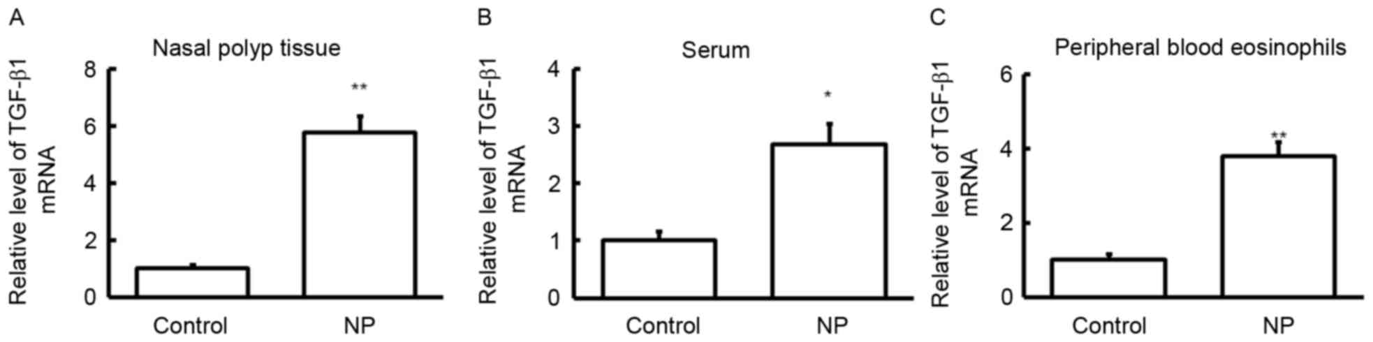
RT-qPCR was performed to detect the expression of
TGF-β1 mRNA in different specimens. The results demonstrated that,
compared with controls, the expression of TGF-β1 mRNA was
significantly upregulated in nasal polyp tissue (P<0.01;
Fig. 1A), serum (P<0.05; Fig. 1B) and peripheral blood eosinophils
(P<0.01; Fig. 1C) of pediatric
patients with nasal polyposis. These results indicate that TGF-β1
may serve an important role in the pathogenesis of nasal polyposis
in children.
Expression of TGF-β1 protein in nasal
polyp tissue and eosinophils
Western blotting was performed to detect the
expression of TGF-β1 protein in nasal polyp tissue and peripheral
blood eosinophils. The results demonstrated that, compared with
controls, expression of TGF-β1 was significantly upregulated in the
nasal polyp tissue (Fig. 2A) and
peripheral blood eosinophils (P<0.01; Fig. 2B) of pediatric patients with nasal
polyposis. To confirm these results, TGF-β1 levels in serum were
measured using ELISA. Compared with controls, levels of TGF-β1
protein were significantly upregulated in the serum of pediatric
patients with nasal polyposis (P<0.05; Fig. 2C). These results suggest that changes
in the expression of TGF-β1 protein are positively associated with
changes in the expression of TGF-β1 mRNA. Thus, upregulated TGF-β1
levels may have regulatory effects on the pathogenesis of nasal
polyposis in children.
Expression of miR-663 expression in
different specimens
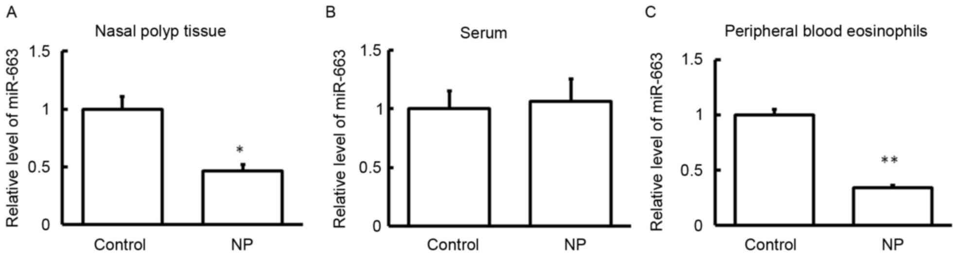
To detect the level of miR-663 in nasal polyp
tissue, serum and peripheral blood eosinophils of pediatric
patients with nasal polyposis, RT-qPCR was performed. As presented
in Fig. 3, the expression of miR-663
was significantly decreased in the nasal polyp tissue (P<0.05;
Fig. 3A) and peripheral blood
eosinophils (P<0.01; Fig. 3C) of
pediatric patients with nasal polyposis compared with controls.
However, there was no significant difference in miR-663 expression
in the serum of patients with nasal polyposis compared with
controls (P>0.05; Fig. 3B). These
results suggest that miR-663 may serve a role in the pathogenesis
of nasal polyposis.
Dual-luciferase reporter assay
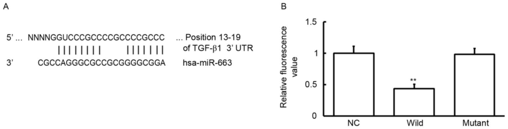
To identify whether TGF-β1 is a target of miR-663, a
bioinformatics prediction was conducted. It was determined that
miR-663 is one of the regulatory genes for TGF-β1 and the binding
sequence is presented (Fig. 4A). A
dual-luciferase reporter assay was conducted to verify this result.
Following co-transfection with miR-663 mimics and wild-type 3′UTR,
the fluorescence values decreased significantly (P<0.05;
Fig. 4B). By contrast, there was no
significant difference in the fluorescence value of the mutant
group compared with the negative control (Fig. 4B). These findings suggest that
miR-663 binds to the 3′-UTR of TGF-β1 to regulate TGF-β1
expression.
Discussion
In the present study, the expression of TGF-β1 and
its upstream gene miR-663 were detected in nasal polyp tissue,
serum and peripheral blood eosinophils of children with nasal
polyposis. A dual-luciferase reporter assay was performed to
confirm the binding of miR-663 and TGF-β1 and to evaluate whether
miR-663 contributes to the pathogenesis of nasal polyposis.
Nasal polyposis is a type of refractory rhinologic
disease in children characterized by an unknown pathogenesis,
persistent activity and difficulty of treatment (22). In the past, it was thought that nasal
polyposis was caused by an allergic reaction (23). However, recent studies suggest that
the clinical features of nasal polyposis are similar to those of
eosinophilic non-allergic rhinitis (24,25). The
concept of eosinophilic non-allergic rhinitis was proposed in 1979
and it was suggested that eosinophils may serve a key role in
inflammation, similar to their role in chronic sinusitis. However,
this hypothesis cannot explain the accumulation of eosinophils that
occurs in nasal polyp tissue. Previous studies have investigated
the mechanism of eosinophil accumulation in nasal polyposis
(26–28), which emphasizes nasal polyps as the
products of inflammatory reactions, regulated by cytokines in the
local microenvironment. Nasal polyp cells, including epithelial
cells and fibroblasts, can secrete cytokines that stimulate and
prolong the cell cycle of inflammatory cells, particularly
eosinophils (29). Furthermore,
eosinophils themselves are able to synthesize and secrete important
inflammatory cytokines and regulatory factors, which in turn can
stimulate inflammatory cell proliferation (30).
TGF-β1 is produced by eosinophils. Studies have
demonstrated that TGF-β1 promotes the production of interleukin
(IL)-6 from human fibroblasts and the growth of fibroblasts and
osteoblasts by regulating the transcription of IL-6 (31–33).
TGF-β1 may promote the formation of the extracellular matrix and
serve an important role in cell morphogenesis, proliferation and
differentiation in muscle tissue (34–36).
In vivo studies have demonstrated that the upregulation of
TGF-β1 in local tissue can promote wound healing and the formation
of typical granulation tissue (37).
In the present study, TGF-β1 expression was upregulated in all
specimens taken from pediatric patients with nasal polyposis. This
upregulation may promote the proliferation of fibroblasts and the
formation of typical granulation tissue; these effects are closely
associated with the formation of nasal polyposis (38,39).
Thus, TGF-β1 may promote the pathogenesis of nasal polyposis.
The regulation mechanism of TGF-β1 expression has
not been fully elucidated. Studies have suggested that nemo-like
kinase and Smad protein may be regulate TGF-β1 (40,41). In
addition, specific miRs can negatively regulate the expression of
TGF-β1 by cleaving TGF-β1 mRNA and inhibiting its translation. This
inhibitory effect is important in maintaining the balance of the
physiological and pathological states of the human body (42,43).
Recent studies have demonstrated that TGF-β1 mRNA regulates miR in
nasal polyposis and it was determined that miR-1343 may regulate
the expression of TGF-β1 receptors (44–46). In
the current study, bioinformatic predictions revealed that miR-663
is an upstream miR, which may have regulatory effects on TGF-β1
expression. The results also demonstrated that levels of miR-663
decrease following the upregulation of TGF-β1 mRNA and protein.
Furthermore, the results of the dual-luciferase reporter assay
determined that miR-663 contributes to the targeted regulation of
TGF-β1 by binding to the 3′-UTR of TGF-β1 and ultimately impacts on
the pathological features of nasal polyposis. However, in the
current study, serum levels of miR-663 did not change markedly,
even though levels of serum TGF-β1 increased. It was thus
speculated that the elevation of TGF-β1 was primarily caused by the
abundant release of inflammatory cells rather than the
downregulation of miR-663.
In conclusion, the present study demonstrated that
miR-663 serves important biological functions in the regulation of
TGF-β1 gene expression and the onset and development of nasal
polyposis. The results of the current study may therefore provide
experimental evidence for the prevention and treatment of pediatric
patients with nasal polyposis in clinical practice.
Acknowledgements
The present study was supported by a grant from the
Natural Science Foundation of Shandong Province of China (grant no.
ZR2013HM024).
References
|
1
|
Bachert C, Zhang N, van Zele T and Gevaert
P: Chronic rhinosinusitis: From one disease to different
phenotypes. Pediatr Allergy Immunol. 23 Suppl 22:S2–S4. 2012.
View Article : Google Scholar
|
|
2
|
Bachert C, Mannent L, Naclerio RM, Mullol
J, Ferguson BJ, Gevaert P, Hellings P, Jiao L, Wang L, Evans RR, et
al: Effect of subcutaneous dupilumab on nasal polyp burden in
patients with chronic sinusitis and nasal polyposis: A randomized
clinical trial. JAMA. 315:469–479. 2016. View Article : Google Scholar : PubMed/NCBI
|
|
3
|
Neubauer PD, Schwam ZG and Manes RP:
Comparison of intranasal fluticasone spray, budesonide atomizer,
and budesonide respules in patients with chronic rhinosinusitis
with polyposis after endoscopic sinus surgery. Int Forum Allergy
Rhinol. 6:233–237. 2016. View Article : Google Scholar : PubMed/NCBI
|
|
4
|
Johansson L, Akerlund A, Holmberg K, Melén
I and Bende M: Prevalence of nasal polyps in adults: The Skövde
population-based study. Ann Otol Rhinol Laryngol. 112:625–629.
2003. View Article : Google Scholar : PubMed/NCBI
|
|
5
|
We J, Lee WH, Tan KL, Wee JH, Rhee CS, Lee
CH, Ahn S, Lee JH and Kim JW: Prevalence of nasal polyps and its
risk factors: Korean national health and nutrition examination
survey 2009–2011. Am J Rhinol Allergy. 29:e24–e28. 2015. View Article : Google Scholar : PubMed/NCBI
|
|
6
|
Feng S, He Q, Fan Y, Mi J, Guo L, Hong H
and Li H: Nasal endoscopic findings and nasal symptoms in patients
with asthma: A clinical study from a rhinological perspective.
Allergol Immunopathol (Madr). 43:42–47. 2015. View Article : Google Scholar : PubMed/NCBI
|
|
7
|
Fetta M, Tsilis NS, Segas JV, Nikolopoulos
TP and Vlastarakos PV: Functional endoscopic sinus surgery improves
the quality of life in children suffering from chronic
rhinosinusitis with nasal polyps. Int J Pediatr Otorhinolaryngol.
100:145–148. 2017. View Article : Google Scholar : PubMed/NCBI
|
|
8
|
Weber SAT, Iyomasa RM, Correa CC,
Florentino WNM and Ferrari GF: Nasal polyposis in cystic fibrosis:
Follow-up of children and adolescents for a 3-year period. Braz J
Otorhinolaryngol. 83:677–682. 2017. View Article : Google Scholar : PubMed/NCBI
|
|
9
|
Veloso-Teles R and Cerejeira R: Endoscopic
sinus surgery for chronic rhinosinusitis with nasal polyps:
Clinical outcome and predictive factors of recurrence. Am J Rhinol
Allergy. 31:56–62. 2017. View Article : Google Scholar : PubMed/NCBI
|
|
10
|
Park IH, Um JY, Hong SM, Cho JS, Lee SH,
Lee SH and Lee HM: Metformin reduces TGF-β1-induced extracellular
matrix production in nasal polyp-derived fibroblasts. Otolaryngol
Head Neck Surg. 150:148–153. 2014. View Article : Google Scholar : PubMed/NCBI
|
|
11
|
Piersma B, Bank RA and Boersema M:
Signaling in Fibrosis: TGF-β, WNT, and YAP/TAZ Converge. Front Med
(Lausanne). 2:592015.PubMed/NCBI
|
|
12
|
Castellone MD and Laukkanen MO: TGF-beta1,
WNT, and SHH signaling in tumor progression and in fibrotic
diseases. Front Biosci (Schol Ed). 9:31–45. 2017. View Article : Google Scholar : PubMed/NCBI
|
|
13
|
Poniatowski LA, Wojdasiewicz P, Gasik R
and Szukiewicz D: Transforming growth factor Beta family: Insight
into the role of growth factors in regulation of fracture healing
biology and potential clinical applications. Mediators Inflamm.
2015:1378232015. View Article : Google Scholar : PubMed/NCBI
|
|
14
|
Vadon-Le Goff S, Hulmes DJ and Moali C:
BMP-1/tolloid-like proteinases synchronize matrix assembly with
growth factor activation to promote morphogenesis and tissue
remodeling. Matrix Biol. 44–46:14–23. 2015. View Article : Google Scholar
|
|
15
|
Richter K, Konzack A, Pihlajaniemi T,
Heljasvaara R and Kietzmann T: Redox-fibrosis: Impact of TGFβ1 on
ROS generators, mediators and functional consequences. Redox Biol.
6:344–352. 2015. View Article : Google Scholar : PubMed/NCBI
|
|
16
|
Lafyatis R: Transforming growth factor
β-at the centre of systemic sclerosis. Nat Rev Rheumatol.
10:706–719. 2014. View Article : Google Scholar : PubMed/NCBI
|
|
17
|
Huang Y, Liu J, Fan L, Wang F, Yu H, Wei W
and Sun G: miR-663 overexpression induced by endoplasmic reticulum
stress modulates hepatocellular carcinoma cell apoptosis via
transforming growth factor beta 1. Onco Targets Ther. 9:1623–1633.
2016. View Article : Google Scholar : PubMed/NCBI
|
|
18
|
Wang Z, Zhang H, Zhang P, Dong W and He L:
MicroRNA-663 suppresses cell invasion and migration by targeting
transforming growth factor beta 1 in papillary thyroid carcinoma.
Tumour Biol. 37:7633–7644. 2016. View Article : Google Scholar : PubMed/NCBI
|
|
19
|
Fokkens W, Lund V and Mullol J: European
Position Paper on Rhinosinusitis and Nasal Polyps Group: EP3OS
2007: European position paper on rhinosinusitis and nasal polyps
2007. A summary for otorhinolaryngologists. Rhinology. 45:97–101.
2007.PubMed/NCBI
|
|
20
|
Qin JJ, Lu ZY, Jiao ZP, Zhu XJ, Wang YX
and Tang H: Modified TRIzol method for RNA and DNA co-extraction
from blood. Fa Yi Xue Za Zhi. 29:209–211. 2013.(In Chinese).
PubMed/NCBI
|
|
21
|
Livak KJ and Schmittgen TD: Analysis of
relative gene expression data using real-time quantitative PCR and
the 2(-Delta Delta C(T)) method. Methods. 25:402–408. 2001.
View Article : Google Scholar : PubMed/NCBI
|
|
22
|
Hulse KE, Stevens WW, Tan BK and Schleimer
RP: Pathogenesis of nasal polyposis. Clin Exp Allergy. 45:328–346.
2015. View Article : Google Scholar : PubMed/NCBI
|
|
23
|
Bernstein JM, Gorfien J and Noble B: Role
of allergy in nasal polyposis: A review. Otolaryngol Head Neck
Surg. 113:724–732. 1995. View Article : Google Scholar : PubMed/NCBI
|
|
24
|
Calus L, Devuyst L, Van Zele T, De Ruyck
N, Derycke L, Bachert C and Gevaert P: The response to nasal
allergen provocation with grass pollen is reduced in patients with
chronic rhinosinusitis with nasal polyposis and grass
sensitization. Clin Exp Allergy. 46:555–563. 2016. View Article : Google Scholar : PubMed/NCBI
|
|
25
|
Becker S, Rasp J, Eder K, Berghaus A,
Kramer MF and Gröger M: Non-allergic rhinitis with eosinophilia
syndrome is not associated with local production of specific IgE in
nasal mucosa. Eur Arch Otorhinolaryngol. 273:1469–1475. 2016.
View Article : Google Scholar : PubMed/NCBI
|
|
26
|
Fokkens W, Lund V and Mullol J: European
Position Paper on Rhinosinusitis and Nasal Polyps group: European
position paper on rhinosinusitis and nasal polyps 2007. Rhinol
Suppl. 20:1–136. 2007.PubMed/NCBI
|
|
27
|
Fundová P, Funda DP, Kovar D, Holy R,
Navara M and Tlaskalová-Hogenová H: Increased expression of
chemokine receptors CCR1 and CCR3 in nasal polyps: Molecular basis
for recruitment of the granulocyte infiltrate. Folia Microbiol
(Praha). 58:219–224. 2013. View Article : Google Scholar : PubMed/NCBI
|
|
28
|
De Corso E, Baroni S, Romitelli F, Luca L,
Di Nardo W, Passali GC and Paludetti G: Nasal lavage CCL24 levels
correlate with eosinophils trafficking and symptoms in chronic
sino-nasal eosinophilic inflammation. Rhinology. 49:174–179.
2011.PubMed/NCBI
|
|
29
|
Pawankar R and Nonaka M: Inflammatory
mechanisms and remodeling in chronic rhinosinusitis and nasal
polyps. Curr Allergy Asthma Rep. 7:202–208. 2007. View Article : Google Scholar : PubMed/NCBI
|
|
30
|
Pawliczak R, Lewandowska-Polak A and
Kowalski ML: Pathogenesis of nasal polyps: An update. Curr Allergy
Asthma Rep. 5:463–471. 2005. View Article : Google Scholar : PubMed/NCBI
|
|
31
|
Janda K, Krzanowski M, Dumnicka P,
Kusnierz-Cabala B, Krasniak A and Sulowicz W: Transforming growth
factor beta 1 as a risk factor for cardiovascular diseases in
end-stage renal disease patients treated with peritoneal dialysis.
Clin Lab. 60:1163–1168. 2014. View Article : Google Scholar : PubMed/NCBI
|
|
32
|
Jin X, Ren S, Macarak E and Rosenbloom J:
Pathobiological mechanisms of peritoneal adhesions: The mesenchymal
transition of rat peritoneal mesothelial cells induced by TGF-β1
and IL-6 requires activation of Erk1/2 and Smad2 linker region
phosphorylation. Matrix Biol. 51:55–64. 2016. View Article : Google Scholar : PubMed/NCBI
|
|
33
|
Xiao K, Jiao L, Cao S, Song Z, Hu C and
Han X: Whey protein concentrate enhances intestinal integrity and
influences transforming growth factor-β1 and mitogen-activated
protein kinase signalling pathways in piglets after
lipopolysaccharide challenge. Br J Nutr. 115:984–993. 2016.
View Article : Google Scholar : PubMed/NCBI
|
|
34
|
Cicha I, Yilmaz A, Klein M, Raithel D,
Brigstock DR, Daniel WG, Goppelt-Struebe M and Garlichs CD:
Connective tissue growth factor is overexpressed in complicated
atherosclerotic plaques and induces mononuclear cell chemotaxis in
vitro. Arterioscler Thromb Vasc Biol. 25:1008–1013. 2005.
View Article : Google Scholar : PubMed/NCBI
|
|
35
|
Chen S, Liu J, Yang M, Lai W, Ye L, Chen
J, Hou X, Ding H, Zhang W, Wu Y, et al: Fn14, a downstream target
of the TGF-β signaling pathway, regulates fibroblast activation.
PLoS One. 10:e01438022015. View Article : Google Scholar : PubMed/NCBI
|
|
36
|
Kanaan RA, Aldwaik M and Al-Hanbali OA:
The role of connective tissue growth factor in skeletal growth and
development. Med Sci Monit. 12:RA277–RA281. 2006.PubMed/NCBI
|
|
37
|
Aström P, Pirilä E, Lithovius R, Heikkola
H, Korpi JT, Hernández M, Sorsa T and Salo T: Matrix
metalloproteinase-8 regulates transforming growth factor-β1 levels
in mouse tongue wounds and fibroblasts in vitro. Exp Cell Res.
328:217–227. 2014. View Article : Google Scholar : PubMed/NCBI
|
|
38
|
Park SK, Jin YD, Park YK, Yeon SH, Xu J,
Han RN, Rha KS and Kim YM: IL-25-induced activation of nasal
fibroblast and its association with the remodeling of chronic
rhinosinusitis with nasal polyposis. PLoS One. 12:e01818062017.
View Article : Google Scholar : PubMed/NCBI
|
|
39
|
Shin JM, Park JH, Kang B, Lee SA, Park IH
and Lee HM: Effect of doxycycline on transforming growth
factor-beta-1-induced matrix metalloproteinase 2 expression,
migration, and collagen contraction in nasal polyp-derived
fibroblasts. Am J Rhinol Allergy. 30:385–390. 2016. View Article : Google Scholar : PubMed/NCBI
|
|
40
|
Xiao Z, Zhang J, Peng X, Dong Y, Jia L, Li
H and Du J: The Notch γ-secretase inhibitor ameliorates kidney
fibrosis via inhibition of TGF-β/Smad2/3 signaling pathway
activation. Int J Biochem Cell Biol. 55:65–71. 2014. View Article : Google Scholar : PubMed/NCBI
|
|
41
|
Shi Y, Ye K, Wu H, Sun Y, Shi H and Huo K:
Human SMAD4 is phosphorylated at Thr9 and Ser138 by interacting
with NLK. Mol Cell Biochem. 333:293–298. 2010. View Article : Google Scholar : PubMed/NCBI
|
|
42
|
Lazzarini R, Sorgentoni G, Caffarini M,
Sayeed MA, Olivieri F, Di Primio R and Orciani M: New miRNAs
network in human mesenchymal stem cells derived from skin and
amniotic fluid. Int J Immunopathol Pharmacol. 29:523–528. 2016.
View Article : Google Scholar : PubMed/NCBI
|
|
43
|
He X, Liu Z, Peng Y and Yu C:
MicroRNA-181c inhibits glioblastoma cell invasion, migration and
mesenchymal transition by targeting TGF-β pathway. Biochem Biophys
Res Commun. 469:1041–1048. 2016. View Article : Google Scholar : PubMed/NCBI
|
|
44
|
Stolzenburg LR, Wachtel S, Dang H and
Harris A: microRNA-1343 attenuates pathways of fibrosis by
targeting the TGF-β receptors. Biochem J. 473:245–256. 2016.
View Article : Google Scholar : PubMed/NCBI
|
|
45
|
Shin JM, Park JH, Park IH and Lee HM:
Pirfenidone inhibits transforming growth factor β1-induced
extracellular matrix production in nasal polyp-derived fibroblasts.
Am J Rhinol Allergy. 29:408–413. 2015. View Article : Google Scholar : PubMed/NCBI
|
|
46
|
Yamin M, Holbrook EH, Gray ST, Busaba NY,
Lovett B and Hamilos DL: Profibrotic transforming growth factor
beta 1 and activin A are increased in nasal polyp tissue and
induced in nasal polyp epithelium by cigarette smoke and Toll-like
receptor 3 ligation. Int Forum Allergy Rhinol. 5:573–582. 2015.
View Article : Google Scholar : PubMed/NCBI
|