Introduction
Obesity is a serious public health issue in the
world related with increase in morbidity, mortality and decrease
lifespan (1). It associated with
metabolic disorders, such as, hypertension, type 2 diabetes,
nonalcoholic fatty liver diseases, atherosclerosis and
cardiovascular diseases (2,3). Obesity is related to an imbalance in
intake of energy, lack of exercise (4) and the excessive accumulation of adipose
tissues (3). The development of
obesity is characterized by adipogenesis, differentiation of
preadipocytes to adipocytes and increased triglycerides deposition
(5,6). Adipogenesis is regulated by
transcription factors, such as, peroxisome proliferation-activated
receptor-gamma (PPAR-γ) and CCAAT/enhancer-binding protein-α
(C/CBPα) (6). In addition, increased
expressions of PPARγ and C/CBPα have been shown to induce
adipogenesis by activating adipocyte-specific proteins, like
leptin, adiponectin (7,8). As a result of these findings, several
studies have targeted adipogenesis as a means of controlling
obesity. Liver is responsible for lipid synthesis and fatty acid
oxidation, and thus, plays an important role in lipid metabolism
(9). Lipid synthesis in liver is
regulated by sterol regulatory element-binding proteins (SREBP),
which control several downstream genes, such as acetyl-CoA
carboxylase (ACC), stearoyl-CoA destaturase (SCD) and fatty acid
synthesis (FAS) (10,11).
Several anti-obesity drugs, like orlistat,
topiramate, sibutraine are commercially available, however their
intolerable side effects such as, insomnia, anorexia, dry mouth and
gastrointestinal indisposition (12)
have limited their uses. This has called scientists to search for
new, effective and more tolerated anti-obesity agents from natural
products (13).
Chrysanthemum indicum (CI) is herb that is
widely used in China and Korea to treat various immune-related
disorders, such as, the symptoms of hypertension and several
infectious diseases (13,14). Previous studies have reported CI
possesses various bioactivities such as, anti-oxidant,
anti-nociceptive, anti-bacterial, anti-viral and anti-inflammatory
effects (14–17). Recently, it was reported that an
extract of CI has anti-oxidant and anti-adipogenic effects in
3T3-L1 cells (18). However, to our
knowledge the inhibitory effect of CIEE in overweight and
obesity-related metabolic diseases have not been studied.
Therefore, the present study investigated the effect of ethanol
extract of Chrysanthemum indicum L. flowers (CIEE) on a
mouse model of obesity induced by high-fat diet (HFD) and
elucidated its underlying mechanism.
Materials and methods
Materials and reagents
Trizol reagent and SuperScript III kit were obtained
from Invitrogen (Thermo Fisher Scientific, Inc., Waltham, MA, USA).
The protein assay kit (RIPA buffer) was from Santa Cruz
Biotechnology, Inc. (Santa Cruz, CA, USA). The primary antibodies,
mouse anti-(PPARγ (sc-7273), CEBPα (sc-166258), β-actin (sc-47778)
and rabbit anti-PPARα (sc-9000) were purchased from Santa Cruz
Biotechnology, Inc., and rabbit anti-FAS (C20G5) from Cell
signaling Technology, Inc. (Danvers, MA, USA). Primary antibodies
at 1:1,000 and secondary antibodies at 1:5,000 dilutions were used.
For rabbit primary antibodies: Goat-anti-rabbit IgG HRP (sc-2030;
Santa Cruz Biotechnology, Inc.) and for mouse primary antibodies:
Goat-anti-mouse IgG HRP (sc-2030; Santa Cruz Biotechnology, Inc.)
were used.
Preparation of the ethanol extract of
CI (CIEE)
CI was obtained from Gungangbogam (Jechon, Korea).
The dried and powdered fruit of CI (500 g) were extracted with 50%
ethanol for 2 h using mantle-reflux at 80°C. After filtration, CIEE
was obtained, as a powder, by removing solvent on a rotary vacuum
evaporator (N-000; Eyela, Tokyo, Japan) (19). The amount of CIEE obtained using this
procedure was 83.5 g (16.7%) with respect to the starting material.
CIEE was stored at 4°C for further use.
Animal treatment
Male C57BL/6 mice (5 weeks old) were purchased from
Samtako Bio Korea (Samtako Bio Korea, Osan, South Korea). The
experimental procedures were conducted using a protocol approved by
the Institutional Animal Care Committee of Chonbuk National
University. Mice were housed at 22±2°C and 50±5% RH and provided a
normal diet. After one week of acclimation, mice were divided into
six groups containing six mice in each group (n=6), as follows. i)
ND, mice fed with normal diet; ii) HFD, mice fed with high-fat
diet; iii) HFD+CIEE (8), mice fed
with HFD and treated with CIEE (8 mg/kg); iv) HFD+CIEE (40), mice
fed with HFD plus CIEE (40 mg/kg) treatment; v) HFD+CIEE (200),
mice fed with HFD plus CIEE (200 mg/kg) treatment; and vi) HFD+GC,
mice fed with HFD plus GC treatment. Garcinia cambogia (GC)
at dose of 200 mg/kg was used as a positive control and was
administered in its powdered form via oral gavage (p.o). The
treatment of CIEE (8, 40 or 200 mg/kg) or GC was administered once
a day for 6 weeks (p.o). Body weight and food consumption were
measured weekly.
Determination of abdominal fat volume
by micro-computed tomography (micro-CT)
Mice were starved for 6 h prior to sacrifice and
anesthetized by intraperitoneal injection of ketamine (75 mg/kg)
and rompun (xylazine) (15 mg/kg) (20). To measure the sign of consciousness,
we used a physical stimulus method, toes were pinched with
atraumatic forceps and the reflex was observed. Images were
acquired using a Skyscan-1076 micro-CT scanner (Skyscan,
Aartselaar, Belgium). CT was performed using a pixel size of 35 µm,
a source voltage of 50 kVp, and a source current of 200 µA. The
X-ray detector contained a 12-bit, water-cooled CCD (charge-coupled
device) camera with 4,000 × 2,300 resolutions and scintillator. The
images were acquired with 0.6 degree. The exposure time was 0.46
sec and a 1 mm aluminum energy filter was used. Abdominal fat
volume was measured using an Olympus SP-500 UZ camera (Olympus
America, Inc., Center Valley, PA, USA). Following micro-CT, under
anesthesia, mice were euthanized through intracardiac puncture.
Blood was collected (0.8–0.9 ml) via cardiac puncture followed by
removal of organs such as liver, kidney and spleen. Following
exsanguination, the death was confirmed by incising the heart and
making sure about no respiratory movement of mice for at least 3
min.
Histological analysis
Liver and abdominal fat tissues were fixed with 10%
neutral buffered formalin, embedded in paraffin wax, and cut
serially into 10 µm-thick sections. Sections were stained with
hematoxylin and eosin (H&E) and histological alterations were
observed under a microscope and photographed (magnification, ×100;
Olympus CX21; Olympus America, Inc.) (21).
Serum lipid profile
Serum samples were analyzed for levels of total
cholesterol (TC), triglyceride (TG), and high-density lipoprotein
cholesterol (HDLc) using a commercially available kit (Asian
Pharmaceutical, Hwaseong-Si, Korea).
Enzyme-linked immunosorbent assay
(ELISA)
Serum leptin and adiponectin levels were measured by
leptin mouse ELISA kit (Enzo Life Sciences, Inc., Farmingdale, NY,
USA) and adiponectin mouse ELISA kit (R&D Systems, Inc.,
Minneapolis, MN, USA), respectively, according to the
manufacturer's instructions.
Western blot analysis
Epididymal white adipose tissue (EWAT) and liver
tissues were lysed in ice-cold RIPA buffer for 40 min and
centrifuged (12,000 × g) for 20 min at 4°C (22,23).
Tissue lysates were run on sodium dodecyl sulfate-polyacrylamide
gel electrophoresis and transferred to polyvinylidene difluoride
(PVDF) membranes (Amersham; GE Healthcare, Chicago, IL, USA), which
were then blocked with 5% skimmed milk in tris-buffered saline
containing 0.1% Tween-20 (TBST) for 1 h at room temperature.
Membranes were probed with primary antibodies at 4°C overnight,
washed with TBST for 4 times, incubated with horseradish
peroxidase-conjugated secondary antibody for 45 min at RT, and
rewashed with TBST 3 times. Proteins were visualized using an
enhanced chemiluminescence detection kit (EMD Millipore, Billerica,
MA, USA).
Ultra pressure liqid chromatography
(UPLC)
UPLC was performed using ACQUITY UPLC® BEH C18
equipped with (2.1×50 mm, 1.7 µm) column and a photodiode array
detector (Waters Corp., Milford, MA, USA) (19). The elution was performed at flow rate
of 0.15 ml/min, using distilled water (DW) containing 0.1% formic
acid (solvent A) and ACN containing 0.1% formic acid (solvent B) in
gradient mode (B 5% from 0 to 1 min, B 5 to 95% from 1 to 16 min, B
95 to 100% from 16 to 18 min, and B 100% from 18 to 26 min; flow
rate was 0.15 ml/min, detection by UV at 330 nm, and temperature at
25°C.
Statistical analysis
Results are expressed as means ± standard error of
mean (SEM) and analyzed using Graph Pad Prism software (version
5.0; GraphPad Software, Inc., La Jolla, CA, USA). One-way analysis
of variance (ANOVA) was used to measure the significant difference
followed by Tukey's post hoc test for comparison of mean. P<0.05
was considered to indicate a statistically significant
difference.
Results
Effect of CIEE on body weight, food
consumption and efficiency in HFD-fed mice
To investigate the anti-obesity effect of CIEE, mice
were fed HFD with or without CIEE (8, 40 and 200 mg/kg, oral gavage
daily) or GC (200 mg/kg, oral gavage daily). No significant
difference in dietary intake was observed between the HFD-fed
control and the CIEE treated groups. HFD induced weight gain as
compared to mice fed the normal diet, whereas CIEE administration
significantly and dose-dependently decreased weight gain (Table I).
 | Table I.Effect of CIEE on body weight gain,
food intake and efficiency (%) in HFD fed mice. |
Table I.
Effect of CIEE on body weight gain,
food intake and efficiency (%) in HFD fed mice.
|
|
|
| CIEE (mg/kg) |
|
|---|
|
|
|
|
|
|
|---|
| Groups | ND | HFD | 8 | 40 | 200 | GC (mg/kg) 200 |
|---|
| Initial body weight
(g) | 21.68±1.10 | 22.32±1.16 | 21.14±0.86 | 22.07±1.30 | 21.96±1.07 | 21.43±0.97 |
| Final body weight
(g) | 26.97±١.٧٥ | 36.73±3.89a | 32.33±2.48 | 31.47±1.84b | 30.11±2.08b | 28.55±1.61b |
| Gain weight
(g) | 5.29±1.65 | 14.41±3.64a | 11.20±2.29 | 9.40±1.68b | 8.15±1.93b | 7.10±0.53b |
| Food intake | 2.67±0.33 | 2.14±0.27 | 2.12±0.21 | 2.08±0.19 | 2.05±0.24 | 1.98±0.22 |
| Food efficiency
(%) | 4.47 | 16.03 | 12.57 | 10.76 | 9.46 | 8.53 |
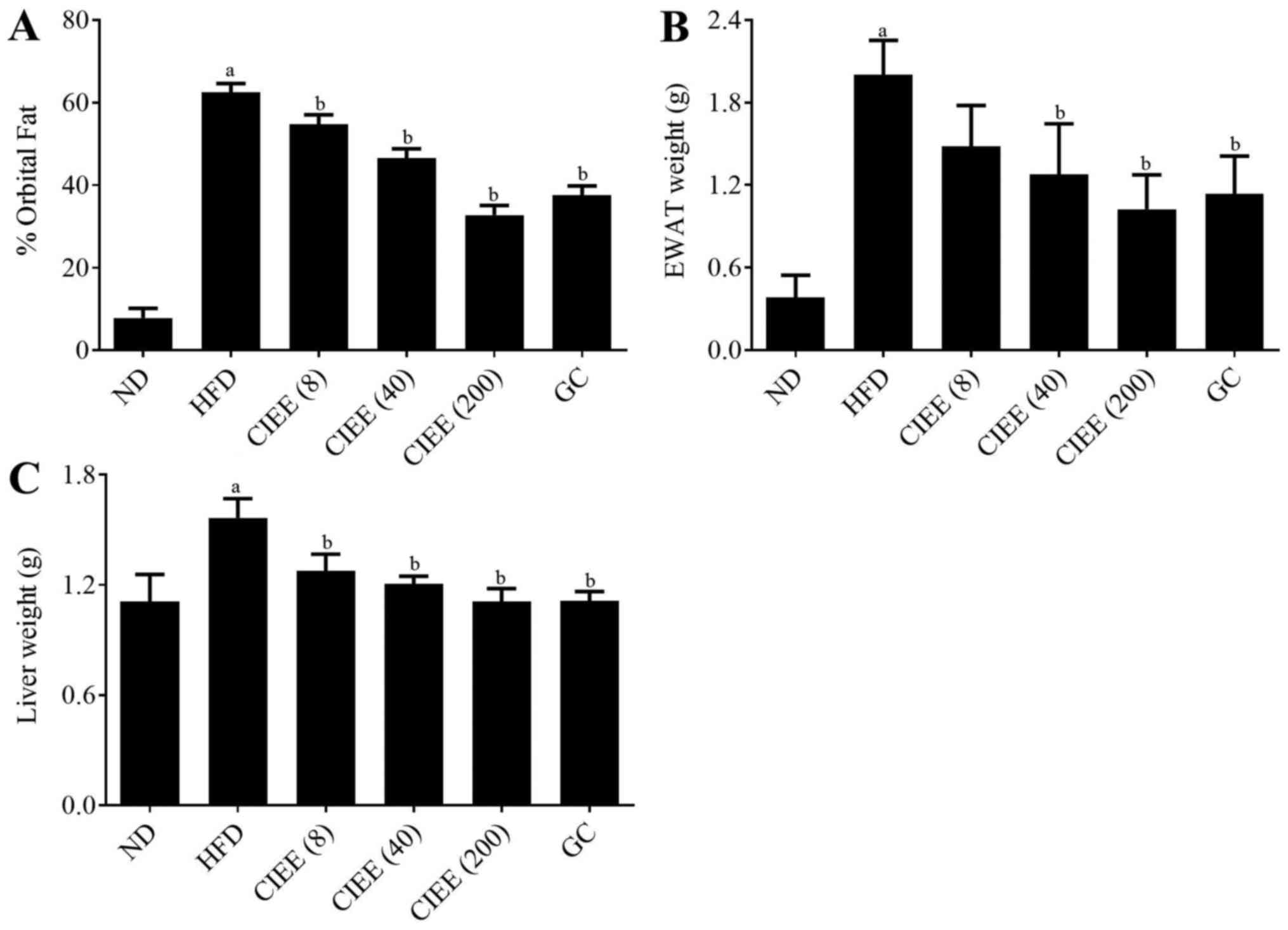
Effect of CIEE on fat deposition (mass
of abdominal WAT, weight of EWAT, and liver tissue) in HFD-fed
mice
To assess the effect of CIEE on adipose tissue
accumulation, body and abdominal fat weight gains were measured in
the study. CIEE dose-dependently diminished orbital fat volume (%)
as determined by micro-CT as compared with the HFD-fed group as
shown in Fig. 1A. CIEE also
significantly reduced EWAT weight in HFD-fed obese mice to a level
below than in GC fed controls (Fig.
1B). Also, CIEE induced significant reductions in liver tissue
weight (Fig. 1C; P<0.05).
Effect of CIEE on histological
observations in HFD-fed mice
Histological analysis of EWAT and liver tissue were
performed by using H&E staining. Results showed enlarged size
of EWAT in the HFD group whereas; CIEE groups reduced the size of
EWAT (Fig. 2A). Also, data revealed
that CIEE significantly reduced vacuolization and lipid droplet
numbers in liver tissue (Fig. 2B;
P<0.05).
 | Figure 2.Effect of CIEE on histopathological
examination of EWAT and liver tissues in experimental groups.
Photograph showing haematoxylin and eosin staining for (A) EWAT and
(B) liver tissue of HFD-fed mice. Representative images of a,
treatment naïve control (ND); b, HFD control; c, HFD+GC; d,
HFD+CIEE (8 mg/kg); e, HFD+CIEE (40 mg/kg) and f, HFD+CIEE (200
mg/kg); ND, normal diet group; HFD, high-fat diet group; CIEE,
ethanol extract of Chrysanthemum indicum L.; GC, Garcinia
cambogia; EWAT; epididymal white adipose tissue. |
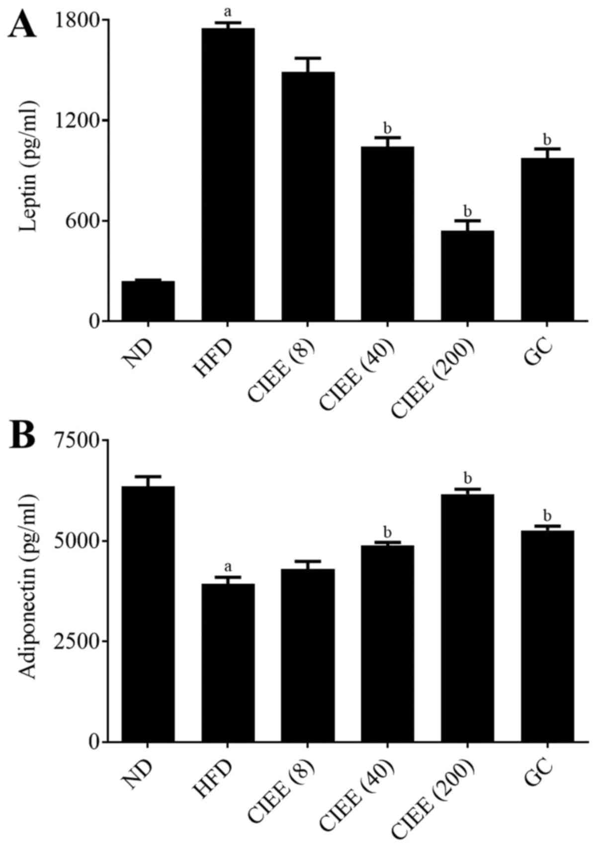
Effect of CIEE on the lipid profiles
of HFD-fed mice
HFD-fed mice had higher serum levels HFD-fed mice
had higher serum levels of TC, low-density lipoprotein cholesterol
(LDLc) and TG, and lower levels of HDLc than mice in the normal
group (P<0.05). CIEE significantly inhibited TC, LDLc and TG
increases, but dose-dependently increased vs. HFD-fed mice
(Table II). Furthermore, CIEE
dose-dependently inhibited leptin levels (Fig. 3A) and increased adiponectin levels as
compared with HFD-fed mice (Fig.
3B).
| Table II.Effect of CIEE on serum lipid
profiles in HFD fed mice. |
Table II.
Effect of CIEE on serum lipid
profiles in HFD fed mice.
|
|
|
| CIEE (mg/kg) |
|
|---|
|
|
|
|
|
|
|---|
| Groups | ND | HFD | 8 | 40 | 200 | GC (mg/kg) 200 |
|---|
| Total cholesterol
(TC, mg/dl) | 125.5±19.6 | 233.1±13.4a | 212.1±14.5b | 183.4±20.9b | 160.8±12.5b | 190.7±38.9b |
| Triglyceride (TG,
mg/dl) | 85.7±8.9 | 142.7±6.5a | 133.7±11.3 | 124.4±18.1 | 109.1±31.4b | 106.3±9.4b |
| LDL-cholesterol
(LDLc, mg/dl) | 54.56±11.7 | 126.4±29.7;a | 111.1±1.6 | 89.8±15.7 | 55.1±16.3b | 86.9±43.2 |
| HDL-cholesterol
(HDLc, mg/dl) | 53.7±11.1 | 78.1±16.7a | 74.3±14.3 | 68.7±18.7 | 87.8±12.4b | 82.6±12.4 |
Effect of CIEE on transcription
factors of adipogenesis and FAS
Western blotting was used to determine whether the
observed increases of EWAT and liver weight in obese mice were
accompanied by changes in the expressions of regulator
transcription factors, PPARα/γ and C/EBPα. HFD-fed mice showed a
significant increase in PPARγ and C/EBPα protein expression in
EWAT. CIEE significantly and dose-dependently inhibited HFD-induced
PPARγ and C/EBPα in EWAT (Fig. 4)
(P<0.05). Furthermore, CIEE dose dependently enhanced the
protein expression of PPARα but decreased C/EBPα in liver tissue
vs. HFD-fed mice (Fig. 5A-C).
PPARα/γ regulates FAS protein during adipogenesis,
and thus we examined the effect of CIEE on FAS protein level in
both EWAT and liver tissues by western blotting. Our results
revealed HFD-fed mice had elevated FAS protein levels and that CIEE
significantly and dose-dependently inhibited this increase in EWAT
(Fig. 4A and D) and liver tissue
(Fig. 5A and D).
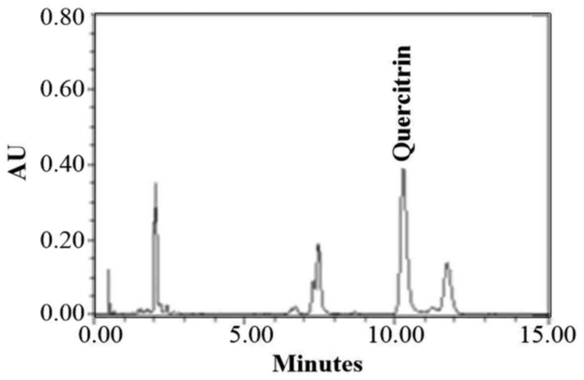
Chemical components of CIEE by
UPLC
UPLC showed that quercitrin was the main component
of CIEE (Fig. 6). However, other
peaks observed were unidentified and further experiments are
necessary to illustrate the other components of CIEE.
Discussion
Obesity is a serious risk factor for hypertension,
cardiovascular disease, type-2 diabetes, and cancers (2–6). Though
anti-obesity drugs, like orlistat and sibutramine, are commercially
available, their adverse effects are abundant, such as, diarrhea,
stomach pain, irregular menstruation, steatorrhea and hypertension
(24,25). Therefore, the identification of
effective natural products with lesser side effects is a topic of
considerable research (26). Several
studies have described the anti-inflammatory and anti-antioxidant
effects of CI. However, the effect of CIEE on HFD-induced obesity
has not been previously studied. In the present study, we
investigated whether CIEE could inhibit obesity in HFD-induced
obese C57BL/6 mice and explored its underlying mechanisms.
Reductions in body weight and fat deposition can
prevent obesity (27), and increased
fat cell numbers and/or sizes are characteristic of obesity
(28). In the present study, food
intakes were similar in the study groups, but efficiency percentage
(body weight gain per unit of food consumption) was reduced by CIEE
treatment in HFD-fed mice. Furthermore, CIEE significantly
decreased body weight gains, EWAT and liver weights, volumes and
lipid accumulation in EWAT and liver tissues in HFD-fed mice, and
reduced HFD-induced increases in serum lipid profiles, that is, TC,
LDLc and TG, but increased serum levels of HDLc. These improved
lipid profiles induced by CIEE may have been associated reduced
fatty liver and hyperlipidemia in HFD-fed mice. Furthermore, these
findings concur with those of a previous study, which showed
Triticumaestivum sprouts had an anti-obesity effect in
HFD-fed mice (19).
The adipokines, leptin and adiponectin secreted by
adipose tissue and regulate lipid metabolism. Leptin plays an
important role in weight control by suppressing food intake and
increasing energy expenditure (29),
and adiponectin is involved in fatty acid oxidation and glucose
regulation in liver (30,31). We found CIEE significantly reduced
serum leptin levels and increased adiponectin levels in HFD-induced
obese mice. Similarly, a previous study reported Poncritus
trifoliate leaf extracts had an anti-obesity effect in HFD-fed
mice (32).
PPARα/γ is ligand-activated transcription factor,
which plays role in glucose and protein metabolism and regulates
preadipocytes proliferation and differentiation (33). C/EBPs are proteins involved in
adipogenesis, and C/EBPα and PPARγ control the biosynthesis of
fatty acids, such as acetyl CoA carboxylase and FAS, are key
regulators of lipogenesis (34).
Also, PPARs regulates lipid metabolism, lipid synthesis, and fatty
acid oxidation in liver tissue. In the present study, CIEE
significantly down-regulated PPARγ and C/EBPα in EWAT but
up-regulated PPARα in liver in HFD fed mice. Moreover, FAS
induction by HFD was significantly reduced by CIEE on both EWAT and
liver tissues. In line with our results was previously reported
anti-obesity effect of germinated brown rice extract through
down-regulation of PPARγ and C/EBPα in HFD fed obese mice (35).
Chemical analysis by UPLC in our study showed the
detection of quercitrin in CIEE. Recent study reported that
quercitrin containing extract of Chrysobalanusicoco leaves
inhibited fat accumulation in HFD-fed mice (36). Also, quercitrin isolated from
Rhododendron oldhamii leaf extract improves fatty liver
syndrome via increase of lipid oxidation and decrease of
lipogenesis (37). Thus, quercitrin
may be one of the compounds that contribute to the anti-obesity
effect of CIEE. The present study demonstrates for the first time
CIEE treatment effectively inhibits adipogenesis, and contains
anti-adipogenic molecules that down-regulate transcription factors
for adipogenesis, such as C/EBPα and PPARα/γ. These results suggest
CIEE possesses anti-obesity effects is a promising agent for the
treatment of obesity and obesity-associated metabolic syndrome.
Acknowledgements
This study was supported by the Ministry of Trade,
Industry and Energy (MOTIE), Korea Institute for Advancement of
Technology (KIAT) through the Encouragement Program for The
Industries of Economic Cooperation Region (R0004885).
References
|
1
|
World Health Organization (WHO): Obesity:
Preventing and managing the global epidemic Series 894. WHO;
Geneva: pp. 2522000
|
|
2
|
Kopelman PG: Obesity as a medical problem.
Nature. 404:635–643. 2000. View
Article : Google Scholar : PubMed/NCBI
|
|
3
|
Kwon TH, Wu YX, Kim JS, Woo JH, Park KT,
Kwon OJ, Seo HJ, Kim T and Park NH: 6,6′-Bieckol inhibits adipocyte
differentiation through downregulation of adipogenesis and
lipogenesis in 3T3-L1 cells. J Sci Food Agric. 95:1830–1837. 2015.
View Article : Google Scholar : PubMed/NCBI
|
|
4
|
Garland T Jr, Schutz H, Chappell MA,
Keeney BK, Meek TH, Copes LE, Acosta W, Drenowatz C, Maciel RC, van
Dijk G, et al: The biological control of voluntary exercise,
spontaneous physical activity and daily energy expenditure in
relation to obesity: Human and rodent perspectives. J Exp Biol.
214:206–229. 2011. View Article : Google Scholar : PubMed/NCBI
|
|
5
|
Poudel B, Nepali S, Xin M, Ki HH, Kim YH,
Kim DK and Lee YM: Flavonoids from Triticum aestivum inhibit
adipogenesis in 3T3-L1 cells by upregulating the insig pathway. Mol
Med Rep. 12:3139–3145. 2015. View Article : Google Scholar : PubMed/NCBI
|
|
6
|
Poudel B, Lim SW, Ki HH, Nepali S, Lee YM
and Kim DK: Dioscin inhibits adipogenesis through the AMPK/MAPK
pathway in 3T3-L1 cells and modulates fat accumulation in obese
mice. Int J Mole Med. 34:1401–1408. 2014. View Article : Google Scholar
|
|
7
|
Fasshauer M and Paschke R: Regulation of
adipocytokines and insulin resistance. Diabetologia. 46:1594–1603.
2003. View Article : Google Scholar : PubMed/NCBI
|
|
8
|
Rocha VZ, Folco EJ, Sukhova G, Shimizu K,
Gotsman I, Vernon AH and Libby P: Interferon-gamma, a Th1 cytokine,
regulates fat inflammation: A role for adaptive immunity in
obesity. Circ Res. 103:467–476. 2008. View Article : Google Scholar : PubMed/NCBI
|
|
9
|
Roberts CK and Sindhu KK: Oxidative stress
and metabolic syndrome. Life Sci. 84:705–712. 2009. View Article : Google Scholar : PubMed/NCBI
|
|
10
|
Strable MS and Ntambi JM: Genetic control
of de novo lipogenesis: Role in diet-induced obesity. Crit Rev
Biochem Mol Biol. 45:199–214. 2010. View Article : Google Scholar : PubMed/NCBI
|
|
11
|
Horton JD, Goldstein JL and Brown MS:
SREBPs: Transcriptional mediators of lipid homeostasis. Cold Spring
Harb Symo Quant Biol. 67:491–498. 2002. View Article : Google Scholar
|
|
12
|
Derosa G, Cicero AF, Murdolo G, Piccinni
MN, Fogari E, Bertone G, Ciccarelli L and Fogari R: Efficacy and
safety comparative evualation of orlistat and sibutramine treatment
in hypertensive obese patients. Diabeted Obes Metab. 7:47–55. 2005.
View Article : Google Scholar
|
|
13
|
Montagut G, Bladé C, Blay M,
Fernández-Larrea J, Pujadas G, Salvadό MJ, Arola L, Pinent M and
Ardévol A: Effects of a grapeseed procyanidin extract (GSPE) on
insulin resistance. J Nutr Biochem. 21:961–967. 2010. View Article : Google Scholar : PubMed/NCBI
|
|
14
|
Cheng W, Li J, You T and Hu C:
Anti-inflammatory and immunomodulatory activities of the extracts
from the inflorescence of Chrysanthemum indicum Linne. J
Ethnopharmacol. 101:334–337. 2005. View Article : Google Scholar : PubMed/NCBI
|
|
15
|
Shunying Z, Yang Y, Huaidong Y, Yue Y and
Guolin Z: Chemical composition and antimicrobial activity of the
essential oils of Chrysanthemum indicum. J Ethnopharmacol.
96:151–158. 2005. View Article : Google Scholar : PubMed/NCBI
|
|
16
|
Kong LD, Cai Y, Huang WW, Cheng CH and Tan
RX: Inhibition of xanthine oxidase by some Chinese medicinal plants
used to treat gout. J Ethnopharmacol. 73:199–207. 2000. View Article : Google Scholar : PubMed/NCBI
|
|
17
|
Shi GB, Zhao MH, Zhao QC, Huang Y and Chen
YF: Mechanisms involved in the antinociception of petroleum ether
fraction from the EtOH extract of Chrysanthemum indicum in mice.
Phytomedicien. 7:609–616. 2011. View Article : Google Scholar
|
|
18
|
Kim RH, Song JH, Shon MS, Chun KS, Choi SU
and Kim GN: Evaluation of water extract prepared from Chrysanthemim
indicum Linne as Nurti-cosmetic and cosmetic material in vitro
model. Asian J beauty Cosmetol. 14:78–88. 2016. View Article : Google Scholar
|
|
19
|
Im JY, Ki HH, Xin M, Kwon SU, Kim YH, Kim
DK, Hong SP, Jin JS and Lee YM: Anti-obesity effect of Triticum
aestivum sprouts extract in high-fat-diet-induced obese mice.
Biosci Biotechnol Biochem. 79:1133–1140. 2015. View Article : Google Scholar : PubMed/NCBI
|
|
20
|
Histing T, Andonyan A, Klein M, Scheuer C,
Stenger D, Holstein JH, Veith NT, Pohlemann T and Menger MD:
Obesity does not affect the healing of femur fractures in mice.
Injury. 47:1435–1444. 2016. View Article : Google Scholar : PubMed/NCBI
|
|
21
|
Nepali S, Ki HH, Lee JH, Cha JY, Lee YM
and Kim DK: Triticum aestivum sprout-derived polysaccharide exerts
hepatoprotective effects against ethanol-induced liver damage by
enhancing the antioxidat system in mice. Int J Mole Med.
40:1243–1252. 2017. View Article : Google Scholar
|
|
22
|
Nepali S, Son JS, Poudel B, Lee JH, Lee YM
and Kim DK: Luteolin is a bioflavonoid that attenuates
adipocyte-derived inflammatory responses via suppression of nuclear
factor-κB/mitogen-activated protein kinases pathway. Pharmacogn
Mag. 11:627–635. 2015. View Article : Google Scholar : PubMed/NCBI
|
|
23
|
Poudel B, Ki HH, Luyen BT, Lee YM, Kim YH
and Kim DK: Triticumoside induces apoptosis via caspase-dependent
mitochondrial pathway and inhibits migration through downregulation
of MMP2/9 in human lung cancer cells. Acta Biochim Biophys Sin
(Shanghai). 48:153–160. 2016. View Article : Google Scholar : PubMed/NCBI
|
|
24
|
Chaput JP, St-Pierre S and Tremblay A:
Currently available drugs for the treatment of obesity: Sibutramine
and orlistat. Mini Rev Med Chem. 1:3–10. 2007. View Article : Google Scholar
|
|
25
|
Siebenhofer A, Jeitler K, Horvath K,
Berghold A, Posch N, Meschik J and Semlitsch T: Long-term effects
of weight-reducing drugs in people with hypertension. Cochrane
Database Syst Rev. 3:CD0076542016.PubMed/NCBI
|
|
26
|
Choudhary M and Grover K: Development of
functional food products in relation to obesity. Funct Foods Health
Dis. 2:188–197. 2012.
|
|
27
|
Hue JJ, Lee KN, Jeong JH, Lee SH, Lee YH,
Jeong SW, Nam SY, Yun YW and Lee BJ: Anti-obesity activity of
diglyceride containing conjugated linoleic acid in C57BL/6J ob/ob
mice. J Vet Sci. 10:189–195. 2009. View Article : Google Scholar : PubMed/NCBI
|
|
28
|
Han LK, Kimura Y and Okuda H: Reduction in
fat storage during chitin-chitosan treatment in mice fed a high-fat
diet. Int J Obes Relat Metab Disord. 23:174–179. 1999. View Article : Google Scholar : PubMed/NCBI
|
|
29
|
Maffei M, Halaas J, Ravussin E, Pratley
RE, Lee GH, Zhang Y, Fei H, Kin S, Lallone R, Ranganathan S, et al:
Leptin levels in human and rodent: Measurement of plasma leptin and
ob RNA in obese and weight-reduced subjects. Nat Med. 1:1155–1161.
1995. View Article : Google Scholar : PubMed/NCBI
|
|
30
|
Combs TP, Berg AH, Obici W, Scherer PE and
Rossetti L: Endogenous glucose production is inhibited by the
adipose-derived protein Acrp30. J Clin Invest. 108:1875–1881. 2001.
View Article : Google Scholar : PubMed/NCBI
|
|
31
|
Pajvani UB and Scherer PE: Adiponectin:
Systemic contributor to insulin sensitivity. Curr Diab Rep.
3:207–213. 2003. View Article : Google Scholar : PubMed/NCBI
|
|
32
|
Jia S, Gao Z, Yan S, Chen Y, Sun C, Li X
and Chen K: Anti-obesity and hypoglycemic effects of poncirus
tridoliata L. Extracts in high-fat diet C57BL/6 mice. Molecules.
21:5432016. View Article : Google Scholar
|
|
33
|
Christodoulides C and Vidal-Puig A: PPARs
and adipocyte function. Mol Cell Endocrinol. 318:61–68. 2010.
View Article : Google Scholar : PubMed/NCBI
|
|
34
|
Motojima K, Passilly P, Peters JM,
Gonzalez FJ and Latruffe N: Expression of putative fatty acid
transporter genes are regulated by peroxisome
proliferator-activated receptor alpha and gamma activators in a
tissue- and inducer-specific manner. J Biol Chem. 27:16710–16714.
1998. View Article : Google Scholar
|
|
35
|
Ho JN, Son ME, Lim WC, Lim ST and Cho HY:
Anti-obesity effects of germinated brown rice extract through
down-regulation of lipogenic genes in high fat diet-induced obese
mice. Biosci Biotechnol Biochem. 76:1068–1074. 2012. View Article : Google Scholar : PubMed/NCBI
|
|
36
|
White PA, Cercato LM, Batista VS, Camargo
EA, De Lucca W Jr, Oliveira AS, Silva FT, Goes TC, Olivera ER,
Moraes VR, et al: Aqueous extract of Chrysobalanus icaco leaves, in
lower doses, prevent fat gain in obese high-fat fed mice. J
Ethnopharmacol. 179:92–100. 2016. View Article : Google Scholar : PubMed/NCBI
|
|
37
|
Liu YL, Lin LC, Tung YT, Ho ST, Chen YL,
Lin CC and Wu JH: Rhododendron oldhamii leaf extract improves fatty
liver syndrome by increasing lipid oxidation and decreasing the
lipogenesis pathway in mice. Int J med Sci. 9:862–870. 2017.
View Article : Google Scholar
|