Introduction
Acute spinal cord injury (SCI) is a serious nervous
system injury, which often results in partial or complete loss of
feeling and motor function below the injury surface. According to a
previous report, the annual incidence of SCI is 15–40/1,000,000
worldwide (1). SCI can be caused by
traffic accidents, falls or sports injuries. Of these, traffic
accidents are the main cause (44.5%) of SCI in the USA, followed by
falls (16.6%) and sports injuries (12.7%) (1). The injured people are commonly of
working age (range, 18–80 years old). Research has indicated that
SCI has brought great economic burden on individuals, families and
society (2). Therefore, treatment
for acute SCI, including nerve damage recovery or reduction, has
great social significance. Nonetheless, SCI treatment remains a
challenge in the medical field.
Acute SCI can be classified into primary SCI and
secondary SCI. Secondary SCI was proposed by Allen in 1911
(3). Primary SCI refers to
mechanical injury to part of the spinal cord. The magnitude of
primary injury is determined by the external force at the moment of
impact. Such injury is irreversible, so it is not an effective
treatment strategy (4). Secondary
injury refers to serious damage within a few minutes of the primary
injury, including edema, inflammation, ischemia, excessively
activated glutamate receptors, lipid peroxidation and calcium
overload. This will result in secondary cell death for a time
period ranging from several days to weeks. Thus, it will cause
massive death of neurons and glia cells after primary injury
(5). The severity of multifactorial
sequence tissue destruction is even greater than that of primary
injury. The injury area will develop serial reactions after SCI. It
will not only damage residual nerve cells, but also cause damage
the spinal cord tissue surrounding the injury center (6).
Previous results have verified that inflammation
aggravates the post-SCI secondary injury (7). Apoptosis is the key manifestation of
secondary injury; thus, inhibition of apoptosis can prevent or
reduce secondary injury, protect nerve function and alleviate nerve
cell loss (7). SCI has allowed the
surviving cells to survive in primary injury and it also allows
more nerve function to be retained (8). Apoptosis, which is also known as
programmed cell death, is an active death process under the
regulation of multiple signaling pathways (9). Energy consumption is required during
cell apoptosis, so as to synthesize new proteins and nucleic acids
(10).
Peroxisome proliferator-activated receptor (PPAR) is
a member of the ligand activated nuclear transcription factor
superfamily (11). PPAR agonists
have been demonstrated in previous studies to exhibit
anti-inflammatory effects and inhibitory effects on brain neural
apoptosis (11,12). Research on PPAR-γ in SCI has focused
little on its effects of protecting spinal neurons and promoting
post-SCI secretion of inflammatory cytokines (12). The current study was thereby
conducted aiming to observe the effects of PPAR-γ agonist
thymoquinone on repairing SCI in rats. Furthermore, its mechanism
was investigated in order to provide therapeutic strategies for
clinical SCI (13).
SCI treatments will promote nerve growth factor
(NGF), activating the phosphoinositide 3-kinase (PI3K)/Akt pathway
to inhibit neuron apoptosis (14).
Therefore, post-SCI, it is of particular importance to adapt proper
methods to induce NGF activation and proliferation, repair the
damaged spinal cord nerve and function of bladder, and activate the
PI3K/Akt pathway to suppress apoptosis, thus improving neural
function (15).
Black cumin, Nigella sativa, belongs to the
family Ranunculaceae, and is an annual herbaceous plant. It is used
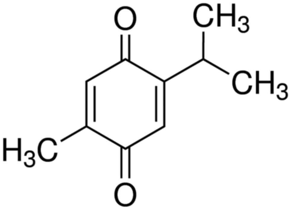
as a traditional natural medicine for numerous diseases (16). Thymoquinone (Fig. 1) is the primary active ingredient of
black cumin seed oil and is commonly used for anti-inflammation,
anti-oxidation and anti-tumor treatment (17). Over the past decade, thymoquinone
could inhibit numerous cancer types, including breast, prostate,
ovarian, liver, pancreatic and colorectal cancer (18–20). The
present study aimed to determine the effect of thymoquinone on
inflammatory response, oxidative stress and apoptosis in SCI rats,
and to investigate its possible molecular mechanisms.
Materials and methods
Animals
Male Sprague Dawley rats (age, 6 weeks; weight,
180±10 g, n=26) were purchased from the Animal Experiment Center of
Shandong University (Shandong, China) and individually housed
(temperature, 23±1°C; 55–60% humidity) and were exposed to a 12 h
light/dark cycle (lights on from 8:00 a.m. to 8:00 p.m.). Rats also
had free access to food and water ad libitum. This study was
performed in accordance with the guidelines of the National
Institutes of Health of Zaozhuang Municipal Hospital as referred to
previously (21), and approved by
Zaozhuang Municipal Hospital of Care and Use Committee.
Surgical procedures and experimental
setup
The rats were randomly divided into three groups:
Sham surgery (sham, n=6), SCI surgery (model, n=10) and SCI +
thymoquinone (thymoquinone, n=10). The rats from the model and
thymoquinone groups were anesthetized with 400 mg/kg of chloral
hydrate, and a laminectomy was performed at the T9-T10 level. The
underlying cord was exposed to contusion injury without disrupting
the dura (22). The thymoquinone
group received thymoquinone at 30 mg/kg once daily by intragastric
administration (23) from 3 weeks
after surgery. The rats from the sham and model groups received an
equal volume of vehicle (PBS) at the same time. In the sham group,
rats were anesthetized with 400 mg/kg of chloral hydrate, and the
surgical procedure was not performed. Furthermore, Sham rats
received normal saline by intragastric administration for 3
weeks.
Histological assessment
Rats were anesthetized using 35 mg/kg pentobarbital
sodium and then was sacrificed using decollation. Spinal cord
tissue was extracted, washed with PBS and fixed in 10% neutral
buffered formalin for 3 days at room temperature. Then, tissue was
decalcified in 10% EDTA for 10 days and embedded into paraffin.
Next, tissue was cut into serial paraffin sections (4 mm), which
were stained with hematoxylin and eosin for 30 min at room
temperature and observed using a microscope (Olympus IX81; Olympus
Corporation, Tokyo, Japan).
Behavioral assessments
The Basso, Beattie and Bresnahan (BBB) scale and
water content in spinal cord tissue were used to assess
neurological function after treatment with thymoquinone. The BBB
score is on a scale from 1 to 21, indicating no hindlimb movement
to normal hindlimb function (24).
Spinal cord tissue samples were extracted after treatment with
thymoquinone and washed with PBS. Tissue samples were weighed as
wet weight and then dried at 72°C for 48 h. Next, tissue samples
were weighed as dry weight. Water content was calculated as
follows: Water content (%) = wet weight/dry weight ×100%.
Measurement of inflammatory response,
oxidative stress and cell apoptosis
Tumor necrosis factor (TNF)-α (cat. no. PT516;
Beyotime Institute of Biotechnology, Haimen, China), interleukin
(IL)-1β (cat. no. PI303; Beyotime Institute of Biotechnology), IL-6
(cat. no. PI328; Beyotime Institute of Biotechnology), IL-18 (cat.
no. E-EL-R0567c; Elabscience, Houston, TX, USA), superoxide
dismutase (SOD; cat. no. S0109; Beyotime Institute of
Biotechnology), catalase (CAT; cat. no. S0051; Beyotime Institute
of Biotechnology), glutathione (GSH; cat. no. S0052; Beyotime
Institute of Biotechnology), PGE2 and malondialdehyde (MDA; cat.
no. S0131; Beyotime Institute of Biotechnology), caspase-3 (cat.
no. C1116; Beyotime Institute of Biotechnology) and caspase-9 (cat.
no. C1158; Beyotime Institute of Biotechnology) activity levels in
the spinal tissue were evaluated using ELISA kits after treatment
with thymoquinone. Absorbency changes were measured using
spectrophotometry at a wavelength of 450 nm (BioTek ELx800
Absorbance Microplate Reader; BioTek Instruments, Inc., Winooski,
VT, USA). Experiments were replicated 6 times.
Western blotting
Spinal cord tissue extracts were extracted after
treatment with thymoquinone, homogenized with
radioimmunoprecipitation assay lysis buffer (Beyotime Institute of
Biotechnology) or 30 min at 4°C, then centrifuged at 12,000 × g for
5 min at 4°C. Protein content was measured using a colorimetric
protein assay kit (Bio-Rad Laboratories, Inc., Hercules, CA, USA).
Protein (50 µg per sample) were loaded onto 12% polyacrylamide gels
and separated by SDS-PAGE, then transferred from the gels to a
nitrocellulose membrane. The membrane was blocked with 5% (w/v)
non-fat milk in Tris-buffered saline containing 0.05% Tween-20 at
37°C for 1 h and incubated with anti-cyclooxygenase 2 (COX-2; cat.
no. sc-7951, dilution 1:1,000; Santa Cruz Biotechnology, Inc.,
Dallas, TX, USA), anti-PPAR-γ (cat. no. sc-9000, dilution 1:1,000;
Santa Cruz Biotechnology, Inc.), anti-PI3K (cat. no. sc-7175,
dilution 1:1,000; Santa Cruz Biotechnology, Inc.), anti-Akt (cat.
no. sc-8312, dilution 1:500; Santa Cruz Biotechnology, Inc.),
anti-p-Akt (cat. no. sc-7985-R, dilution 1:1,000; Santa Cruz
Biotechnology, Inc.) and anti-GAPDH (cat. no. sc-25778, dilution
1:2,000; Santa Cruz Biotechnology, Inc.) at 4°C overnight. The
membrane was incubated with horseradish peroxidase-conjugated goat
anti-rabbit secondary antibody (dilution 1:5,000, cat. no. sc-2004;
Santa Cruz Biotechnology, Inc.) for 1 h at 37°C and visualized with
an enhanced chemiluminescence system using sodium Image_Lab_3.0
(Bio-Rad Laboratories, Inc.). Experiments were replicated three
times.
Statistical analysis
Data are expressed as the mean ± standard deviation
using SPSS 17.0 (SPSS, Inc., Chicago, IL, USA). Statistical
differences were determined using one-way ANOVA followed by Tukey's
test. P<0.05 was considered to indicate a statistically
significant difference.
Results
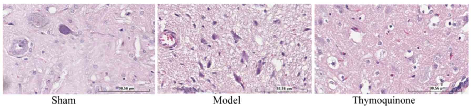
Thymoquinone reduces symptoms of
SCI
To investigate the in vivo effects of
thymoquinone on SCI, SCI rats were treated with thymoquinone from 3
weeks after surgery. SCI model promoted necrosis in the SCI model
group compared with the sham group (Fig.
2). Furthermore, the administration of thymoquinone reduced
necrosis in SCI rats as compared with the SCI model group (Fig. 2).
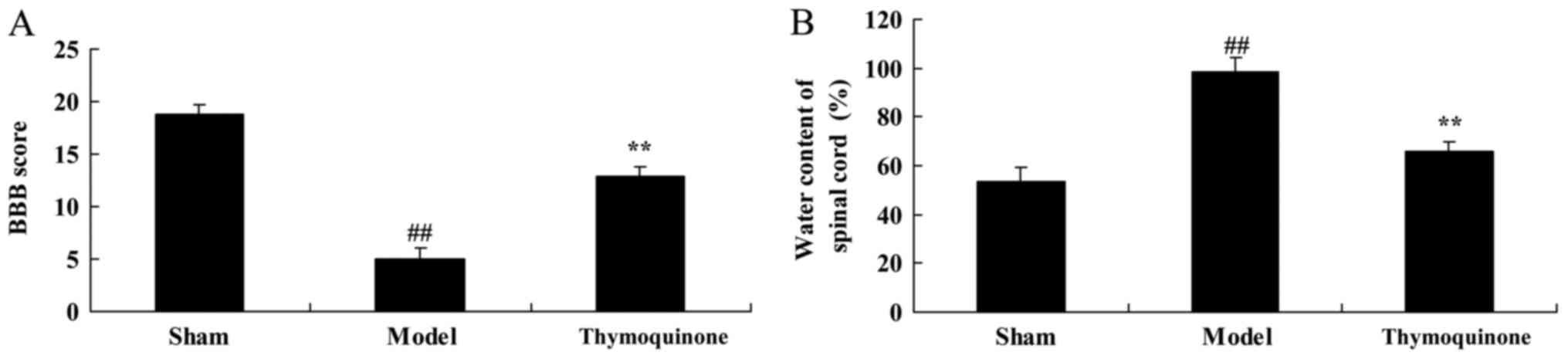
Thymoquinone increases BBB score and
reduces water content in spinal cord tissue
Next, BBB score and water content in spinal cord
tissue were analyzed in SCI rats treated with thymoquinone. As
shown in Fig. 3A, a significant
decrease in BBB score was observed in the SCI model group compared
with the sham group (P<0.01). As shown in Fig. 3B, a significant increase of water
content was observed in the SCI model group tissue, compared with
the sham group (P<0.01). Treatment with thymoquinone
significantly increased BBB score and significantly reduced spinal
cord tissue water content in SCI rats as compared with the SCI
model (P<0.01; Fig. 3A and B).
These results demonstrated that thymoquinone could prevent SCI, but
its mechanism required further elucidation.
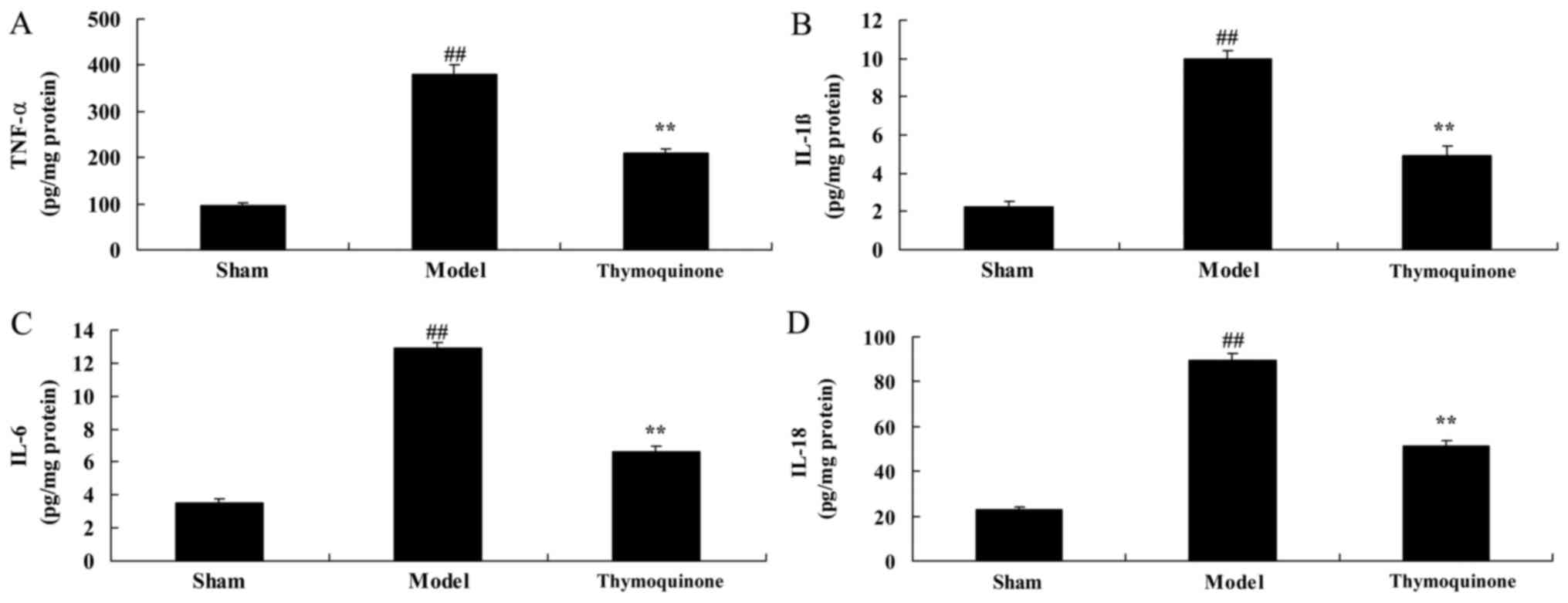
Thymoquinone decreases inflammatory
responses in SCI rats
The levels of inflammatory factors were analyzed to
determine whether thymoquinone affected the inflammatory response
in SCI rats. As shown in Fig. 4,
TNF-α, IL-1β, IL-6 and IL-18 activity levels in the SCI model group
were significantly higher compared with the sham group (P<0.01).
However, TNF-α, IL-1β, IL-6 and IL-18 activity levels were
significantly decreased in SCI rats treated with thymoquinone
compared with the SCI model group (Fig.
4). These results indicated that thymoquinone exhibits
anti-inflammatory effects in the treatment of SCI.
Thymoquinone decreases oxidative
stress in SCI rats
In order to determine whether thymoquinone affects
oxidative stress in SCI rats, SOD, CAT, GSH and MDA activity levels
were measured using ELISA kits. As shown in Fig. 5, SOD, CAT and GSH levels were
significantly decreased and MDA levels were significantly increased
in SCI model rats compared with the sham group (P<0.01).
Thymoquinone treatment significantly increased the SOD, CAT and GSH
activity levels and significantly decreased the MDA activity level
in SCI rats compared with the model group (P<0.01). These
results indicated that thymoquinone inhibits SCI-induced oxidative
stress.
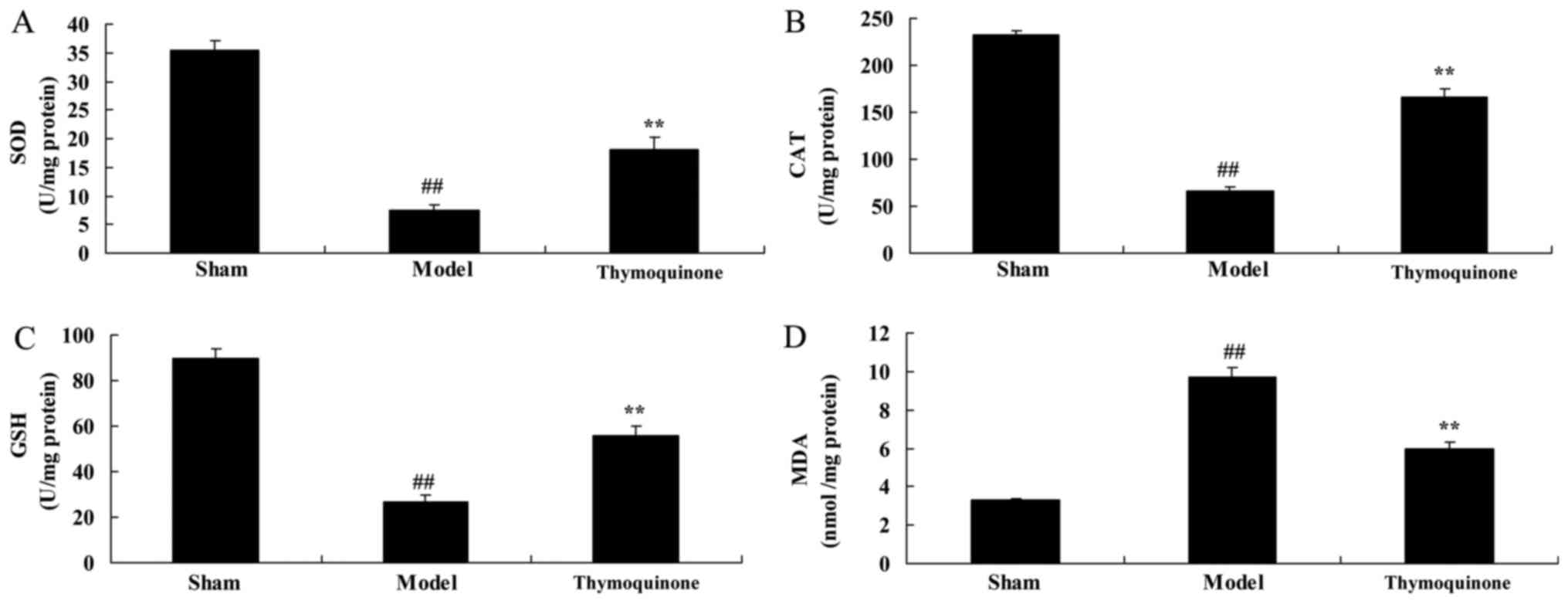 | Figure 5.Effect of thymoquinone on oxidative
stress in SCI rats. Activity levels of (A) SOD, (B) CAT, (C) GSH
and (D) MDA were measured using ELISA kits. ##P<0.01
vs. Sham; **P<0.01 vs. Model. SCI, spinal cord injury; Sham,
sham surgery group; Model, SCI surgery group; Thymoquinone, SCI +
thymoquinone group; SOD, superoxide dismutase; CAT, catalase; GSH,
glutathione; MDA, malondialdehyde. |
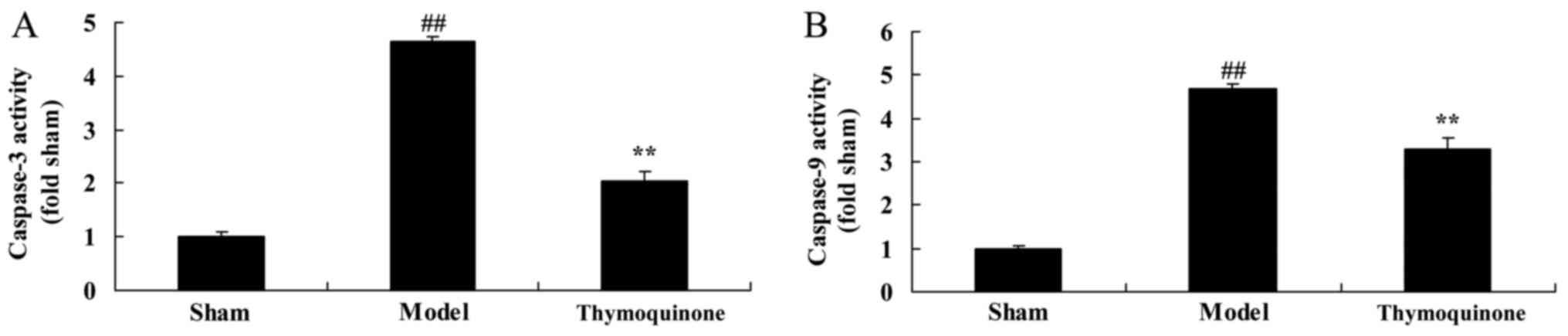
Thymoquinone decreases cell apoptosis
in SCI rats
In order to determine whether thymoquinone regulates
cell apoptosis in SCI rats, caspase-3 and −9 activity levels were
measured using ELISA kits. As shown in Fig. 6, caspase-3 and −9 activity levels
were significantly higher in SCI model rats compared with the sham
group (P<0.01). Treatment with thymoquinone significantly
decreased caspase-3 and −9 activity levels in SCI rats as compared
with the SCI model (P<0.01). These results indicated that
thymoquinone inhibits apoptosis in the treatment of SCI.
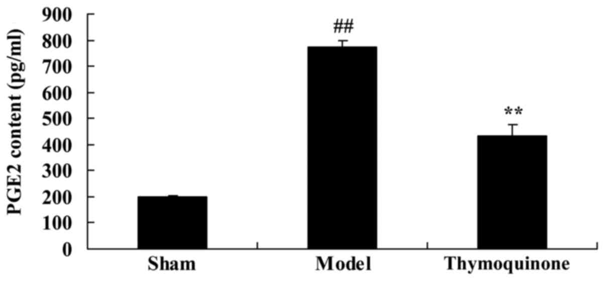
Thymoquinone inhibits prostaglandin E2
(PGE2) activity in SCI rat
To characterize the mechanism of thymoquinone on
SCI, PGE2 activity was analyzed in SCI rats. A significant increase
in PGE2 activity was observed in SCI model rats compared with the
sham group (P<0.01; Fig. 7).
Administration with thymoquinone significantly inhibited PGE2
activity in SCI rats as compared with the SCI model (P<0.01;
Fig. 7). These results indicated
that thymoquinone reduces PGE2 activity to inhibit inflammation in
SCI.
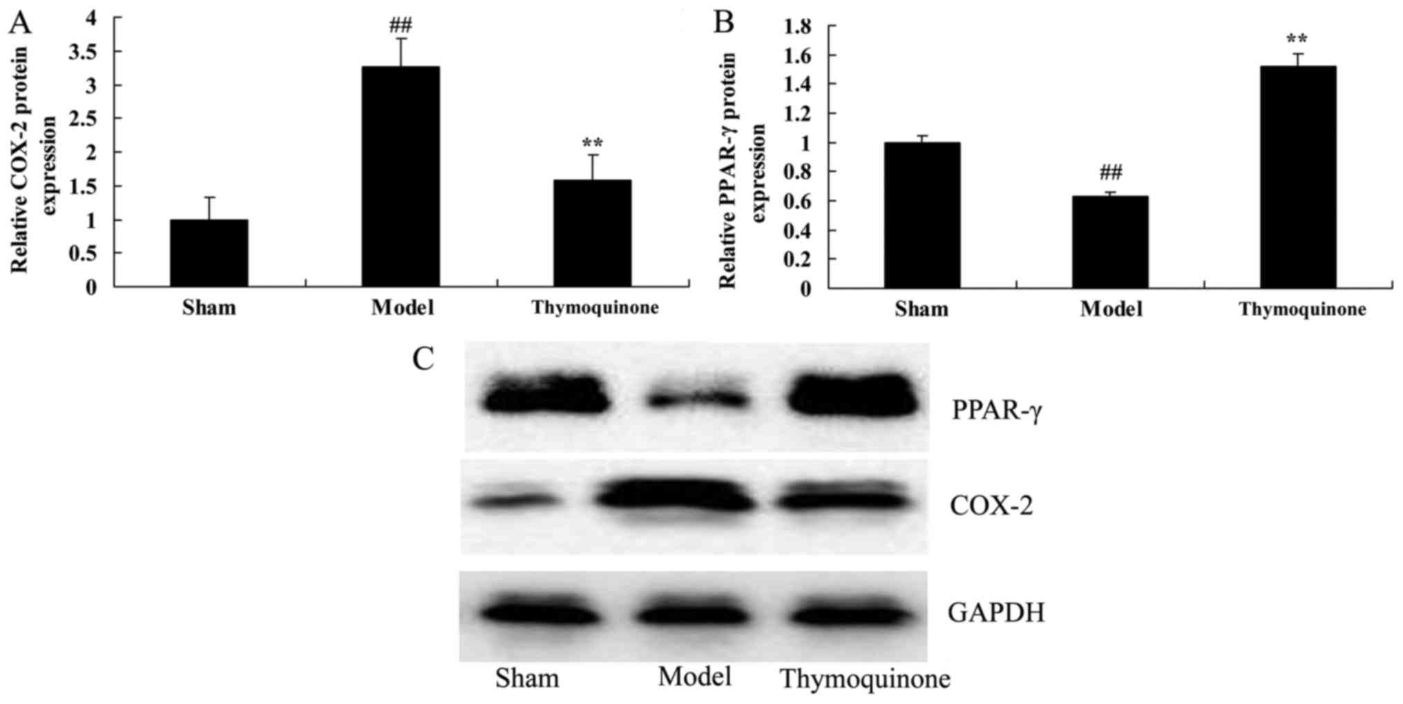
Thymoquinone suppresses COX-2 and
activates PPAR-γ protein expression in SCI rats
To further characterize the mechanism of
thymoquinone in SCI, COX-2 and PPAR-γ protein expression were
analyzed. As shown in Fig. 8, COX-2
protein expression was significantly increased and PPAR-γ protein
expression was significantly decreased in SCI model rats as
compared with the sham group (P<0.01). Thymoquinone
significantly suppressed COX-2 and increased PPAR-γ protein
expression in SCI rats as compared with the SCI model rats
(P<0.01; Fig. 8). These results
indicated that thymoquinone reduces COX-2 activity to inhibit
inflammation, and increases PPAR-γ to reduce apoptosis in SCI.
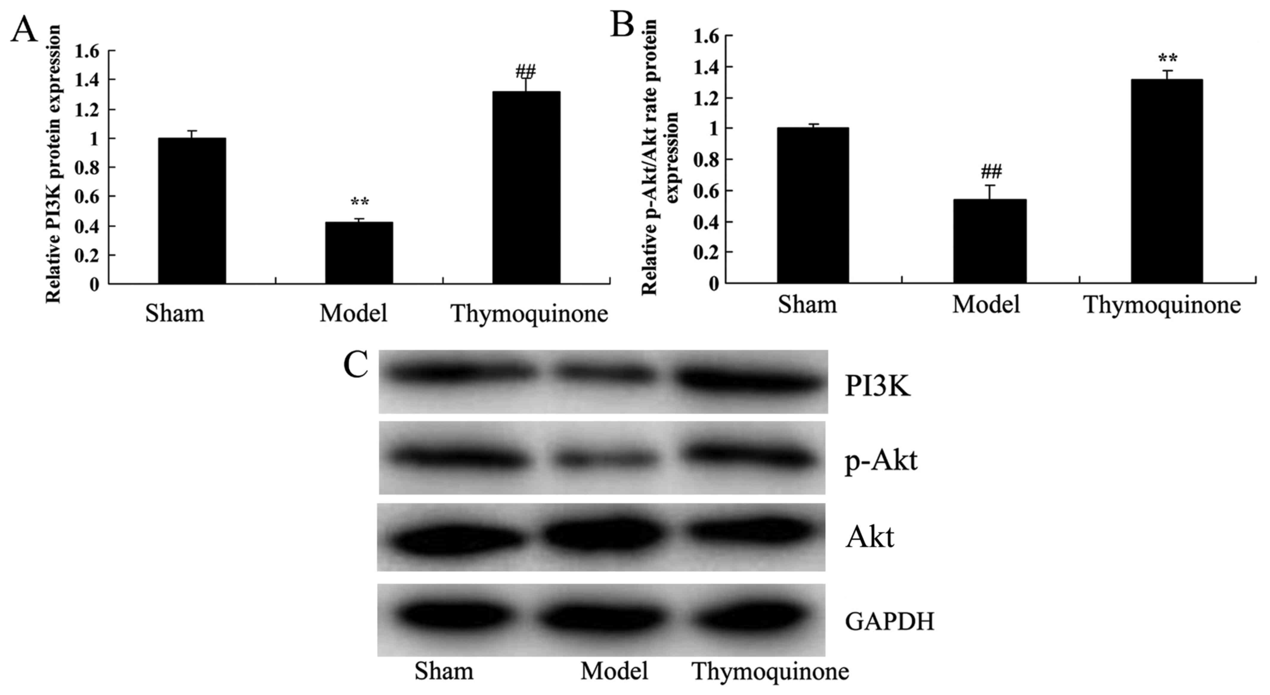
Thymoquinone activates PI3K and
p-Akt/Akt protein expression in SCI rats
In order to evaluate whether the PI3K/Akt pathway
was downregulated early after SCI and whether thymoquinone could
regulate the PI3K/Akt pathway, PI3K and p-Akt/Akt protein
expression were analyzed. There was a significant suppression of
PI3K and p-Akt/Akt protein expressionin the SCI model group
compared with the sham group (P<0.01; Fig. 9). Notably, thymoquinone treatment
significantly promoted the PI3K and p-Akt/Akt protein expression in
SCI rats as compared with the SCI model (P<0.01; Fig. 9). These results suggest that the
PI3K/Akt pathway is involved in the effects of thymoquinone in
SCI.
Discussion
SCI is a serious nervous system injury, which can
cause motor dysfunction and severe disability, thus causing a large
economic burden to individuals, families and society (25). Therefore, it is of great significance
to treat acute SCI and recover or alleviate nerve damage. Acute SCI
is currently classified into primary and secondary SCI (26). Secondary SCI is reversible and can be
controlled. Consequently, secondary SCI determines the patient's
final outcome (27). At present,
treatment of secondary SCI is the key strategy for acute SCI
treatment. Inflammatory response is the primary component of
secondary injury of the spinal cord (25). It was identified in the present study
that thymoquinone can reduce SCI, increase BBB score and decrease
water content in spinal cord tissue in SCI rat.
Cytokines, inflammation, free radicals, excitatory
toxins and other factors can trigger apoptosis (28). Researchers have identified that
apoptosis associated genes are involved. For instance, genes
including caspase-3, B-cell lymphoma 2 (Bcl-2) and Bcl-2-associated
X protein are associated with apoptosis (29). In particular, caspase-3 is closely
correlated with apoptosis regulation (29). In addition, caspase-3 activity has
formed the positive and negative regulation of apoptosis, while the
ratio between the two decides cell apoptosis (29,30).
Massive neuron apoptosis has been identified in numerous central
nervous system injury models (31).
The present data indicate that thymoquinone can decrease
inflammatory response, oxidative stress and cell apoptosis in an
SCI rat model. Dur et al (17) demonstrated that thymoquinone could
prevent inflammation and oxidative stress in rat acute
pancreatitis. These results suggest that thymoquinone exerts an
anti-inflammatory, anti-oxidative and anti-apoptotic effect on
SCI.
Previous studies on PPAR-γ have primarily focused on
lipid metabolism and internal environment stability. It has been
identified that PPAR-γ is involved in numerous physiological and
pathological processes (32). In
addition, PPAR-γ agonists have been demonstrated to reduce marked
neuron loss after being injected into the cerebral cortex (33). During pretreatment of neural and
glial cells, upregulated PPAR-γ expression can upregulate GLT1/EAAT
mRNA expression, which can also be observed in PPAR-γ
agonist-cultured cells. Thus, a nerve protective effect can be
achieved (34). PPAR-γ activation
contributes to reducing the injury effect of free radicals on
nerves through multiple pathways. It has been demonstrated that
PPAR-γ activation can inhibit the expression of free radicals in
patients with progressive spinal muscular atrophy, multiple
sclerosis and inflammation of ischemia-based nervous system
(11). In inflammatory responses,
PPAR-γ can inhibit related inflammatory signal pathways in a
competitive manner for the formation of inflammatory mediators
(33). The current results suggest
that thymoquinone treatment can inhibit PGE2 activity, suppress
COX-2 protein expression and promote PPAR-γ protein expression in
SCI rats. In addition, Pei et al (35) demonstrated that thymoquinone
inhibited angiotens in II-induced vascular smooth muscle cell
proliferation through the PPAR-γ/PPAR-γ coactivator-1 pathway.
These results suggested that treadmill exercise could promote the
protective effect of thymoquinone on SCI by stimulating the
expression of PPAR-γ.
The PI3K/Akt signaling pathway responds to
extracellular signals, growth factors and the energy status of the
cell, as well as cell growth, proliferation, survival and
differentiation of SCI (36). This
pathway serves a key function in neural physiological and
pathological processes (36). As
previously demonstrated, the PI3K/Akt pathway is vital in neural
cell proliferation, development, differentiation, axonal
regeneration, myelin formation, apoptosis and plasticity of
synapses (37,38). The results of the current study
demonstrate that thymoquinone treatment contributes to activating
PPAR-γ and PI3K/Akt protein expression in SCI rats. Liu et
al (39)suggested that
thymoquinone improves cardiovascular function, and inhibits
inflammation, oxidative stress and apoptosis via the PI3K/Akt
pathway in diabetic rats. These results suggest that thymoquinone
induces activation of the PI3K/Akt signaling pathway, which may be
associated with its protective effect against SCI.
The present study demonstrated that thymoquinone can
reduce SCI, increase BBB score and decrease water content in the
spinal cord tissue of an SCI rat model. The protective effects of
thymoquinone on SCI may be attributed to its activation of PPAR-γ
and the PI3K/Akt pathway.
Acknowledgements
Not applicable.
Funding
No funding was received.
Availability of data and materials
The datasets used and/or analyzed during the current
study are available from the corresponding author on reasonable
request.
Authors' contributions
YC and BW conceived and designed the experiments. YC
and HZ performed the experiments. YC and BW analyzed the data and
wrote paper.
Ethics approval and consent to
participate
Not applicable.
Consent for publication
Not applicable.
Competing interests
The authors declare that they have no competing
interests.
References
|
1
|
Arora M, Harvey LA, Hayes AJ, Chhabra HS,
Glinsky JV, Cameron ID, Lavrencic L, Arumugam N, Hossain S and Bedi
PK: Effectiveness and cost-effectiveness of telephone-based support
versus usual care for treatment of pressure ulcers in people with
spinal cord injury in low-income and middle-income countries: Study
protocol for a 12-week randomised controlled trial. BMJ Open.
5:e0083692015. View Article : Google Scholar : PubMed/NCBI
|
|
2
|
Dudley-Javoroski S and Shields RK:
Active-resisted stance modulates regional bone mineral density in
humans with spinal cord injury. J Spinal Cord Med. 36:191–199.
2013. View Article : Google Scholar : PubMed/NCBI
|
|
3
|
Harper LA, Coleman JA, Perrin PB, Olivera
SL, Perdomo JL, Arango JA and Arango-Lasprilla JC: Comparison of
mental health between individuals with spinal cord injury and
able-bodied controls in Neiva, Colombia. J Rehabil Res Dev.
51:127–136. 2014. View Article : Google Scholar : PubMed/NCBI
|
|
4
|
Lόpez-Larraz E, Antelis JM, Montesano L,
Gil-Agudo A and Minguez J: Continuous decoding of motor attempt and
motor imagery from EEG activity in spinal cord injury patients.
Conf Proc IEEE Eng Med Biol Soc. 2012:1798–1801. 2012.PubMed/NCBI
|
|
5
|
Jia C, Liao LM, Chen G and Sui Y: Detrusor
botulinum toxin A injection significantly decreased urinary tract
infection in patients with traumatic spinal cord injury. Spinal
Cord. 51:487–490. 2013. View Article : Google Scholar : PubMed/NCBI
|
|
6
|
Laubacher M, Perret C and Hunt KJ:
Work-rate-guided exercise testing in patients with incomplete
spinal cord injury using a robotics-assisted tilt-table. Disabil
Rehabil Assist Technol. 10:433–438. 2015. View Article : Google Scholar : PubMed/NCBI
|
|
7
|
Rosety-Rodriguez M, Camacho A, Rosety I,
Fornieles G, Rosety MA, Diaz AJ, Bernardi M, Rosety M and Ordonez
FJ: Low-grade systemic inflammation and leptin levels were improved
by arm cranking exercise in adults with chronic spinal cord injury.
Arch Phys Med Rehabil. 95:297–302. 2014. View Article : Google Scholar : PubMed/NCBI
|
|
8
|
Nelissen S, Vangansewinkel T, Geurts N,
Geboes L, Lemmens E, Vidal PM, Lemmens S, Willems L, Boato F,
Dooley D, et al: Mast cells protect from post-traumatic spinal cord
damage in mice by degrading inflammation-associated cytokines via
mouse mast cell protease 4. Neurobiol Dis. 62:260–272. 2014.
View Article : Google Scholar : PubMed/NCBI
|
|
9
|
Amin B, Abnous K, Motamedshariaty V and
Hosseinzadeh H: Attenuation of oxidative stress, inflammation and
apoptosis by ethanolic and aqueous extracts of Crocus sativus L.
stigma after chronic constriction injury of rats. An Acad Bras
Cienc. 86:1821–1832. 2014. View Article : Google Scholar : PubMed/NCBI
|
|
10
|
Yu WR and Fehlings MG: Fas/FasL-mediated
apoptosis and inflammation are key features of acute human spinal
cord injury: Implications for translational, clinical application.
Acta Neuropathol. 122:747–761. 2011. View Article : Google Scholar : PubMed/NCBI
|
|
11
|
Yi JH, Park SW, Brooks N, Lang BT and
Vemuganti R: PPARgamma agonist rosiglitazone is neuroprotective
after traumatic brain injury via anti-inflammatory and
anti-oxidative mechanisms. Brain Res. 1244:164–172. 2008.
View Article : Google Scholar : PubMed/NCBI
|
|
12
|
McTigue DM: Potential therapeutic targets
for PPARgamma after spinal cord injury. PPAR Res. 2008:5171622008.
View Article : Google Scholar : PubMed/NCBI
|
|
13
|
Yan J, Li B, Chen JW, Jiang SD and Jiang
LS: Spinal cord injury causes bone loss through peroxisome
proliferator-activated receptor-γ and Wnt signalling. J Cell Mol
Med. 16:2968–2977. 2012. View Article : Google Scholar : PubMed/NCBI
|
|
14
|
Chae CH and Kim HT: Forced,
moderate-intensity treadmill exercise suppresses apoptosis by
increasing the level of NGF and stimulating phosphatidylinositol
3-kinase signaling in the hippocampus of induced aging rats.
Neurochem Int. 55:208–213. 2009. View Article : Google Scholar : PubMed/NCBI
|
|
15
|
Kim Y, Seger R, Babu Suresh CV, Hwang SY
and Yoo YS: A positive role of the PI3-K/Akt signaling pathway in
PC12 cell differentiation. Mol Cells. 18:353–359. 2004.PubMed/NCBI
|
|
16
|
Randhawa MA, Alenazy AK, Alrowaili MG and
Basha J: An active principle of Nigella sativa L.,
thymoquinone, showing significant antimicrobial activity against
anaerobic bacteria. J Intercult Ethnopharmacol. 6:97–101. 2016.
View Article : Google Scholar : PubMed/NCBI
|
|
17
|
Dur A, Kose H, Kocyigit A, Kocaman O,
Ismayilova M and Sonmez FC: The anti-inflammatory and antioxidant
effects of thymoquinone on ceruleine induced acute pancreatitis in
rats. Bratisl Lek Listy. 117:614–618. 2016.PubMed/NCBI
|
|
18
|
Zhang L, Bai Y and Yang Y: Thymoquinone
chemosensitizes colon cancer cells through inhibition of NF-κB.
Oncol Lett. 12:2840–2845. 2016. View Article : Google Scholar : PubMed/NCBI
|
|
19
|
Liu X, Dong J, Cai W, Pan Y, Li R and Li
B: The effect of thymoquinone on apoptosis of SK-OV-3 ovarian
cancer cell by regulation of Bcl-2 and Bax. Int J Gynecol Cancer.
27:1596–1601. 2017. View Article : Google Scholar : PubMed/NCBI
|
|
20
|
Barkat MA, Abul H, Ahmad J, Khan MA, Beg S
and Ahmad FJ: Insights into the targeting potential of thymoquinone
for therapeutic intervention against triple-negative breast cancer.
Curr Drug Targets. 19:70–80. 2018. View Article : Google Scholar : PubMed/NCBI
|
|
21
|
Jiang ZS, Pu ZC and Hao ZH: Carvacrol
protects against spinal cord injury in rats via suppressing
oxidative stress and the endothelial nitric oxide synthase pathway.
Mol Med Rep. 12:5349–5354. 2015. View Article : Google Scholar : PubMed/NCBI
|
|
22
|
Liu L, Moody J and Gall A: A quantitative,
pooled analysis and systematic review of controlled trials on the
impact of electrical stimulation settings and placement on pressure
ulcer healing rates in persons with spinal cord injuries. Ostomy
Wound Manage. 62:16–34. 2016.PubMed/NCBI
|
|
23
|
Üstün N, Aras M, Ozgur T, Bayraktar HS,
Sefil F, Ozden R and Yagiz AE: Thymoquinone attenuates trauma
induced spinal cord damage in an animal model. Ulus Travma Acil
Cerrahi Derg. 20:328–332. 2014. View Article : Google Scholar : PubMed/NCBI
|
|
24
|
Foroughi Asl H, Talukdar HA, Kindt AS,
Jain RK, Ermel R, Ruusalepp A, Nguyen KD, Dobrin R, Reilly DF,
Schunkert H, et al: Expression quantitative trait Loci acting
across multiple tissues are enriched in inherited risk for coronary
artery disease. Circ Cardiovasc Genet. 8:305–315. 2015. View Article : Google Scholar : PubMed/NCBI
|
|
25
|
Tian DS, Liu JL, Xie MJ, Zhan Y, Qu WS, Yu
ZY, Tang ZP, Pan DJ and Wang W: Tamoxifen attenuates
inflammatory-mediated damage and improves functional outcome after
spinal cord injury in rats. J Neurochem. 109:1658–1667. 2009.
View Article : Google Scholar : PubMed/NCBI
|
|
26
|
Paterniti I, Genovese T, Crisafulli C,
Mazzon E, Di Paola R, Galuppo M, Bramanti P and Cuzzocrea S:
Treatment with green tea extract attenuates secondary inflammatory
response in an experimental model of spinal cord trauma. Naunyn
Schmiedebergs Arch Pharmacol. 380:179–192. 2009. View Article : Google Scholar : PubMed/NCBI
|
|
27
|
Jiang S, Bendjelloul F, Ballerini P,
D'Alimonte I, Nargi E, Jiang C, Huang X and Rathbone MP: Guanosine
reduces apoptosis and inflammation associated with restoration of
function in rats with acute spinal cord injury. Purinergic Signal.
3:411–421. 2007. View Article : Google Scholar : PubMed/NCBI
|
|
28
|
Uchida K, Nakajima H, Watanabe S, Yayama
T, Guerrero AR, Inukai T, Hirai T, Sugita D, Johnson WE and Baba H:
Apoptosis of neurons and oligodendrocytes in the spinal cord of
spinal hyperostotic mouse (twy/twy): Possible pathomechanism of
human cervical compressive myelopathy. Eur Spine J. 21:490–497.
2012. View Article : Google Scholar : PubMed/NCBI
|
|
29
|
Zhang SQ, Wu MF, Gu R, Liu JB, Li Y, Zhu
QS and Jiang JL: Senegenin inhibits neuronal apoptosis after spinal
cord contusion injury. Neural Regen Res. 11:657–663. 2016.
View Article : Google Scholar : PubMed/NCBI
|
|
30
|
Mizuno A, Miyauchi K, Nishizaki Y, Yamazoe
M, Komatsu I, Asano T, Mitsuhashi H, Nishi Y, Niwa K and Daida H:
Impact of the augmentation time ratio on direct measurement of
central aortic pressure in the presence of coronary artery disease.
Hypertens Res. 38:684–689. 2015. View Article : Google Scholar : PubMed/NCBI
|
|
31
|
Xu GY, Liu S, Hughes MG and McAdoo DJ:
Glutamate-induced losses of oligodendrocytes and neurons and
activation of caspase-3 in the rat spinal cord. Neuroscience.
153:1034–1047. 2008. View Article : Google Scholar : PubMed/NCBI
|
|
32
|
Griggs RB, Donahue RR, Morgenweck J, Grace
PM, Sutton A, Watkins LR and Taylor BK: Pioglitazone rapidly
reduces neuropathic pain through astrocyte and nongenomic PPARγ
mechanisms. Pain. 156:469–482. 2015. View Article : Google Scholar : PubMed/NCBI
|
|
33
|
Paterniti I, Impellizzeri D, Crupi R,
Morabito R, Campolo M, Esposito E and Cuzzocrea S: Molecular
evidence for the involvement of PPAR-δ and PPAR-γ in
anti-inflammatory and neuroprotective activities of
palmitoylethanolamide after spinal cord trauma. J
Neuroinflammation. 10:202013. View Article : Google Scholar : PubMed/NCBI
|
|
34
|
Park SW, Yi JH, Miranpuri G, Satriotomo I,
Bowen K, Resnick DK and Vemuganti R: Thiazolidinedione class of
peroxisome proliferator-activated receptor gamma agonists prevents
neuronal damage, motor dysfunction, myelin loss, neuropathic pain,
and inflammation after spinal cord injury in adult rats. J
Pharmacol Exp Ther. 320:1002–1012. 2007. View Article : Google Scholar : PubMed/NCBI
|
|
35
|
Pei X, Li X, Chen H, Han Y and Fan Y:
Thymoquinone inhibits angiotensin II-induced proliferation and
migration of vascular smooth muscle cells through the
AMPK/PPARγ/PGC-1α pathway. DNA Cell Biol. 35:426–433. 2016.
View Article : Google Scholar : PubMed/NCBI
|
|
36
|
Isele NB, Lee HS, Landshamer S, Straube A,
Padovan CS, Plesnila N and Culmsee C: Bone marrow stromal cells
mediate protection through stimulation of PI3-K/Akt and MAPK
signaling in neurons. Neurochem Int. 50:243–250. 2007. View Article : Google Scholar : PubMed/NCBI
|
|
37
|
Zhang P, Zhang L, Zhu L, Chen F, Zhou S,
Tian T, Zhang Y, Jiang X, Li X, Zhang C, et al: The change tendency
of PI3K/Akt pathway after spinal cord injury. Am J Transl Res.
7:2223–2232. 2015.PubMed/NCBI
|
|
38
|
Felix MS, Bauer S, Darlot F, Muscatelli F,
Kastner A, Gauthier P and Matarazzo V: Activation of Akt/FKHR in
the medulla oblongata contributes to spontaneous respiratory
recovery after incomplete spinal cord injury in adult rats.
Neurobiol Dis. 69:93–107. 2014. View Article : Google Scholar : PubMed/NCBI
|
|
39
|
Liu H, Liu HY, Jiang YN and Li N:
Protective effect of thymoquinone improves cardiovascular function,
and attenuates oxidative stress, inflammation and apoptosis by
mediating the PI3K/Akt pathway in diabetic rats. Mol Med Rep.
13:2836–2842. 2016. View Article : Google Scholar : PubMed/NCBI
|