|
1
|
Sadaka F, Doerr D, Hindia J, Lee KP and
Logan W: Continuous electroencephalogram in comatose postcardiac
arrest syndrome patients treated with therapeutic hypothermia:
Outcome prediction study. J intensive care med. 30:292–296. 2015.
View Article : Google Scholar : PubMed/NCBI
|
|
2
|
Deakin CD, Nolan JP, Soar J, Sunde K,
Koster RW, Smith GB and Perkins GD: European resuscitation council
guideline for resuscitation 2010 section 4. Adult advanced life
support. Resuscitation. 81:1305–1352. 2010. View Article : Google Scholar : PubMed/NCBI
|
|
3
|
Pell JP, Sirel JM, Marsden AK, Ford I,
Walker NL and Cobbe SM: Presentation, management and outcome of out
of hospital cardiopulmonary arrest: Comparison by underlying
aetiology. Heart. 89:839–842. 2003. View Article : Google Scholar : PubMed/NCBI
|
|
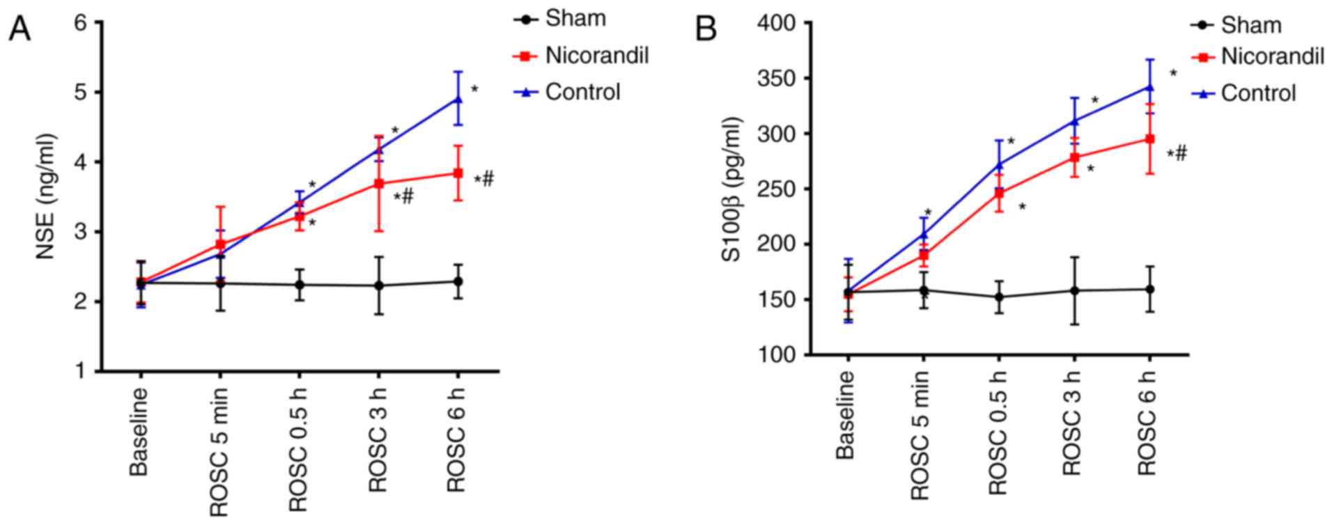
4
|
Nolan JP, Soar J, Wenzel V and Paal P:
Cardiopulmonary resuscitation andmanagement of cardiac arrest. Nat
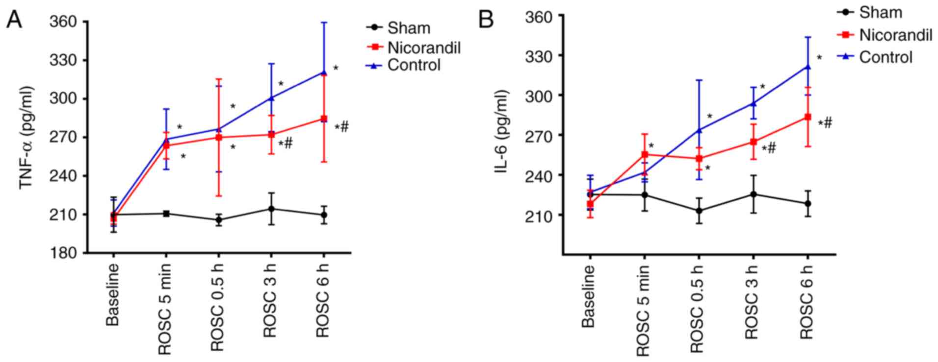
Rev Cardiol. 9:499–511. 2012. View Article : Google Scholar : PubMed/NCBI
|
|
5
|
Neumar RW, Nolan JP, Adrie C, Aibiki M,
Berg RA, Böttiger BW, Callaway C, Clark RS, Geocadin RG, Jauch EC,
et al: Post-cardiac arrest syndrome: Epidemiology, pathophysiology,
treatment and prognostication. A consensus statement from the
international liaison committee on resuscitation (American Heart
Association, Australian and New Zealand Council on Resuscitation,
European Resuscitation Council, Heart and Stroke Foundation of
Canada, Inter American Heart Foundation, Resuscitation Council of
Asia and the Resuscitation Council of Southern Africa); the
American Heart Association Emergency Cardiovascular Care Committee;
the Council on Cardiovascular Surgery and Anesthesia; the Council
on Cardiopulmonary, Perioperative and Critical Care; the Council on
Clinical Cardiology; and the Stroke Council. Circulation.
118:2452–2483. 2008. View Article : Google Scholar : PubMed/NCBI
|
|
6
|
Adrie C, Haouache H, Saleh M, Memain N,
Laurent I, Thuong M, Dargues L, Geurrini P and Monchi M: An
underrecognized source of organ donors: Patients with brain death
after successfully resuscitated cardiac arrest. Intensive Care Med.
34:132–137. 2008. View Article : Google Scholar : PubMed/NCBI
|
|
7
|
Safar P, Behringer W, Bottiger BW and
Sterz F: Cerebral resuscitation potentials for cardiac arrest. Crit
Care Med. 30 4 Suppl:S140–S144. 2002. View Article : Google Scholar : PubMed/NCBI
|
|
8
|
Krumholz A, Stern BJ and Weiss HD: Outcome
from coma after cardiopulmonary resuscitation: Relation to seizures
and myoclonus. Neurology. 38:401–405. 1988. View Article : Google Scholar : PubMed/NCBI
|
|
9
|
Pusswald G, Fertl E, Faltl M and Auff E:
Neurological rehabilitation of severely disabled cardiac arrest
survivors, part II: life situation of patients and families after
treatment. Resuscitation. 47:241–248. 2000. View Article : Google Scholar : PubMed/NCBI
|
|
10
|
Groswasser Z, Cohen M and Costeff H:
Rehabilitation outcome after anoxic brain damage. Arch Phys Med
Rehabil. 70:186–188. 1989.PubMed/NCBI
|
|
11
|
Taraszewska A, Zelman IB, Ogonowska W and
Chrzanowska H: The pattern of irreversible brain changes after
cardiac arrest in humans. Folia Neuropathol. 40:133–141.
2002.PubMed/NCBI
|
|
12
|
Lipton P: Ischemic cell death in brain
neurons. Physiol Rev. 79:1431–1568. 1999. View Article : Google Scholar : PubMed/NCBI
|
|
13
|
Horinaka S: Use of nicorandil in
cardiovascular disease and its optimization. Drugs. 71:1105–1119.
2011. View Article : Google Scholar : PubMed/NCBI
|
|
14
|
Wu H, Ye M, Yang J, Ding J, Yang J, Dong W
and Wang X: Nicorandil protects the heart from ischemia/reperfusion
injury by attenuating endoplasmic reticulum response-induced
apoptosis through PI3K/Akt signaling pathway. Cell Physiol Biochem.
35:2320–2332. 2015. View Article : Google Scholar : PubMed/NCBI
|
|
15
|
Nagata K, Obata K, Odashima M, Yamada A,
Somura F, Nishizawa T, Ichihara S, Izawa H, Iwase M, Hayakawa A, et
al: Nicorandil inhibits oxidative stress-induced apoptosis in
cardiac myocytes through activation of mitochondrial ATP-sensitive
potassium channels and a nitrate-like effect. J Mol Cell Cardiol.
35:1505–1512. 2003. View Article : Google Scholar : PubMed/NCBI
|
|
16
|
Raveaud S, Verdetti J and Faury G:
Nicorandil protects ATP-sensitive potassium channels against
oxidation-induced dysfunction in cardiomyocytes of aging rats.
Biogerontology. 10:537–547. 2009. View Article : Google Scholar : PubMed/NCBI
|
|
17
|
Lacza Z, Snipes JA, Kis B, Szabó C, Grover
G and Busija DW: Investigation of the subunit composition and the
pharmacology of the mitochondrial ATP-dependent K+ channel in the
brain. Brain Res. 994:27–36. 2003. View Article : Google Scholar : PubMed/NCBI
|
|
18
|
Bajgar R, Seetharaman S, Kowaltowski AJ,
Garlid KD and Paucek P: Identification and properties of a novel
intracellular (mitochondrial) ATP-sensitive potassium channel in
brain. J Biol Chem. 276:33369–33374. 2001. View Article : Google Scholar : PubMed/NCBI
|
|
19
|
Teshima Y, Akao M, Baumgartner WA and
Marbán E: Nicorandil prevents oxidative stress-induced apoptosis in
neurons by activating mitochondrial ATP-sensitive potassium
channels. Brain Res. 990:45–50. 2003. View Article : Google Scholar : PubMed/NCBI
|
|
20
|
Kurihara J, Ochiai N and Kato H:
Protection by nicorandil against the dysfunction of the central
vagal baroreflex system following transient global cerebral
ischemia in dogs. Br J Pharmacol. 109:1263–1267. 1993. View Article : Google Scholar : PubMed/NCBI
|
|
21
|
Xia YF, Wang ZP, Zhou YC, Yan T and Li ST:
Cerebral protective effect of nicorandil premedication on patients
undergoing liver transplantation. Hepatobiliary Pancreat Dis Int.
11:132–136. 2012. View Article : Google Scholar : PubMed/NCBI
|
|
22
|
National Research Council of The National
Academies: Guide for the Care and Use of Laboratory Animals. 8th
edition. The National Academies Press; Washington, DC: 2010
|
|
23
|
Marangos PJ, Schmechel DE, Parma AM and
Goodwin FK: Developmental profile of neuron-specific (NSE) and
non-neuronal (NNE) enolase. Brain Res. 190:185–193. 1980.
View Article : Google Scholar : PubMed/NCBI
|
|
24
|
Heizmann CW, Fritz G and Schäfer BW: S100
proteins: Structure, functions and pathology. Front Biosci.
7:d1356–d1368. 2002. View
Article : Google Scholar : PubMed/NCBI
|
|
25
|
Cavus E, Bein B, Dörges V, Stadlbauer KH,
Wenzel V, Steinfath M, Hanss R and Scholz J: Brain tissue oxygen
pressure and cerebral metabolism in an animal model of cardiac
arrest and cardiopulmonary resuscitation. Resuscitation. 71:97–106.
2006. View Article : Google Scholar : PubMed/NCBI
|
|
26
|
Ekmektzoglou KA, Xanthos T and
Papadimitriou L: Biochemical markers (NSE, S-100, IL-8) as
predictors of neurological outcome in patients after cardiac arrest
and return of spontaneous circulation. Resuscitation. 75:219–228.
2007. View Article : Google Scholar : PubMed/NCBI
|
|
27
|
Rundgren M, Karlsson T, Nielsen N,
Cronberg T, Johnsson P and Friberg H: Neuron specific enolase and
S-100B as predictors of outcome after cardiac arrest and induced
hypothermia. Resuscitation. 80:784–789. 2009. View Article : Google Scholar : PubMed/NCBI
|
|
28
|
Vereczki V, Martin E, Rosenthal RE, Hof
PR, Hoffman GE and Fiskum G: Normoxic resuscitation after cardiac
arrest protects against hippocampal oxidative stress, metabolic
dysfunction and neuronal death. J Cereb Blood Flow Metab.
26:821–835. 2006. View Article : Google Scholar : PubMed/NCBI
|
|
29
|
Richards EM, Fiskum G, Rosenthal RE,
Hopkins I and McKenna MC: Hyperoxic reperfusion after global
ischemia decreases hippocampal energy metabolism. Stroke.
38:1578–1584. 2007. View Article : Google Scholar : PubMed/NCBI
|
|
30
|
Xing J and Lu J: HIF-1α activation
attenuates IL-6 and TNF-α pathways in hippocampus of rats following
transient global ischemia. Cell Physiol Biochem. 39:511–520. 2016.
View Article : Google Scholar : PubMed/NCBI
|
|
31
|
Kleffner I, Bungeroth M, Schiffbauer H,
Schäbitz WR, Ringelstein EB and Kuhlenbäumer G: The role of
aquaporin-4 polymorphisms in the development of brain edema after
middle cerebral artery occlusion. Stroke. 39:1333–1335. 2008.
View Article : Google Scholar : PubMed/NCBI
|
|
32
|
Ho JD, Yeh R, Sandstrom A, Chorny I,
Harries WE, Robbins RA, Miercke LJ and Stroud RM: Crystal structure
of human aquaporin 4 at 1.8 Å and its mechanism of conductance.
Proc Natl Acad Sci. 106:7437–7442. 2009. View Article : Google Scholar : PubMed/NCBI
|
|
33
|
Xiao F, Arnold TC, Zhang S, Brown C,
Alexander JS, Carden DL and Conrad SA: Cerebral cortical
aquaporin-4 expression in brain edema following cardiac arrest in
rats. Acad Emerg Med. 11:1001–1007. 2004. View Article : Google Scholar : PubMed/NCBI
|
|
34
|
Wang H, Wang X and Guo Q: The correlation
between DTI parameters and levels of AQP-4 in the early phases of
cerebral edema after hypoxic-ischemic/reperfusion injury in
piglets. Pediatr Radiol. 42:992–999. 2012. View Article : Google Scholar : PubMed/NCBI
|
|
35
|
Nunes C, Barbosa RM, Almeida L and
Laranjinha J: Nitric oxide and DOPAC-induced cell death: from GSH
depletion to mitochondrial energy crisis. Mol Cell Neurosci.
48:94–103. 2011. View Article : Google Scholar : PubMed/NCBI
|
|
36
|
Radak D, Resanovic I and Isenovic ER: Link
between oxidative stress and acute brain ischemia. Angiology.
65:667–676. 2014. View Article : Google Scholar : PubMed/NCBI
|
|
37
|
Dunn-Meynell AA and Rawson NE:
Distribution and phenotype of neurons containing the ATP-sensitive
K+ channel in rat brain. Brain Res. 814:41–54. 1998. View Article : Google Scholar : PubMed/NCBI
|
|
38
|
Garlid KD: Mitochondrial potassium
transport: The K(+) cycle. Biochim Biophys Acta. 1606:23–41. 2003.
View Article : Google Scholar : PubMed/NCBI
|
|
39
|
Tarkin J M and Kaski JC: Vasodilator
therapy: Nitrates and nicorandil. Cardiovasc Drugs Ther.
30:367–378. 2016. View Article : Google Scholar : PubMed/NCBI
|