Introduction
The incidence of kidney stones has been increasing
over the years (1). What's more, the
incidence rate of kidney stone disease increases with aging
(2,3). Senile urolithiasis belongs to complex
renal calculi disease, since the patients usually have many
comorbidities, such as chronic heart, lung, liver, kidney
disfuction or severe urinary tract infection (4), adding extra difficulty to clinical
treatment. Patients with kidney stones usually presented with
hydronephrosis, back pain and fever and other symptoms, and need
active and effective surgical treatment (5). Safe, timely and effective treatment of
kidney stones can reduce serious complications such as kidney
failure in senile patients, and significantly improve quality of
life. This has brought urologists new requirements and
challenges.
Nowadays, operation methods to treat kidney stones
include open surgery, extracorporeal shock wave lithotripsy (ESWL),
percutaneous nephroscope lithotripsy (PCNL) and ureteroscopic
lithotripsy (URL). Traditional open surgery has lots of
complications with considerate influence to renal function. The
elderly with low body reserve capacity, reduced tolerance to
surgery, slow postoperative recovery may develop many complications
as the result of prolonged bed rest after open surgery (6,7). In the
past, limited by the hardware, kidney stones were treated by ESWL
or PCNL with limited safety and efficacy (8). In recent years, with the development of
flexible ureteroscope technical and auxiliary equipment (9), the flexible ureteroscopy (fURS)
combined with holium laser lithotripsy is becoming mature and has
gradually become an ideal choice for kidney stone. Compared with
the ESWL or PCNL, especially in the treatment of senile kidney
stone, its advantages are higher stone-free rate (SFR) and safer
operation and fewer intraoperative and postoperative complications
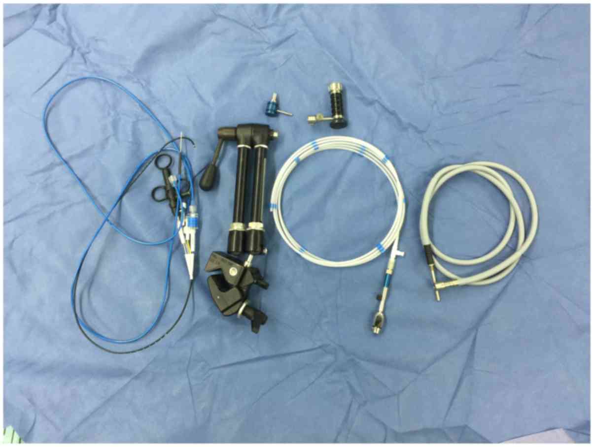
(10,11). PolyScope is a novel removable modular
flexible ureteroscope manufactured by Polydiagnost GmbH
(Pfaffenhofen, Germany) (Fig. 1).
The core components are the independent, optical, imaging modules,
convenient to assemble or disassemble. The optical system has a
soft yet tough synthetic metal wire net wrapped around for
protection. The outer casing of the flexible ureteroscope is
replaceable at a relatively low price. This eradicates the risk of
cross-infection between operations, and enables the ureteroscope be
used in successive procedures. The main advantage of PolyScope is
its convenience for the replacement of consumable components to
reduce the maintenance costs (12).
We had already evaluated PolyScope against its conventional
counterpart earlier (13). Yet its
safety and effectiveness in treating senile patients, a special
group of patients with urolithiasis, has not been reported so far.
In the present study, from December 2013 to December 2016, 157
cases of senile patients with kidney stones were treated with
PolyScope in Xin Hua Hospital Affiliated to Shanghai Jiao Tong
University, School of Medicine (Shanghai, China). The results were
satisfactory and are reported in the following sections.
Patients and methods
Patient characteristics
This is a retrospective study. A total of 157
patients (88 males and 69 females, aged from 60 to 82 years, mean
age 69.1±6.1 years old) with kidney stone who were admitted to Xin
Hua Hospital Affiliated to Shanghai Jiao Tong University, School of
Medicine from December 2013 to December 2016 were recruited for
this study. The body mass index (BMI) ranged from 16.3 to 32.5 and
the mean BMI was 23.4±2.9. Nineteen patients had I degree of
obesity (25≤ BMI <30) and 5 patients had with II degree of
obesity (BMI ≥30). A total of 86 patients presented with
hypertension, 26 with cardiovascular disease, 21 with
anticoagulants or antiplatelet medications, 15 with diabetes, 38
with hydronephrosis, 13 with creatinine abnormality, 27 with COPD,
2 with horseshoe kidney, 2 with congenital solitary kidney.
Thirty-two of the patients had been hospitalized and treated with
ESWL, 17 with PCNL, 12 with fURS. Nineteen patients had previously
had an indwelling double-J stent placed with ureteroscopic guidance
in other hospital. All patients had been diagnosed by plain film of
kidney-ureter-bladder (KUB) intravenous urography (IVU) and
computed tomography (CT). In addition, 149 patients exhibited
one-sided calculi, whereas 8 patients exhibited two-sided calculi.
The total number of renal calculi was 245 and mean number of each
side was 1.5±1.0, including 61 renal pelvic calculi, 43 upper
calyceal calculi, 55 middle calyceal calculi, 86 lower calyceal
calculi. Digital surface area of each patient ranged from 55.8 to
478.3 mm2 (mean, 154.6±57.7 mm2). All
patients' middle segment urine samples were collected for the
germiculture and sensitivity test. The result was that 148 cases
presented with no bacterial growth, 7 with Escherichia coli, 2 with
Proteus. The information of 332 adult patients (185 males and 147
females, aged from 18 to 59 years, mean age 41.5±9.9 years old) of
the same period, who were also treated with PolyScope, were also
analyzed and compared with that of the senile patients (Table I). The two groups had difference in
comorbidity rate, including hypertension (P<0.01),
cardiovascular disease (P<0.05), anti-coagulation (P<0.05)
and COPD (P<0.01). Other criteria were statistically similar
between two groups.
 | Table I.Demographical characteristics. |
Table I.
Demographical characteristics.
| Variable | Senile | Non-senile | P-value |
|---|
| Age
(years)a | 69.1±6.1 | 41.5±9.9 | <0.01 |
| Sex (n) |
| Male | 88 | 185 | NS |
|
Female | 69 | 147 |
|
| Body mass index
(kg/m2)a | 23.4±2.9 | 22.9±3.6 | NS |
| Intervention history
(%) |
| ESWL | 32
(20.4) | 59
(17.8) | NS |
| PCNL | 17
(10.8) | 45
(13.6) | NS |
| fURS | 12 (7.6) | 31
(9.3) | NS |
| Preoperative stenting
(%) | 19
(12.1) | 20
(6.0) | NS |
| Comorbidity (%) |
|
Hypertension | 86
(54.8) | 74
(22.3) | <0.01 |
|
Cardiovascular disease | 26
(16.6) | 35
(10.5) | <0.05 |
|
Anticoagulation | 21
(13.4) | 23
(6.9) | <0.05 |
| Chronic
obstructive pulmonary disease | 27
(17.2) | 18
(5.4) | <0.01 |
| Morbid
obesity | 24
(15.3) | 73
(22.0) | NS |
| Diabetes
mellitus | 15
(9.6) | 19
(5.7) | NS |
|
Hydronephrosis | 38
(24.2) | 70
(21.1) | NS |
| Urinary
tract infection | 21
(13.4) | 31 (9.3) | NS |
| Abnormal
creatine | 13 (8.3) | 17
(5.1) | NS |
| Horseshoe
kidney | 2
(1.3) | 14
(4.2) | NS |
| Solitary
kidney | 2
(1.3) | 3
(0.9) | NS |
| Stone side (n) |
|
Unilateral | 149 | 305 | NS |
|
Bilateral | 8 | 27 | NS |
| Stone location
(n) |
|
Upper | 43 | 129 | NS |
|
Middle | 55 | 114 | NS |
|
Lower | 86 | 181 | NS |
|
Pelvis | 61 | 147 | NS |
| Number of stones
per side (n)a |
1.5±1.0 |
1.6±0.8 | NS |
| Mean stone burden
(mm)a | 13.2±4.9 | 13.7±5.8 | NS |
| Mean digitized
surface area (mm2)a | 154.6±57.7 | 163.5±69.3 | NS |
| UAS successful
insertion rate (%) | 138 (87.9) | 296 (89.2) | NS |
| Operative time
(min)a |
54.6±13.2 |
67.1±16.5 | NS |
| Single session SFR
(%) | 128 (81.5) | 246 (74.1) | NS |
| Overall SFR
(%) | 140 (89.2) | 284 (85.5) | NS |
| Hospitalization
stay (days)a |
1.2±1.6 |
1.4±1.2 | NS |
| Complication rate
(%) |
|
| NS |
|
Persistent hematuria | 3 (1.9) | 5 (1.5) | NS |
|
Ureteral perfortion | 0 (0) | 0 (0) | NS |
|
Fever | 24 (15.3) | 43 (13.0) | NS |
Operational parameters
All patients were placed under spinal or general
anesthesia (153 spinal anesthesia, 4 general anesthesia) in a
lithotomy position with head tilted down. After location of the
ureter, a zebra guide wire was inserted and a Wolf F8/9.8 rigid
ureteroscope (Richard Wolf GmbH, Knittlingen, Germany) was placed
into the ureter to reach the level of the pelvis. Following the
removal of the rigid ureteroscope, a Cook F12/14 ureteral access
sheath (UAS; Cook Medical, Inc., Bloomington, IN, USA) was
positioned along the guide wire, as close to the level of the
uretero-pelvic junction as possible, then PolyScope was inserted
into the pelvis through the UAS. The power of the 220 mm holmium
laser lithotripsy was set at 1.0–2.0 J. The ‘nibble’ approach was
used to break and crumble the stones into fine granules measuring
2–3 mm. Some stones with the diameter of 4–5 mm were removed with a
stone basket, while some lower calyceal stones were translocated
with the basket from the lower to the upper or middle calyx for
improving the efficiency of lithotripsy and also for protection of
the optical fiber.
A F6 double-J stent was typically indwelled for 4
weeks following the surgery and a Foley urethral catheter was
indwelled for 1–2 days. Plain film of KUB was performed 1 day
subsequent to the surgery in order to determine the result of the
lithotripsy and the position of the double-J stent. Postoperative
follow-up to the hospital in a month was required. KUB or CT was
performed and double-J stent removed.
Ethics approval and consent to
participate
The Ethics Committee of Xin Hua Hospital Affiliated
to Shanghai Jiao Tong University School of Medicine has approved
the study and permitted the waiver of written informed consent from
patients. The study protocol is also in accordance with the
Declaration of Helsinki.
Statistical analysis
The data were analyzed using the SPSS 17.0 (SPSS,
Inc., Chicago, IL, USA). Continuous data were presented as mean ±
standard deviation, and compared using Student's t-test. For
categorical data, the values outside the brackets are the absolute
patient number and the values inside the brackets are the
percentage, and were compared using Chi-square or Fisher's exact
test, and P<0.05 was considered statistically significant. All
tests were performed as a 2-tailed test.
Results
Lithotripsy was performed successfully in all 157
patients. 139 patients underwent one-stage lithotripsy and 18
patients two-stage. The duration of the surgery ranged from 37 to
124 min (mean duration, 54.6±13.2 min). Little bleeding was
recorded and no ureteral perforation or damage was encountered. A
total of 24 patients presented with fever following the surgery,
their body temperature all returned to normal following
anti-inflammatory treatment with either empirical antibiotics or
according to the susceptibility results of preoperative urine
culture. Three patients' urine color remained pink in one week
after surgery, yet all became normal following the use of
hemostatic drugs. The SFR was 81.5% for one-stage lithotripsy and
89.2% for two-stage lithotripsy. The remaining 17 patients had
postoperative residual stones (diameter ranged from 4 to 5 mm,
lower calyceal calculi) and did not take further ESWL treatment.
The postoperative follow-up period was 3–24 months. All symptoms of
back pain and abdominal pain were eliminated or greatly relieved
and hydronephrosis was improved markedly according to KUB or CT.
The operation data of the senile group was compared to that of the
non-senile group. Operative time and SFR were similar between two
groups, so was the hospitalization length and complication rates
(Table I).
Within the senile group, between the patients with
and without fever, the diabetes mellitus ratio (P<0.05),
hydronephrosis ratio (P<0.05) and urinary tract infection ratio
(P<0.01), was significantly higher, and the operative time was
also significantly longer (P<0.01) (Table II).
| Table II.Comparison between febrile and
afebrile senile patients. |
Table II.
Comparison between febrile and
afebrile senile patients.
| Characteristic | Febrile senile | Afebrile
senile | P-value |
|---|
| Age (years) | 68.5±5.8 | 69.2±6.0 | NS |
| Body mass index
(kg/m2) | 23.7±3.0 | 23.3±2.8 | NS |
| Preoperative
stenting (%) | 4 (16.7) | 15 (11.3) | NS |
| Diabetes mellitus
(%) | 6 (25.0) | 9 (6.8) | <0.05 |
| Hydronephrosis
(%) | 10 (41.7) | 28 (21.1) | <0.05 |
| Urinary tract
infection (%) | 8 (33.3) | 13 (9.8) | <0.01 |
| Mean stone burden
(mm) | 13.9±5.4 | 13.1±4.6 | NS |
| Mean digitized
surface area (mm2) | 170.1±74.6 | 151.8±54.9 | NS |
| Operative time
(min) |
62.6±17.4 | 53.2±11.9 | <0.05 |
| Total number
(n) | 24 | 133 |
|
Discussion
The world's elderly population is growing at a rapid
rate. In 2015, people aged ≥65 years accounted for 8.5% of the
world's population, and this proportion is expected to rise to 17%
by the year 2050 (14). Chronic
disease, such as hypertension, diabetes and cardiovascular disease,
has high prevalence among the elderly. And they are also weaker
than the young in cardiopulmonary function, immunity, restorability
and so on. On the other hand, percentage of obese patients in the
elderly is also increasing gradually with the rising of the living
standards. Now a days, the selection of treatment for renal calculi
with which kind of minimally invasive method become a prominent
question. ESWL or PCNL used to be the choice for the majority of
senile patients with kidney stones. ESWL is a kind of non-invasive
treatment exhibiting desirable efficacy and safety. However, its
SFR is influenced by many factors, such as the size, the location
and the texture of stone, the obesity degree of patient and so on.
Also, larger stones are often too large to be shattered and require
several times of treatment to reach the target size. The reported
clearance rate of ESWL for lower calyceal stone is 25–85% (15). From our experience, it is often very
difficult, if not impossible, for the lower calyceal stone to be
cleared by ESWL. And the SFR of ESWL for obese patients is also
greatly reduced (16). It is
reported that, in comparison with the non-senile group treated by
ESWL, there was no significant difference in SFR in senile group
(17). However, the incidence of
complications was higher in senile group than that in non-senile
group. Compared with ESWL, the SFR of PCNL is higher, especially
for large stones (18). PCNL is an
invasive method which is not suitable for relatively weak senile
patient with coagulation disorders or poor heart and lung function.
The hospitalization time of PCNL is also longer than that of ESWL
and URL (19,20). The difficulty of puncture of PCNL in
obese patients increases and effect of operation decreases. Berkan
used to score and grade on senile patients with kidney stone before
PCNL according to Charlson Comorbidity Index. He found that the
incidence of complications rose with the score increasing and
suggested to take conservative treatment instead of operation for
those asymptomatic kidney stone patients with high scores (21).
The technology of fURS has developed rapidly since
it was reported by Bagley in 1987 for the first time. fURS has
higher SFR for lower calyceal calculi and stone larger than 1 cm
compared with ESWL (22). fURS
rarely cause severe arrhythmia which may be induced by ESWL in
senile patients with basic rhythm problems and renal colic caused
by gravels clogging the ureter after ESWL in some patients
(23). fURS has comparable SFR to
PCNL, however it has lots of advantages, such as fewer
contraindications with a wider range of application, smaller trauma
without puncture, less blood loss without potential demand for
blood transfusion, shorter hospital stay with quicker recovery
(24). Since it is unnecessary to
puncture, there are no special requirements for the patient's body
shape in fURS. Its therapeutic effect in obese patient is similar
to that in patient with normal BMI (25). In our study, all obese patients,
including 19 patients with I degree of obesity (25≤ BMI <30) and
5 patients with II degree of obesity (BMI ≥30), underwent
successful operation and there was no significant difference in
operation time with patients with normal BMI. Although some
patients had poor cardiopulmonary function, there was no relevant
accident or complication due to less influence of spinal anesthesia
on circulatory/respiratory system compared with general
anesthesia.
PolyScope has the advantages of simple operation,
clear visibility and a reliable efficacy (12,26).
However, the most obvious weakness of this type of flexible
ureteroscope is its capability of being able to flex in just one
direction. In an empty working channel, the lens barrel has been
demonstrated to bend at an angle of 265; the bending angle is
reduced by 10 and 2% respectively, following the insertion of a
3.0F stone basket or a 200 µm laser fiber (12). Before the insertion of the modular
flexible ureteroscope, it was necessary for the UAS to be
positioned near the level of the ureteropelvic junction. This aids
to reduce the renal pelvic pressure during surgery, serve as
protection for the lens barrel and provide a convenient access for
the extraction of stone fragments (26). In case of ureteral stenosis, it is
necessary to indwell an F6 double-J stent for a second operation.
It is also necessary for the end of the flexible ureteroscope to be
maintained in a stretched state during the insertion of the 220 µm
holmium laser optical fiber in order to avoid damaging the work
channel of the lens barrel and the kidney. When losing the sense of
direction in the intracavity, just take out the ureteroscope for
reorientation.
For senile patients with kidney stones, the
following tips need to be remembered: i) As the majority of senile
patients have commodities of hypertension and/or diabetes, their
blood pressure and blood sugar need to be monitored closely with
systolic pressure under 140–160 mmHg and blood sugar between 6–10
mmol/l. ii) Operation time need to be controlled strictly, as the
immune system of the senile is relatively weak. The plan of stage
operation should be made for those patients with large stone. Avoid
long-time lithotripsy might reduce the probability of postoperative
fever. It is necessary to do the germiculture and sensitivity test
before operation and take postoperative prophylactic antibiotic
therapy. iii) As many senile patients take antiplatelet drugs or
anticoagulant drugs like aspirin and clopidogrel, they need to stop
taking the drug for one week to restore their coagulation function.
Timely restoration of the use of antiplatelet or anticoagulant
agents after the surgery according to the urine color is also
necessary. Usually, there is no problem to restore their use 3 days
after operation. The duration should be longer if the urine is
still red. In particular, urologists should be cautious with the
patients with diabetes for many years. Operations should be gentle
and meticulous to avoid ureteral mucosa injury, hemorrhage and
postoperative infection.
Our experience and suggestions are as follows: i) In
consideration of the relatively weak immunity of senile patients,
the use of PolyScope can avoid cross infection as it is disposable.
ii) The 8F diameter lens barrel means the gap between lens barrel
and UAS in the PolyScope is larger than that in the traditional
fURS with 8.5F diameter lens barrel. The large gap brings many
advantages, such as sufficient drainage which can reduce
intrapelvic pressure effectively, great vortex effects which can
accelerate stone extraction and decreased fraction between the lens
barrel and the stone which can reduce the damage of lens barrel.
iii) We recommend upgrading the PolyScope by adding a ‘handle’ just
like the one used in SMP (27),
consisting of a straight and an oblique bifurcated tube. The
straight tube can be contiguous with the access sheath and has a
receptacle for a silicone or rubber cap at the proximal end. A
longitudinal slit can be designed along the axis of the oblique
tube to be used as a pressure vent through which the negative
aspiration pressure can be adjusted. The end of the oblique tube
can be connected to a continuous negative pressure aspirator
through clear flexible tubing with the same or larger lumen. A
specimen collection bottle can be added between the handle and the
aspirator to facilitate stone fragment collection.
Although the quality of fURS has been greatly
improved in recent years, it is still not endurable enough to deal
with the numerous patents with urolithiasis, especially in a
country with a huge population like China. In a multi-center
clinical trial, Knudsen et al found that flexible
ureteroscope required fixing after its use in 5.3–18 patients
(28–30). In addition, according to statistics
from multiple articles, the main reasons for repair were the
decreased capacity of active bending, the decreased image quality
caused by fiber cuts and the sparking of laser in lens barrel
(28–32). It is necessary to protect the
flexible ureteroscope during operation. The indwelling UAS provides
a convenient access point and protection for the lens barrel,
benefits for the extraction of stone fragments, also helps to
reduce the renal pelvic pressure during surgery which can reduce
the intraoperative absorption of lavage fluid and the incidence of
postoperative fever and bacteremia (32,33). It
is of great significance to adjust the position and curvature of
lens barrel according to the ureteral walk, kidney shape, calculi
position and calculi size by carefully reading preoperative CTU/IVU
(26). The protection of flexible
ureteroscope is particularly important in the treatment of lower
calyceal calculi as in such circumstance, the flexible ureteroscope
needs to maintain a relatively large angle bending state for a long
time. In our study, we all take measures of adjusting patients'
position and trying to translocate lower calyceal calculi with the
stone basket from the lower to the upper or middle calyx to improve
the efficiency of the stone breaking. On the one hand, this method
can prevent calculi running to the place that the flexible
ureteroscope can't reach; on the other hand, it can extend the life
of the flexible ureteroscope and reduce equipment losses and
maintenance costs by reducing the time of maximal bending.
The removable modular flexible ureteroscopic
management with holmium laser lithotripsy has high SFR in senile
patients with kidney stone. Given the many advantages of PolyScope
such as extensive application, minimal invasion, no blood
preparation or transfusion, short hospital stay and reduced
maintenance costs, urologists may take into consideration the
removable modular flexible ureteroscopic management with holmium
laser lithotripsy as the preferred treatment for senile patients
with kidney stone.
Acknowledgements
Not applicable.
Funding
This study was funded by the Shanghai Municipal
Commission of Health and Family Planning Grant (grant no.
201440314) and Shanghai Shenkang Three Year Action Project (grant
no. 16CR2030B) and the Shanghai Sailing Program (grant no.
18YF1415200).
Availability of data and materials
The datasets used and/or analyzed during the current
study are available from the corresponding author on reasonable
request.
Authors' contributions
YH was involved in project supervision. BS, KX, JD
and YW collected and analyzed the data. YH performed surgical
procedures. KX, BS and YW wrote the manuscript. YW and JD corrected
the language of the paper.
Ethics approval and consent to
participate
The Ethics Committee of Xin Hua Hospital Affiliated
to Shanghai Jiao Tong University School of Medicine has approved
the study and permitted the waiver of written informed consent from
patients. The study protocol is also in accordance with the
Declaration of Helsinki.
Consent for publication
Publication of the data in our study does not
compromise anonymity or confidentiality or breach local data
protection laws.
Competing interests
The authors declare no potential conflicts of
interest.
References
|
1
|
Sorokin I, Mamoulakis C, Miyazawa K,
Rodgers A, Talati J and Lotan Y: Epidemiology of stone disease
across the world. World J Urol. 35:1301–1320. 2017. View Article : Google Scholar : PubMed/NCBI
|
|
2
|
Indridason OS, Birgisson S, Edvardsson VO,
Sigvaldason H, Sigfusson N and Palsson R: Epidemiology of kidney
stones in Iceland: A population-based study. Scand J Urol Nephrol.
40:215–220. 2006. View Article : Google Scholar : PubMed/NCBI
|
|
3
|
Scales CD Jr, Smith AC, Hanley JM and
Saigal CS: Urologic Diseases in America Project: Urologic diseases
in america, prevalence of kidney stones in the united states. Eur
Urol. 62:160–165. 2012. View Article : Google Scholar : PubMed/NCBI
|
|
4
|
Joo Kb, Bae SH and Park YS: Causality of
burnout in families with senile dementia elderly using LISREL. Int
J Consul Psychol Patients. 1:1–8. 2017. View Article : Google Scholar
|
|
5
|
Akman T, Binbay M, Sari E, Yuruk E,
Tepeler A, Akcay M, Muslumanoglu AY and Tefekli A: Factors
affecting bleeding during percutaneous nephrolithotomy: Single
surgeon experience. J Endourol. 25:327–333. 2011. View Article : Google Scholar : PubMed/NCBI
|
|
6
|
Eremenko M, Pink C, Biffar R, Schmidt CO,
Ittermann T, Kocher T and Meisel P: Cross-sectional association
between physical strength, obesity, periodontitis and number of
teeth in a general population. J Clin Periodontol. 43:401–407.
2016. View Article : Google Scholar : PubMed/NCBI
|
|
7
|
Astrand PO: Human physical fitness with
special reference to sex and age. Physiol Rev. 36:307–335. 1956.
View Article : Google Scholar : PubMed/NCBI
|
|
8
|
Michel MS, Trojan L and Rassweiler JJ:
Complications in percutaneous nephrolithotomy. Eur Urol.
51:899–906. 2007. View Article : Google Scholar : PubMed/NCBI
|
|
9
|
Hussain M, Acher P, Penev B and Cynk M:
Redefining the limits of flexible ureterorenoscopy. J Endourol.
25:45–49. 2011. View Article : Google Scholar : PubMed/NCBI
|
|
10
|
Yang B, Ning H, Liu Z, Zhang Y, Yu C,
Zhang X, Pan D and Ding K: Safety and efficacy of flexible
ureteroscopy in combination with holmium laser lithotripsy for the
treatment of bilateral upper urinary tract calculi. Urol Int.
98:418–424. 2017. View Article : Google Scholar : PubMed/NCBI
|
|
11
|
Cocuzza M, Colombo JR Jr, Cocuzza AL,
Mascarenhas F, Vicentini F, Mazzucchi E and Srougi M: Outcomes of
flexible ureteroscopic lithotripsy with holmium laser for upper
urinary tract calculi. Int Braz J Urol. 34:143–150. 2008.
View Article : Google Scholar : PubMed/NCBI
|
|
12
|
Bader MJ, Gratzke C, Walther S, Schlenker
B, Tilki D, Hocaoglu Y, Sroka R, Stief CG and Reich O: The
PolyScope: A modular design, semidisposable flexible
ureterorenoscope system. J Endourol. 24:1061–1066. 2010. View Article : Google Scholar : PubMed/NCBI
|
|
13
|
Ding J, Xu D, Cao Q, Huang T, Zhu Y, Huang
K, Chen Y, Liang C, Qi J and Huang Y: Comparing the efficacy of a
multimodular flexible ureteroscope with its conventional
counterpart in the management of renal stones. Urology. 86:224–229.
2015. View Article : Google Scholar : PubMed/NCBI
|
|
14
|
He W, Goodkind D and Kowal P: An aging
world: 2015. Washington, DC: US Government Publishing Office;
2016
|
|
15
|
Mugiya S: Guidelines on urolithiasis:
Update of diagnosis and treatment. Hinyokika Kiyo. 58:703–706.
2012.(In Japanese). PubMed/NCBI
|
|
16
|
Javanmard B, Razaghi MR, Jafari Ansari A
and Mazloomfard MM: Flexible ureterorenoscopy versus extracorporeal
shock wave lithotripsy for the treatment of renal pelvis stones of
10–20 mm in obese patients. J Lasers Med Sci. 6:162–166. 2015.
View Article : Google Scholar : PubMed/NCBI
|
|
17
|
Chen YZ, Lin WR, Lee CC, Sun FJ, Chow YC,
Tsai WK, Chiang PK, Lin TP, Chen M and Chiu AW: Comparison of
safety and outcomes of shock wave lithotripsy between elderly and
non-elderly patients. Clin Interv Aging. 12:667–672. 2017.
View Article : Google Scholar : PubMed/NCBI
|
|
18
|
De S, Autorino R, Kim FJ, Zargar H,
Laydner H, Balsamo R, Torricelli FC, Di Palma C, Molina WR, Monga M
and De Sio M: Percutaneous nephrolithotomy versus retrograde
intrarenal surgery: A systematic review and meta-analysis. Eur
Urol. 67:125–137. 2015. View Article : Google Scholar : PubMed/NCBI
|
|
19
|
Mousavi-Bahar SH, Mehrabi S and Moslemi
MK: Percutaneous nephrolithotomy complications in 671 consecutive
patients: A single-center experience. Urol J. 8:271–276.
2011.PubMed/NCBI
|
|
20
|
de la Rosette JJ, Opondo D, Daels FP,
Giusti G, Serrano A, Kandasami SV, Wolf JS Jr, Grabe M and Gravas
S: CROES PCNL Study Group: Categorisation of complications and
validation of the Clavien score for percutaneous nephrolithotomy.
Eur Urol. 62:246–255. 2012. View Article : Google Scholar : PubMed/NCBI
|
|
21
|
Resorlu B, Diri A, Atmaca AF, Tuygun C,
Oztuna D, Bozkurt OF and Unsal A: Can we avoid percutaneous
nephrolithotomy in high-risk elderly patients using the Charlson
comorbidity index? Urology. 79:1042–1047. 2012. View Article : Google Scholar : PubMed/NCBI
|
|
22
|
El-Nahas AR, Ibrahim HM, Youssef RF and
Sheir KZ: Flexible ureterorenoscopy versus extracorporeal shock
wave lithotripsy for treatment of lower pole stones of 10–20 mm.
BJU Int. 110:898–902. 2012. View Article : Google Scholar : PubMed/NCBI
|
|
23
|
Fankhauser C, Hermanns T, Lieger L,
Diethelm O, Müntener M, Umbehr M, Luginbühl T, Sulser T and Poyet
C: Comparison of success and complication rates between
extracorporeal shock wave lithotripsy (ESWL) and flexible
ureterorenoscopy (URS) for untreated renal calculi. Eur Urol Suppl.
16:e7322017. View Article : Google Scholar
|
|
24
|
Zhang Y, Yu CF, Jin SH, Zhu H and Na YQ: A
prospective comparative study between minimally invasive
percutaneous nephrolithotomy in supine position and flexible
ureteroscopy in the management of single large stone in the
proximal ureter. Urology. 83:999–1002. 2014. View Article : Google Scholar : PubMed/NCBI
|
|
25
|
Chew BH, Zavaglia B, Paterson RF, Teichman
JM, Lange D, Zappavigna C, Matlaga BR, Nunez-Nateras R, Bruhn A,
Altamar HO, et al: A multicenter comparison of the safety and
effectiveness of ureteroscopic laser lithotripsy in obese and
normal weight patients. J Endourol. 27:710–714. 2013. View Article : Google Scholar : PubMed/NCBI
|
|
26
|
Gu SP, Huang YT, You ZY, Zhou X, Lu YJ, He
CH and Qi J: Clinical effectiveness of the PolyScope™ endoscope
system combined with holmium laser lithotripsy in the treatment of
upper urinary calculi with a diameter of less than 2 cm. Exp Ther
Med. 6:591–595. 2013. View Article : Google Scholar : PubMed/NCBI
|
|
27
|
Zeng GH, Wan SP, Zhao ZJ, Zhu J, Tuerxun
A, Song C, Zhong L, Liu M, Xu K, Li H, et al: Super-mini
percutaneous nephrolithotomy (SMP): A new concept in technique and
instrumentation. BJU Int. 117:655–661. 2016. View Article : Google Scholar : PubMed/NCBI
|
|
28
|
Kramolowsky E, McDowell Z, Moore B, Booth
B and Wood N: Cost analysis of flexible ureteroscope repairs:
Evaluation of 655 procedures in a community-based practice. J
Endourol. 30:254–256. 2016. View Article : Google Scholar : PubMed/NCBI
|
|
29
|
Chi T, Usawachintachit M, Chu C, Allen I,
Xu A, Duty B, Sur R, Zaid U, Ramaswamy K, Sorensen M, et al: 1029
Durability of flexible ureteroscopy and predictors of repair: A
prospective multi-center study. Eur Urol Suppl. 15:e10292016.
View Article : Google Scholar
|
|
30
|
Knudsen B, Miyaoka R, Shah K, Holden T,
Turk TM, Pedro RN, Kriedberg C, Hinck B, Ortiz-Alvarado O and Monga
M: Durability of the next-generation flexible fiberoptic
ureteroscopes: A randomized prospective multi-institutional
clinical trial. Urology. 75:534–538. 2010. View Article : Google Scholar : PubMed/NCBI
|
|
31
|
Sooriakumaran P, Kaba R, Andrews HO and
Buchholz NP: Evaluation of the mechanisms of damage to flexible
ureteroscopes and suggestions for ureteroscope preservation. Asian
J Androl. 7:433–438. 2005. View Article : Google Scholar : PubMed/NCBI
|
|
32
|
Traxer O, Dubosq F, Jamali K, Gattegno B
and Thibault P: New-generation flexible ureterorenoscopes are more
durable than previous ones. Urology. 68:276–281. 2006. View Article : Google Scholar : PubMed/NCBI
|
|
33
|
Kaplan AG, Lipkin ME, Scales CD Jr and
Preminger GM: Use of ureteral access sheaths in ureteroscopy. Nat
Rev Urol. 13:135–140. 2016. View Article : Google Scholar : PubMed/NCBI
|