Introduction
Biological tissues are very complex and
intrinsically three-dimensional. Since most biological tissues are
not transparent, and imaging processes are limited by light
scatter, imaging deep within the tissues is problematic (1–3).
Scientists have made multiple attempts to reduce the impacts of
light scatter, including the development of various light
microscopes (such as confocal/multi-photon, light sheet,
volume-illumination and Bessel-beam illumination microscopes)
(1,4), as well as tissue clearing techniques
(Scale (5), ScaleS (6), iDISCO (7), 3DISCO (8,9), SeeDB
(10), ClearT (11), CUBIC (12), FRUIT (13), CLARITY (14)). Tissue clearing techniques preserve
both the molecular and structural information with minimal
disassembly of the sample. Combined with labeling approaches, this
method enables the integration of molecular, cellular and systems
biology across different scales (2).
Thus, the increasing interest for tissue clearing techniques is
driving the development of many new techniques from laboratories
around the world (2,15).
Clear Lipid-exchanged Acrylamide-hybridized Rigid
Imaging/Immunostaining/In situ hybridization-compatible
Tissue-hYdrogel, or CLARITY (14),
is a novel tissue clearing technique proposed in 2013. By infusing
hydrogel monomers (acrylamide and bis-acrylamide), formaldehyde and
thermally triggered initiators into tissues at 4°C, this technique
transforms tissues into a hydrogel-hybridized form after heat
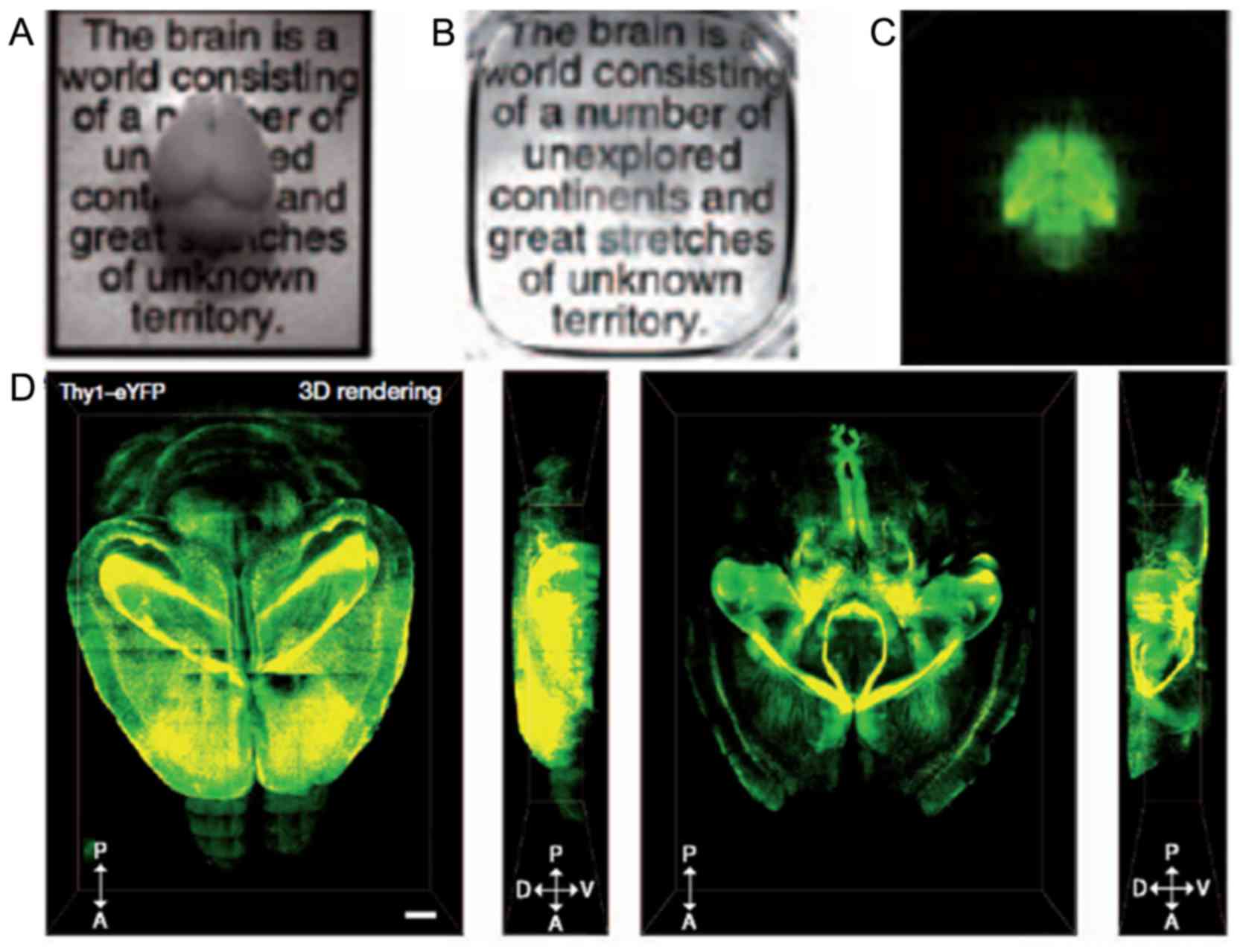
initiation. The 3D hydrogel network is relatively stable, optically
transparent and macromolecular-permeable (Fig. 1). Biological molecules are combined
with the hydrogel network except lipids. Thus, proteins and nucleic
acids are preserved after removal of lipids by detergent. CLARITY
was selected as one of ten notable breakthroughs in 2013 by
Science. As reported, CLARITY leaves tissues sufficiently
sturdy to repeatedly infiltrate with different labels, unlike other
methods, which render see-through brains too fragile (16). Therefore, it could be sped up by many
fold tasks such as counting all the neurons in a given brain
region.
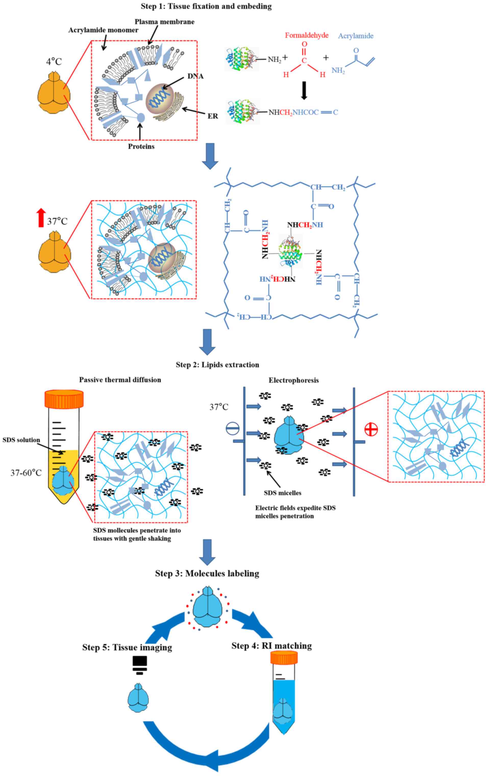
The original CLARITY technique includes the
following steps: Fixation of tissues; hydrogel monomer infusion;
hydrogel-tissue hybridization; lipid extraction; molecular labeling
(if needed); and refractive index matching and imaging (Fig. 2) (14,17).
Although it may seem difficult to implement CLARITY effectively due
to the complexity of the tissue clearing process, this technique
has several advantages over other techniques. CLARITY can make
tissues highly transparent without severe tissue deformation
(tissue expansion occurs during lipid removal but is reversible
after immersing in refractive index (RI) matching solutions), and
it preserves both anatomical and molecular information well (only
~8% protein loss was observed) (14,18). In
addition, as proteins and nucleic acids are bound to the hydrogel
mesh with chemical tethering, it permits several rounds of
molecular labeling and elution. If the sample has endogenous
transgenic expression of fluorophores, it's also compatible with
molecular labeling of other targets (14,17,19).
After tissue clearing, the samples can be stored for weeks to
months (17,20). In the years following its invention,
CLARITY and its improved techniques have been widely used in many
fields, especially neuroscience. Furthermore, this technique seems
compatible with a variety of tissues and organs, such as brain,
bone, pancreas, liver, spleen, kidney, lung, heart, intestine and
even the whole body (21–25). It has also been reported that CLARITY
is suitable for the 3D molecular imaging of plant organs, termed
PEA-CLARITY (26).
However, there are several limitations and
challenges facing CLARITY. For example, too much time is required
from tissue clearing to the acquisition of imaging data. Also,
electrophoretic tissue clearing (ETC) requires a custom-designed
chamber and continuous exchange of detergent (SDS solution)
(17). Moreover, during
electrophoresis, the temperature of the solution increases
gradually requiring temperature control or the sample will become
brown due to the heat (14,17). In addition, the RI-matching solution
(Focus Clear, Cell Explorer Labs) in the original protocols is not
commonly affordable (17). These
problems make the implementation of this technique difficult.
Therefore, many groups are attempting to simplify and improve
CLARITY. Many other clearing techniques based on hydrogel-tissue
hybridization have emerged since the introduction of CLARITY (such
as PACT, PARS, ACT-PRESTO and MAP) (18,27,28).
As all CLARITY-based tissue clearing techniques
involve similar procedures, in this study, we introduce
improvements for the CLARITY technique by dividing its component
techniques into five implementation procedures: i) tissue fixation
and embedding; ii) lipid extraction (including ETC and passive
thermal diffusion); iii) molecule labeling; iv) refractive index
matching; and v) tissue imaging. Thus, we provide a new perspective
on this technique allowing for the integration of different
methods. Additionally, this approach helps to develop a method for
large-scale sample clearing.
Improvements for CLARITY technique
Tissue fixation and embedding
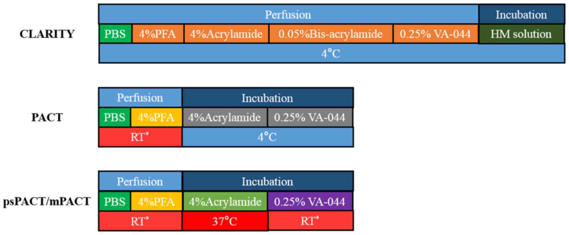
The first step in CLARITY is tissue fixation. In the
original CLARITY protocol, animals were transcardially perfused
with HM solution (including 4% PFA, 4% acrylamide, 0.05%
bis-acrylamide and 0.25% VA-044 initiator in PBS) after removing
blood. Next, the tissue is incubated in HM solution at 4°C for 2–3
days followed by hydrogel polymerization with heating (14). In a separate protocol, the incubation
time was shortened to 1 day (17).
PFA, acrylamide, proteins and nucleic acids
chemically bond together to form a hydrogel mesh. This binding
preserves intrinsic molecules (proteins and nucleic acids) and
permits the penetration of labeling dyes. However, the speed of
clearing and the immunolabeling process depend on the pore size of
hydrogel mesh. High concentrations of PFA and acrylamide result in
a dense hydrogel mesh. This mesh creates a stable hydrogel-tissue
hybridization and reduced the loss of proteins. However, a dense
hydrogel mesh slows both tissue clearing and antibody perfusion
(17,21,28,29).
Therefore, many researchers have adjusted the mixture ratio for
different tissues and clearing approaches to maintain a balance
between hydrogel rigidity and porosity with minimal protein loss
and a faster speed for clearing and immunolabeling (19).
In ETC, the original solution (4% PFA, 4%
acrylamide, 0.05% bis-acrylamide) is the most common ratio to date.
Epp et al systematically researched the influence of
different ETC temperatures and hydrogel composition on tissue
transparency and expansion using an intact mouse brain (21). These researchers recommended a 4%
formaldehyde and 4% acrylamide mixture for examining endogenous
fluorescent markers and a composition of 3% acrylamide and 3%
formaldehyde for immunohistochemistry. For passive CLARITY, the
process of tissue fixation is the same as ETC in the original
article (17). Next, Yang et
al proposed PACT, which adopted a different perfusion method
(28). Compared with the passive
CLARITY protocol, PACT fixes tissues with 4% PFA followed by
incubation at 4°C overnight in the hydrogel monomer solution (A4P0,
4% acrylamide in PBS supplemented with VA-044 initiator). These
researchers also demonstrated the structural integrity of tissues
throughout PACT processing using 1-mm-thick Thy1-eYFP tissue
sections. The pore of the hydrogel mesh in tissues processing with
PACT is larger than that observed with HM solution because of the
obstruction of lipid bilayer when incubating in A4P0 (23). Thus, tissues fixed with A4P0 would be
more easily deformed compared with those in original protocol.
However, this provides advantages in certain dense tissues, such as
bone (PACT-deCAL) (23,25). This perfusion method for PACT is also
used in an improved ETC technique termed ACT-PRESTO (18). Lee et al optimized the
original PACT procedure, creating psPACT (process-separated PACT)
and mPACT (modified PACT: psPACT with added α-thioglycerol)
(30). In psPACT, fixed tissues were
incubated in A4P0 (4% acrylamide in PBS without VA-044) at 37°C for
24 h (the original protocol incubated tissues with A4P0 within
VA-044 initiator and stored tissues at 4°C). Next, tissues were
covered with 0.25% VA-044 at room temperature for 6 to 24 h. The
authors compared psPACT, mPACT with PACT in the rat central nervous
system (spinal cord and brain), as well as in other internal
organs. These researchers concluded that this tissue fixation
method could accelerate tissue clearing, with mPACT being faster
than psPACT. We compare these different fixation methods in
Fig. 3.
We have observed that there is no obvious limitation
to the perfusion method used in different tissue clearing
approaches (ETC vs. passive CLARITY). The most important challenge
is to shorten the time of tissue clearing by adjusting the pore
diameter of hydrogel mesh using different concentrations of
ingredients and perfusion methods while keeping tissue structure
stable. Passive CLARITY and PACT requires more time to extract
lipids from tissue-hydrogel hybrids with shaking with a higher
temperature than would be used in ETC when handling relatively
large tissues (ETC adopted 37°C; the original passive CLARITY
protocol recommended 37°C-60°C; PACT first adopted 37°C; another
study concluded 42°C-47°C is a better temperature range for PACT)
(17,28,31).
Thus, the requirement for tissue stability in PACT is higher than
that of ETC, and the concentration of acrylamide and PFA is of
vital importance when implementing passive CLARITY and PACT.
The following step after tissue fixation is hydrogel
polymerization. In the original protocol, nitrogen and vacuum pump
are used to extract and replace air in the tissue container (as
oxygen impedes hydrogel formation) (14). Notably, oxygen must be completely
removed; otherwise, hydrogel formation in the inner parts of tissue
would be incomplete and result in the emergence of cavities after
lipids extraction. It was also reported that adding a thick layer
of mineral oil over the top of the hydrogel solution before
polymerization could minimize the exposure of samples to the air
(32).
Lipid extraction
Since membrane lipids are the main cause of light
diffraction, lipid extraction becomes a key process in CLARITY
(1,20,33).
Tissue transparency primarily depends on the degree of lipid
elution. In general, there are two methods for lipid extraction:
passive thermal diffusion and electrophoresis. Passive thermal
diffusion is a way to transport SDS micelles into tissues via
heat-induced diffusion. And electrophoresis applies electric fields
to accelerate the penetrating of SDS micelles. All modified CLARITY
techniques elute membrane lipids based on these two methods (PACT,
PARS, ETC, ACT-PRESTO- and stochastic electrotransport) (14,17,18,14).
In this study, we compared strengths and weaknesses of these two
methods in Table I. Next, we
reviewed the modified CLARITY techniques based on the two
methods.
 | Table I.Comparison of passive thermal
diffusion and electrophoresis lipids extraction. |
Table I.
Comparison of passive thermal
diffusion and electrophoresis lipids extraction.
| Methods | Principle | Elution time | Complexity of
implementation | Refresh SDS
buffer | Volume of SDS
buffer | Circulation |
|---|
| Passive thermal
diffusion | Transporting
surfactant micelles into tissues via heat-induced diffusion | Weeks to
months | Easy | Occasionally | Small (several ten
milliliters) | No |
|
Electrophoresis | Transporting
surfactant micelles into tissues via electric fields | Several days | Difficult | Frequently | Large (several
liters for each circulation) | Yes (to control the
temperature) |
Passive thermal diffusion
Regarding passive thermal diffusion in-passive
CLARITY, tissues are incubated in 4% SDS at pH 8.5 with gentle
shaking at 37°C with an increased temperature of up to 60°C for
faster clearing (17). Because of
dense hydrogel mesh structure and low efficiency of SDS
transportation, this method is time-consuming and may not reach a
high degree of tissue transparency for larger tissues, except at
higher temperatures (34). However,
higher temperatures may cause potential tissue deformation and
damage to natural molecules. Therefore, in order to accelerate SDS
transportation and to keep tissue structure stable, Yang et
al proposed new methods, termed PACT and PARS (23,28).
PACT adopted 8% SDS buffer at pH 8.5 and samples are gently shaking
at 37°C for tissue clearing. These researchers emphasized that only
an 8% SDS concentration could achieve uniform tissue clearing
throughout the entire 3-mm block, while the SDS concentration in
ETC and most other modified techniques is 4% (14,17).
PARS is a method for whole-body clearing and labeling (23,28).
After fixing tissues with PFA and A4P0, tissues of interest are not
removed from animal's body and the clearing process is achieved
in vivo. PARS utilized the intact vasculature to deliver and
circulate the SDS buffer. For different tissues, different parts of
vasculature could be selected. For brain or spinal cord clearing,
PACT regents could be delivered with a subdural cannula inserted
above the region of interest, and the buffer circulation would be
similar to that of cerebrospinal fluid (28). The principle of PARS is also based on
passive diffusion. The difference is that PARS increased the
contact area between the clearing buffer and tissues using the
intrinsic vasculature; therefore, the tissue clearing speed becomes
faster. Additionally, PARS minimizes tissue expansion during
clearing, as the skull and other physiologic structures limit the
available space. However, implementation of PARS is more
complicated than PACT (a cannula needs to be set and kept stable as
tissue vasculature is easily destroyed and obstructed during buffer
circulation). Thus, the application of PARS is limited. After that,
several modified methods based on PACT emerged. For mPACT, the
author added 0.5% α-thioglycerol to 8% SDS buffer for lipid
extraction with gentle shaking, and it showed faster clearing speed
than PACT. The use of α-thioglycerol was also reported in other
articles to avoid browning and autofluorescence accumulation as a
result of the Maillard reaction (10,18). The
α-thioglycerol works via the sulfhydryl, suggesting that other
chemical materials with sulfhydryl, such as β-mercaptoethanol, may
also work (10). It has also been
reported that 0–50 mM sodium sulfite could exhibit an anti-browning
effect in other tissue clearing methods, such as SWITCH (35). Lee et al also combined mPACT
and PARS as a whole body perfusion method (PARS-mPACT) (30). PARS-mPACT fixed tissues via the
transcardial perfusion of separated mPACT regents. Next,
polymerized tissues were isolated and passively cleared according
to the mPACT protocol. However, PARS-mPACT and mPACT showed no
differences in tissue transparency. Another method, named CLARITY2,
adopted a different way to accelerate tissue clearing (36). After tissue fixation and hydrogel
polymerization, brain tissue was cut to 1–1.5-mm-thick coronal
slices and underwent passive tissue clearing. This approach enabled
us to accelerate and simplify the clearing, staining and imaging
steps compared with original protocol. However, CLARITY2 did not
avoid damaging the tissues and is similar to a traditional tissue
slice. However, this technique is still an ideal time-saving method
for methodological studies (32,37,38). In
addition, it's reported that with pancreatic lipase breaking down
lipid droplets, lipid-rich tissues were better cleared when
performing passive clearing (39).
And there were no obvious tradeoffs in the quality
or characteristics of cleared tissues with the SDS buffer at
different pH, except for a slight enhancement in the rate of
clearance at a more alkaline pH (23). Thus PACT, PARS and other passive
CLARITY techniques could dissolve SDS using 1× PBS at pH 7.5 for
convenience, adjusting the SDS concentration according to the needs
of the study to maintain a balance between tissue integrity and
transparency.
Electrophoresis
Electrophoresis is faster than passive thermal
diffusion, as electric fields accelerate the transport of SDS.
First, the ETC system is composed of fourelements: i) A chamber for
containing samples and electrodes; ii) a power supply; iii) a
circulator to control flow rate and buffer temperature, which is
combined with the chamber; and iv) a filter to remove
byproducts.
In the original protocol, a range of conditions
(10–60 V) were applied across the tissue at 37–50°C for several
days with clearing solution circulating through ETC chamber
(14). However, the high temperature
and voltage could cause a higher risk of tissue deformation, as
well as epitope and fluorescence loss (17,22).
Therefore, the voltage and circulation temperature should be
precisely controlled to produce better outcomes. It was also
recommended that ETC be completed at 25 V and 37°C for an adult
mouse brain (17). The limitations
to the application of high electric fields in the ETC system was
compounded by the fact that the electrical properties in different
regions of a tissue are not homogeneous, leading to regions with
concentrated electric fields. Thus, high electric fields may cause
deformation of these regions. This suggests that electrophoresis is
ineffective for promoting the transportation of SDS into large,
dense samples only if high electric fields are used. Stochastic
electrotransport, created by Kim et al could promote the
migration of freely moving molecules with high electromobility
while suppressing the displacement of endogenous biological
molecules with low electromobility within the sample. This
technique may be an ideal way to solve this high-voltage ETC
problem (22). Compared with the
stationary samples and electrodes in original ETC chamber, the
author implemented stochastic electrotransport by creating a
continuously rotating sample chamber with respect to two parallel
electrodes (22). Next, these
researchers made a contrast among mouse brains cleared by
stochastic electrotransport, static electrophoresis and thermal
diffusion. The mouse brain cleared with stochastic electrotransport
was remarkably transparent within 3 days, while others did not show
the same results (22). Lee et
al created a modified clearing method termed ACT (18). ACT alternated the platinum wires to
platinum plate to generate a dense regular current in the ETC
chamber and adopted a two-step fixation protocol as mentioned in
PACT. Therefore, ACT could clear tissues faster than ETC (2 h of
ACT could achieve complete optical transparency in 1-mm brain
sections and 15 h was sufficient to clear entire adult rat brain)
(18). However, this method has a
high risk of tissue deformation because of the strong electric
fields generated and relatively unstable tissue-hydrogel
hybridization structure compared to HM solution fixed tissues.
Moreover, modifications to ETC were proposed via
changing the parameters of ETC (current or temperature) to achieve
better results. One study reported that different optimized current
of ETC for various organs (including brain, pancreas, kidney,
liver, intestine and lung) (24),
demonstrating that the current and voltage of electrophoresis
should correspond with the needs of the study. Epp et al
tested effects of different ETC temperatures on tissue transparency
and proposed a combined 37/55°C clearing protocol, in which they
ran ETC at 20 V for the first 4 days at 37°C and increased the
temperature to 55°C on the final day (21). Using this method, tissues showed
better transparency and structural integrity than those produced by
running ETC at only 37°C or 55°C.
There are additional problems when repeating the
original protocol. First, the by-products formed during
electrophoresis would discolor tissues and colored particles may
deposit on the surface of tissues (24). To solve this problem, the author in
stochastic electrotransport used a temperature controller to
maintain SDS buffer temperature at ~15°C to prevent browning
tissues due to heat. Apart from that step, nanoporous membranes
were used to contain samples and mechanically divide the
circulating solution into an inner portion and outer portion
(22). The lower concentrations in
outer portion were designed to slow down electro-oxidation of the
surfactant molecules (22). Second,
the continuously emerging bubbles during electrophoresis could
interrupt electrophoresis if the ETC chamber was filled (14,17). In
ACT (18), the author designed a
long ETC chamber, which allowed bubbles float to the top where they
were removed through the top outlet. Bastrup and Larsen (29) also designed an adjustable ETC chamber
to fit variable tissue sizes.
Molecules labeling
In general, there are three methods to visualization
interest molecules: i) Genetic introduction of fluorescent markers;
ii) in vivo labeling of cells or regions with viruses or
chemicals; and iii) chemical/antibody staining (40). For the first two methods,
fluorescence may quench throughout clearing process and require
amplification (2,19). Therefore, chemical/antibody staining
is an alternative way to label cleared tissues. With this method,
multiple rounds of staining [three rounds of staining with up to 4
channels were reported (14)] are
possible without damaging the preserved structures as proteins and
nucleic acids are chemically bound to the hydrogel mesh (19). The clearing solution SDS buffer can
be used to wash antibodies and other molecular labels out of the
hydrogel-tissue hybridization in preparation for another round of
staining.
In the original protocol, tissues were immunostained
by incubating them at 37°C with a high concentration of primary and
secondary antibodies (dilution, 1:50-1:100) and gentle shaking.
Itrequires between 2 days and 2 weeks, depending on the size of the
tissue sample (14). The principle
of this immunostaining method is the same with passive tissue
clearing, expediting the penetration of antibodies by passive
thermal diffusion. Some other methods to provide external forces
for accelerating the transportation and penetration of antibodies
were applied to labeling tissues with a large volume. Similar to
lipid extraction, the improvement of immunostaining is also based
on two aspects: passive diffusion and electrophoresis (41).
Passive diffusion labeling
Many labeling methods are based on passive diffusion
as it's simple to implement. It has been reported that PARS was
suitable for immunostaining and that all immunohistochemical
solutions were delivered through the PARS circulation system
(including blocking reagent, antibodies or fluorescently labeled
molecules and wash buffers) (23,28).
This delivery is target-specific and uniformly distributed
throughout organs with a low background (28). However, similar to original protocol,
it cannot expedite the labeling process (23). To solve this problem, another method
termed PRESTO (pressure related efficient and stable transfer of
macromolecules into organs) emerged (18). This method was composed of c-PRESTO
(centrifugal PRESTO) and s-PRESTO (syringe PRESTO). The c-PRESTO
technique applied a centrifugal force with a standard table top
centrifuge (600 rcf) to expedite antibody diffusion. For example,
kidney tissues incubated for 3 h with antibodies showed labeled
structures 10–30 µm deep, whereas for normal tissues, 3 h was
sufficient to label structures 120 µm deep (18). The s-PRESTO created a convection flow
with a syringe pump, which infused labeling reagents into the
specimen. This technique showed a labeling depth that was four
times greater than that obtained in original passive labeling
(41).
Almost all cleared tissues are applied a
fluorescence-based phenotype to label and image interest tissues.
But one study used a colorimetric, non-fluorescent method based on
the conversion of horseradish peroxidase to diaminobenzidine to
label PACT cleared tissues (42).
However, this technique was only applied to 50 and 100 µm sections
of adult mouse brain tissue; therefore, it did not contain the
anatomical structure information of an intact brain. Furthermore,
non-fluorescence labeling cannot achieve 3D reconstruction,
limiting further applications in tissueimaging.
Electrophoresis labeling
As antibodies are charged molecules in solution with
certain pH, applying external electric fields can expedite the
transportation of them into tissues. A study by Li et al
applied a simple constant electric field across a 500-µm-thick
brain section, and it decreased the delivery time of antibodies by
more than 800-fold over simple diffusion without incurring
structural damage (43). In this
study, the 500-µm-thick brain section was stained in only 30 min at
an external voltage of 25 V. However, when applying this to larger
tissues (whole brains or animal bodies), heat damage caused by
electrodes may occur necessitating buffer circulation to control
the temperature. To ensure the concentration of dyes, the staining
solution and circulated buffer should be separated. Similar to
lipid extraction with ETC, static electrophoresis resulted in
substantial tissue damage in large-volume sample, as it needed a
longer time for electrophoresis. This problem was solved with
stochastic electrotransport (22).
Similar to the device for clearing in stochastic electrotransport,
antibodies were confined inside of the sample chamber with
nanoporous membranes and PBS was circulated in the outer chamber to
control the temperature.
Non-protein molecules labeling
However, these techniques have been poorly explored
with RNA studies. Yang et al first used single-molecule
fluorescence in situ hybridization (sm-FISH) to detect
single RNA molecules in 100-µm-thick PACT-processed mouse brain
sections (28). When applying a
single-molecule hybridization chain reaction (smHCR), single mRNAs
could be detected within 500-µm-thick PACT-processed brain slices
(44). Additionally, EDC-CLARITY
used EDC (1-Ethyl-3-3-dimethyl-aminopropyl carbodiimide) to link
5′-phosphate groups with surrounding amine-containing proteins to
stably retain RNAs in clarified tissues (45). With the hairpin chain reaction (HCR)
amplification system, it presented validation for selecting
microRNAs, cell-type markers and immediate-early genes.
CLARITY achieved tissue transparency by eluting
membrane lipids and lipophilic dyes. Therefore, lipophilic
fluorescent dyes, such as DiI, which stains cellular membranes, is
unable to be applied with CLARITY (46,47). A
study by Jensen and Berg altered the molecular structure of the dye
to adhere to both membranes and proteins such that the dye remained
in the tissue after tissue clearing (47). These researchers tested three
Dil-analogue dyes, CM-DiI, SP-DiI and FM 1-43FX in PACT-processed
spinal cords of adult rats and mice. All three dyes remained in the
tissue after lipid-clearing, but CM-DiI had the sharpest and FM
1-43FX had the strongest fluorescent signals. This modification
provided a new way to label neurons (retrograde or anterograde) and
mark the position of extracellular electrodes after
electrophysiology.
Refractive index matching
Before imaging, cleared tissues need to be incubated
in solutions that match the average RI of the tissue (~1.46) to
achieve RI homogenization. Afterward, tissues would be optically
transparent and photon scattering from both the excitation light
and the emitted fluorescence signal would be reduced. In theory,
the RI of each solution can be matched to the cleared tissue or
microscope objective by adjusting concentration of the main
ingredient. However, the anti-swelling of cleared tissues should
also be considered an important factor affecting the solution. In
this study, we contrasted several solutions used in CLARITY-based
techniques.
In original protocol, the author applied FocusClear
(CelExplorer Labs) and 85–87% glycerol to match RI (14). These reagents both promoted tissue
transparency, but the best results were achieved with FocusClear.
However, FocusClear is expensive and not suitable for long-term
storage (long-term storage resuls in the formation of an
irreversible white precipitate) (14,17).
Yang et al used a solution termed RIMS (refractive index
matching solution) to achieve tissue transparency (28). It contains 88% Histodenz in 0.02 M
phosphate buffer with an RI of 1.47. RIMS is stable with long-term
storage and preserves fluorescent markers over months. Their group
had also derived sRIMS, containing 70% sorbitol in 0.02 M phosphate
buffer and cRIMS, containing 88% histodenz in 0.005 M phosphate
buffer (23). Next, one study found
that 63% 2,2′-thiodiethanol (TDE) in PBS was more suitable for RI
matching in CLARITY cleared tissues (48). A different solutuion, 80% Nycodenz
solution (nRIMS), was applied with mPACT (30). In ACT-PRESTO, a CUBIC-mount solution
(50% Sucrose, 25% urea and 25% N, N, N', N'-tetrakis
(2-hydroxypropyl) ethylenediamine) was used to match the RI of the
tissues (18). The comparison of
these RI-matching solutions can be seen in Table II. However, in a study by
Poguzhelskaya et al imaging without an RI-matching solution
but in PBST was also possible (36).
| Table II.Comparison of different RI matching
solutions. |
Table II.
Comparison of different RI matching
solutions.
| Solution | Focus clear | RIMS | sRIMS | cRIMS | nRIMS | THE | CUBIC-mount
solution |
|---|
| Main
ingredients | Unknown | 88% Histodenz in
0.02 M phosphate buffer | 70% sorbitol in
0.02 M phosphate buffer | 88% histodenz in
0.005 M phosphate buffer | 80% Nycodenz (TDE)
in PBS | 63% 2,
2′-thiodiethanol | 50% Sucrose, 25%
urea and 25% N, N, N', N'- tetrakis (2-hydroxypropyl)
ethylenediamine |
Although all of these solutions were intended to
match the RI of cleared tissues, for different samples, it may
still fail to match the RI well after tissue clearing, as various
elements differ in inner tissues. For example, a study by Liu et
al reported that 47% TDE in phosphate buffer (not PBS) was more
suitable for human brain tissues (37). Therefore, if the concentrations
reported in articles do not make the interest tissue ideally
transparent, different concentrations and solvents (PBS, phosphate
buffer or normal saline) should be tested. Moreover, solutions with
a high concentration could reduce tissue swelling. When imaging
without an RI-matching solution but in simple purified water or
PBST, the tissue will remain swollen, and fail to get the
structural information. Therefore, RI-matching and anti-swelling
should both be considered when choosing RI-matching solutions.
Tissue imaging
Finding an optimized, high-resolution deep-imaging
approach for large-scale samples is still a major challenge.
Furthermore, imaging a 3D volume is a time-consuming task.
Therefore, when selecting a microscope and objective, imaging time,
resolution and sample size should all be taken into account. In
this study, we compared different microscopes and objectives
applied to cleared samples.
There are three primary microscopes used for
three-dimensional fluorescence imaging: i) The standard confocal
microscope; ii) the two-photon microscope; and iii) the light sheet
microscope. The standard confocal microscope achieves optical
sectioning by using a pinhole in front of the photomultiplier tubes
and scans samples point to point. Therefore, this microscope's
processing time is notably slow, particularly in large samples, and
long-time imaging can lead to photobleaching (20,49). A
two photon microscope results in lower photobleaching of
fluorophores and provides greater imaging depth than a confocal
microscope, but point-by-point scanning is still slow. For the
light sheet microscope, it uses fast sCMOS or CCD cameras to image
a selectively illuminated focal plane and provides a high imaging
speed (2–3 orders of magnitude faster than point-scanning methods),
high signal-to-noise ratio and low levels of photobleaching
(17,50). Tomer et al developed SPED
(SPherical-aberration-assisted Extended Depth-of-field) light sheet
microscopy, which combined an extension of the depth-of-field with
the optical sectioning of a light sheet microscope, thereby
eliminating the need to physically scan detection objectives for
volumetric imaging (51). It enabled
the scanning of thousands of volumes per-second, limited only by
the camera acquisition rate.
The key microscope objective parameters are working
distance (WD), numerical aperture (NA) and RI. WD is the distance
between the objective lens and the focal plane. A long WD avoids
the sample directly contacting with lens and ensures imaging of
large samples (52). The NA of an
objective relates to the collection of emitted signal, and higher
NA enables higher resolution (17).
The RI of the cleared tissues is ~1.46; therefore, it is better to
choose objectives designed with an RI near 1.46 when using a
non-optimized objective.
Challenges and opportunities
Though many efforts have been spared on the
improvement for CLARITY, the primary issues still remain. First, we
acknowledge that the implementation of CLARITY has limitations due
to the factors mentioned above. A more simplified protocol is
needed and the processing time should be shortened for examining
samples more quickly.
The largest challenge for tissue clearing, as with
other tissue clearing techniques, is the clearing of large-volume
samples, such as a monkey brain (3).
Though tissue clearing techniques are widely used in neuroscience,
information acquired in this way concerning the human brain is
still limited. Therefore, animals similar to humans are ideal
models for studying the human brain. Therefore, integration of
current techniques or the development of new methods for clearing
large samples are of vital importance. Furthermore, in terms of
imaging, large sample is not feasible due to the limitations of
microscopes and data analysis. Thus, long WD, high NA microscopes
and fast data analysis software are also in demand.
However, there are several advantages from other
clearing and labeling techniques from which we can learn. SWITCH
(System-Wide control of Interaction Time and kinetics of Chemicals)
produces tissue-glutaraldehyde hybrids which are heat- and
chemical-resistant and permit multiple rounds (>20) of labeling
(35). When applying antibody
labeling, SWITCH enables scalable and uniform labeling by
controlling probe-target binding kinetics with a low concentration
(0.5 to 1.0 mM) SDS. Therefore, it may also be accessible to
control the binding kinetics in CLARITY in order to achieve
homogeneity and more rounds of labeling. In CLARITY, we use
RI-matching solutions to resist tissue swelling and maintain a
normal size (17). In contrast, a
method called MAP (magnified analysis of the proteome), which is
also based on hydrogel-tissue hybridization, linearly expands
entire organs four-fold while preserving their overall architecture
and three-dimensional proteome organization (27). Similarly, Chen et al reported
a method termed expansion microscopy (ExM) that can linearly expand
tissues ~4.5-fold to enable imaging with conventional confocal
microscopes to examine the subcellular architecture (53). Then, their group developed iterative
expansion microscopy (iExM), in which a sample can be expanded
~20-fold. These methods provide us with new ways to utilize tissue
expansion to study subcellular architecture, and even for
expediting antibodies penetration during labeling (expanded tissues
have bigger pore size of hydrogel network).
Conclusions
Compared with other tissue clearing techniques,
CLARITY is more compatible with different samples and provides
tissues with higher transparency, despite having several
limitations. However, CLARITY still faces many challenges. High
resolution and multi-level imaging of large-scale samples require
the integration of different clearing methods and imaging
techniques. Additionally, massive volumetric data sets acquired
from samples with a larger volume present a challenge for data
analysis. However, with future studies focused on these problems,
tissue clearing can open the door to many new discoveries and
provide a new view of the inner interactions among cells, organs
and systems. The combination of existing clearing methods and
development of new techniques may help us to address these
challenges.
Acknowledgements
Not applicable.
Funding
This work was supported b the National Natural
Science Foundation of China (61471367).
Availability of data and materials
Not applicable.
Authors' contributions
HD was a major contributor in writing the
manuscript. PH wrote the ‘molecular labeling’ part of the
manuscript and designed the illustrations. BZ contributed to the
conception that divides different methods into five procedures and
made the tables. QL designed the study and made important revisions
to the manuscript. All authors contributed to assess and revise the
manuscript.
Ethics approval and consent to
participate
Not applicable.
Patient consent for publication
Not applicable.
Competing interests
The authors declare that they have no conflict of
interest.
References
|
1
|
Zhu X, Xia Y, Wang X, Si K and Gong W:
Optical brain imaging: A powerful tool for neuroscience. Neurosci
Bull. 33:95–102. 2017. View Article : Google Scholar : PubMed/NCBI
|
|
2
|
Seo J, Choe M and Kim SY: Clearing and
labeling techniques for large-scale biological tissues. Mol Cells.
39:439–446. 2016. View Article : Google Scholar : PubMed/NCBI
|
|
3
|
Richardson DS and Lichtman JW: Clarifying
tissue clearing. Cell. 162:246–257. 2015. View Article : Google Scholar : PubMed/NCBI
|
|
4
|
Planchon TA, Gao L, Milkie DE, Davidson
MW, Galbraith JA, Galbraith CG and Betzig E: Rapid
three-dimensional isotropic imaging of living cells using Bessel
beam plane illumination. Nat Methods. 8:417–423. 2011. View Article : Google Scholar : PubMed/NCBI
|
|
5
|
Hama H, Kurokawa H, Kawano H, Ando R,
Shimogori T, Noda H, Fukami K, Sakaue-Sawano A and Miyawaki A:
Scale: A chemical approach for fluorescence imaging and
reconstruction of transparent mouse brain. Nat Neurosci.
14:1481–1488. 2011. View
Article : Google Scholar : PubMed/NCBI
|
|
6
|
Hama H, Hioki H, Namiki K, Hoshida T,
Kurokawa H, Ishidate F, Kaneko T, Akagi T, Saito T, Saido T and
Miyawaki A: ScaleS: An optical clearing palette for biological
imaging. Nat Neurosci. 18:1518–1529. 2015. View Article : Google Scholar : PubMed/NCBI
|
|
7
|
Renier N, Wu Z, Simon DJ, Yang J, Ariel P
and Tessier-Lavigne M: iDISCO: A simple, rapid method to
immunolabel large tissue samples for volume imaging. Cell.
159:896–910. 2014. View Article : Google Scholar : PubMed/NCBI
|
|
8
|
Ertürk A and Bradke F: High-resolution
imaging of entire organs by 3-dimensional imaging of solvent
cleared organs (3DISCO). Exp Neurol. 242:57–64. 2013. View Article : Google Scholar : PubMed/NCBI
|
|
9
|
Ertürk A, Becker K, Jährling N, Mauch CP,
Hojer CD, Egen JG, Hellal F, Bradke F, Sheng M and Dodt HU:
Three-dimensional imaging of solvent-cleared organs using 3DISCO.
Nat Protoc. 7:1983–1995. 2012. View Article : Google Scholar : PubMed/NCBI
|
|
10
|
Ke MT, Fujimoto S and Imai T: SeeDB: A
simple and morphology-preserving optical clearing agent for
neuronal circuit reconstruction. Nat Neurosci. 16:1154–1161. 2013.
View Article : Google Scholar : PubMed/NCBI
|
|
11
|
Kuwajima T, Sitko AA, Bhansali P, Jurgens
C, Guido W and Mason C: ClearT: A detergent- and solvent-free
clearing method for neuronal and non-neuronal tissue. Development.
140:1364–1368. 2013. View Article : Google Scholar : PubMed/NCBI
|
|
12
|
Susaki EA, Tainaka K, Perrin D, Kishino F,
Tawara T, Watanabe TM, Yokoyama C, Onoe H, Eguchi M, Yamaguchi S,
et al: Whole-brain imaging with single-cell resolution using
chemical cocktails and computational analysis. Cell. 157:726–739.
2014. View Article : Google Scholar : PubMed/NCBI
|
|
13
|
Hou B, Zhang D, Zhao S, Wei M, Yang Z,
Wang S, Wang J, Zhang X, Liu B, Fan L, et al: Scalable and
DiI-compatible optical clearance of the mammalian brain. Front
Neuroanat. 9:192015. View Article : Google Scholar : PubMed/NCBI
|
|
14
|
Chung K, Wallace J, Kim SY,
Kalyanasundaram S, Andalman AS, Davidson TJ, Mirzabekov JJ,
Zalocusky KA, Mattis J, Denisin AK, et al: Structural and molecular
interrogation of intact biological systems. Nature. 497:332–337.
2013. View Article : Google Scholar : PubMed/NCBI
|
|
15
|
Vigouroux RJ, Belle M and Chédotal A:
Neuroscience in the third dimension: Shedding new light on the
brain with tissue clearing. Mol Brain. 10:332017. View Article : Google Scholar : PubMed/NCBI
|
|
16
|
2013 runners-up. CLARITY makes it
perfectly clear. Science. 342:1434–1435. 2013.
|
|
17
|
Tomer R, Ye L, Hsueh B and Deisseroth K:
Advanced CLARITY for rapid and high-resolution imaging of intact
tissues. Nat Protoc. 9:1682–1697. 2014. View Article : Google Scholar : PubMed/NCBI
|
|
18
|
Lee E, Choi J, Jo Y, Kim JY, Jang YJ, Lee
HM, Kim SY, Lee HJ, Cho K, Jung N, et al: ACT-PRESTO: Rapid and
consistent tissue clearing and labeling method for 3-dimensional
(3D) imaging. Sci Rep. 6:186312016. View Article : Google Scholar : PubMed/NCBI
|
|
19
|
Jensen KHR and Berg RW: Advances and
perspectives in tissue clearing using CLARITY. J Chem Neuroanat.
86:19–34. 2017. View Article : Google Scholar : PubMed/NCBI
|
|
20
|
Kim SY, Chung K and Deisseroth K: Light
microscopy mapping of connections in the intact brain. Trends Cogn
Sci. 17:596–599. 2013. View Article : Google Scholar : PubMed/NCBI
|
|
21
|
Epp JR, Niibori Y, Hsiang Liz HL, Mercaldo
V, Deisseroth K, Josselyn SA and Frankland PW: Optimization of
CLARITY for clearing whole-brain and other intact organs. eNeuro.
2:ENEURO. 0022. –15. 2015. View Article : Google Scholar : PubMed/NCBI
|
|
22
|
Kim SY, Cho JH, Murray E, Bakh N, Choi H,
Ohn K, Ruelas L, Hubbert A, McCue M, Vassallo SL, et al: Stochastic
electrotransport selectively enhances the transport of highly
electromobile molecules. Proc Natl Acad Sci USA. 112:E6274–E6283.
2015. View Article : Google Scholar : PubMed/NCBI
|
|
23
|
Treweek JB, Chan KY, Flytzanis NC, Yang B,
Deverman BE, Greenbaum A, Lignell A, Xiao C, Cai L, Ladinsky MS, et
al: Whole-body tissue stabilization and selective extractions via
tissue-hydrogel hybrids for high-resolution intact circuit mapping
and phenotyping. Nat Protoc. 10:1860–1896. 2015. View Article : Google Scholar : PubMed/NCBI
|
|
24
|
Lee H, Park JH, Seo I, Park SH and Kim S:
Improved application of the electrophoretic tissue clearing
technology, CLARITY, to intact solid organs including brain,
pancreas, liver, kidney, lung and intestine. Bmc Dev Biol.
14:482014. View Article : Google Scholar : PubMed/NCBI
|
|
25
|
Greenbaum A, Chan KY, Dobreva T, Brown D,
Balani DH, Boyce R, Kronenberg HM, McBride HJ and Gradinaru V: Bone
CLARITY: Clearing, imaging and computational analysis of
osteoprogenitors within intact bone marrow. Sci Transl Med.
9:eeah65182017. View Article : Google Scholar
|
|
26
|
Palmer WM1, Martin AP, Flynn JR, Reed SL,
White RG, Furbank RT and Grof CP: PEA-CLARITY: 3D molecular imaging
of whole plant organs. Sci Rep-Uk. 5:134922015. View Article : Google Scholar
|
|
27
|
Ku T, Swaney J, Park JY, Albanese A,
Murray E, Cho JH, Park YG, Mangena V, Chen J and Chung K:
Multiplexed and scalable super-resolution imaging of
three-dimensional protein localization in size-adjustable tissues.
Nat Biotechnol. 34:973–981. 2016. View Article : Google Scholar : PubMed/NCBI
|
|
28
|
Yang B, Treweek JB, Kulkarni RP, Deverman
BE, Chen CK, Lubeck E, Shah S, Cai L and Gradinaru V: Single-cell
phenotyping within transparent intact tissue through whole-body
clearing. Cell. 158:945–958. 2014. View Article : Google Scholar : PubMed/NCBI
|
|
29
|
Bastrup J and Larsen PH: Optimized CLARITY
technique detects reduced parvalbumin density in a genetic model of
schizophrenia. J Neurosci Meth. 283:23–32. 2017. View Article : Google Scholar
|
|
30
|
Lee M, Seo JM, Park HS and Cho YE:
Optimization of the optical transparency of rodent tissues by
modified PACT-based passive clearing. Exp Mol Med. 48:e2742016.
View Article : Google Scholar : PubMed/NCBI
|
|
31
|
Yu T, Qi Y, Zhu J, Xu J, Gong H, Luo Q and
Zhu D: Elevated-temperature-induced acceleration of PACT clearing
process of mouse brain tissue. Sci Rep. 7:388482017. View Article : Google Scholar : PubMed/NCBI
|
|
32
|
Zheng H and Rinaman L: Simplified CLARITY
for visualizing immunofluorescence labeling in the developing rat
brain. Brain Struct Funct. 221:2375–2383. 2016. View Article : Google Scholar : PubMed/NCBI
|
|
33
|
Yushchenko DA and Schultz C: Tissue
clearing for optical anatomy. Angew Chem Int Ed. 52:10949–10951.
2013. View Article : Google Scholar
|
|
34
|
El-Sherbiny IM and Yacoub MH: Hydrogel
scaffolds for tissue engineering: Progress and challenges. Glob
Cardiol Sci Pract. 2013:316–342. 2013.PubMed/NCBI
|
|
35
|
Murray E, Cho JH, Goodwin D, Ku T, Swaney
J, Kim SY, Choi H, Park YG, Park JY, Hubbert A, et al: Simple,
scalable proteomic imaging for high-dimensional profiling of intact
systems. Cell. 163:1500–1514. 2015. View Article : Google Scholar : PubMed/NCBI
|
|
36
|
Poguzhelskaya E, Artamonov D, Bolshakova
A, Vlasova O and Bezprozvanny I: Simplified method to perform
CLARITY imaging. Mol Neurodegener. 9:192014. View Article : Google Scholar : PubMed/NCBI
|
|
37
|
Liu AK, Hurry ME, Ng OT, DeFelice J, Lai
HM, Pearce RK, Wong GT, Chang RC and Gentleman SM: Bringing CLARITY
to the human brain: Visualization of Lewy pathology in three
dimensions. Neuropath Appl Neuro. 42:573–587. 2016. View Article : Google Scholar
|
|
38
|
Magliaro C, Callara AL, Mattei G,
Morcinelli M, Viaggi C, Vaglini F and Ahluwalia A: Clarifying
CLARITY: Quantitative optimization of the diffusion based
delipidation protocol for genetically labeled tissue. Front
Neurosci. 10:1792016. View Article : Google Scholar : PubMed/NCBI
|
|
39
|
Lai M, Li X, Li J, Hu Y, Czajkowsky DM and
Shao Z: Improved clearing of lipid droplet-rich tissues for
three-dimensional structural elucidation. Acta Bioch Bioph Sin.
49:465–467. 2017. View Article : Google Scholar
|
|
40
|
Susaki EA and Ueda HR: Whole-body and
whole-organ clearing and imaging techniques with single-cell
resolution: Toward organism-level systems biology in mammals. Cell
Chem Biol. 23:137–157. 2016. View Article : Google Scholar : PubMed/NCBI
|
|
41
|
Tainaka K, Kuno A, Kubota SI, Murakami T
and Ueda HR: Chemical principles in tissue clearing and staining
protocols for whole-body cell profiling. Annu Rev Cell Dev Biol.
32:713–741. 2016. View Article : Google Scholar : PubMed/NCBI
|
|
42
|
Sung K, Ding Y, Ma J, Chen H, Huang V,
Cheng M, Yang CF, Kim JT, Eguchi D, Di Carlo D, et al: Simplified
three-dimensional tissue clearing and incorporation of colorimetric
phenotyping. Sci Rep. 6:307362016. View Article : Google Scholar : PubMed/NCBI
|
|
43
|
Li J, Czajkowsky DM, Li X and Shao Z: Fast
immuno-labeling by electrophoretically driven infiltration for
intact tissue imaging. Sci Rep. 5:106402015. View Article : Google Scholar : PubMed/NCBI
|
|
44
|
Shah S, Lubeck E, Schwarzkopf M, He TF,
Greenbaum A, Sohn CH, Lignell A, Choi HM, Gradinaru V, Pierce NA
and Cai L: Single-molecule RNA detection at depth by hybridization
chain reaction and tissue hydrogel embedding and clearing.
Development. 143:2862–2867. 2016. View Article : Google Scholar : PubMed/NCBI
|
|
45
|
Sylwestrak EL, Rajasethupathy P, Wright
MA, Jaffe A and Deisseroth K: Multiplexed intact-tissue
transcriptional analysis at cellular resolution. Cell. 164:792–804.
2016. View Article : Google Scholar : PubMed/NCBI
|
|
46
|
Lai HM, Ng WL, Gentleman SM and Wu W:
Chemical probes for visualizing intact animal and human brain
tissue. Cell Chem Biol. 24:659–672. 2017. View Article : Google Scholar : PubMed/NCBI
|
|
47
|
Jensen KH and Berg RW: CLARITY-compatible
lipophilic dyes for electrode marking and neuronal tracing. Sci
Rep. 6:326742016. View Article : Google Scholar : PubMed/NCBI
|
|
48
|
Costantini I, Ghobril JP, Di Giovanna AP,
Mascaro Allegra AL, Silvestri L, Müllenbroich MC, Onofri L, Conti
V, Vanzi F, Sacconi L, et al: A versatile clearing agent for
multi-modal brain imaging. Sci Rep. 5:98082015. View Article : Google Scholar : PubMed/NCBI
|
|
49
|
Ji N, Freeman J and Smith SL: Technologies
for imaging neural activity in large volumes. Nat Neurosci.
19:1154–1164. 2016. View Article : Google Scholar : PubMed/NCBI
|
|
50
|
Krzic U, Gunther S, Saunders TE, Streichan
SJ and Hufnagel L: Multiview light-sheet microscope for rapid in
toto imaging. Nat Methods. 9:730–733. 2012. View Article : Google Scholar : PubMed/NCBI
|
|
51
|
Tomer R, Lovett-Barron M, Kauvar I,
Andalman A, Burns VM, Sankaran S, Grosenick L, Broxton M, Yang S
and Deisseroth K: SPED light sheet microscopy: Fast mapping of
biological system structure and function. Cell. 163:1796–1806.
2015. View Article : Google Scholar : PubMed/NCBI
|
|
52
|
Keller PJ and Ahrens MB: Visualizing
whole-brain activity and development at the single-cell level using
light-sheet microscopy. Neuron. 85:462–483. 2015. View Article : Google Scholar : PubMed/NCBI
|
|
53
|
Chen F, Tillberg PW and Boyden ES:
Expansion microscopy. Science. 347:543–548. 2015. View Article : Google Scholar : PubMed/NCBI
|