Introduction
Hypertensive disorder complicating pregnancy (HDCP)
refers to relevant diseases, in which pregnancy coexists with
hypertension. It is estimated that the global incidence rate of
HDCP is approximately 12.5%, and it has been >10% in pregnant
women during pregnancy in China, among which patients with
preeclampsia (PE) account for approximately 7%. PE is a symptom of
pregnant women after 20 weeks of gestation, which is mainly
characterized by new-onset hypertension and urinary protein,
seriously threatening the maternal and infant health and safety. At
present, it is also one of the major causes of increased mortality
rates of pregnant women and perinatal infants (1–5). Studies
have shown that PE is among the top three reasons for the death of
pregnant women in developing countries, and severe HDCP will lead
to serious complications in patients, such as heart failure,
cerebral hemorrhage, placental abruption and coagulation disorders,
and increase the risks of cardiovascular and cerebrovascular
diseases in the long term after pregnancy (6–8). The
pathogenesis of PE remains unclear so far, but it certain that the
abnormal placental function plays an important role in the
occurrence and development of PE.
As a newly-discovered ribonucleic acid (RNA) in
recent years, microRNA (miRNA) is a kind of endogenous non-coding
small RNA, which inhibits or promotes the cleavage of messenger RNA
(mRNA) through the specific sequence translation to regulate the
expression of target genes, resulting in the biological behaviors
of diseases (9–11). Some studies have shown (12) that the differentially-expressed miRNA
exists in PE in placental tissues of pregnant women, which may lead
to the cell apoptosis and transcription in placental tissues, and
some miRNAs are stably expressed in the blood and are easily
detected. As mentioned in the articles of Mayor-Lynn et al
(13), the comparison of miRNA and
target gene expression between PE and normal placenta showed that
miRNA and target gene expression are involved in the occurrence of
PE. The high and low expression of 25-hydroxyvitamin D (25-OH-VD),
an indispensable vitamin of human body, may directly reflect the
storage level of VD (14). Wei et
al (15) described in their
articles that the serum 25-OH-VD level in PE patients is
significantly decreased compared with that in normal pregnant
women. Baker and Delles (16)
reported that miRNA-376c is expressed in PE patients. Whether there
is a relationship between the expression of 25-OH-VD and miRNA-376c
remains unknown. Therefore, the relationship between miRNA-376c and
25-OH-VD was investigated in this study, so as to provide a basis
for studying the occurrence and development of PE.
Patients and methods
Clinical data of patients
A total of 199 pregnant women in Jining First
People's Hospital (Jining, China) from September 2014 to May 2016
were collected, including 67 normal pregnant women aged 28.4±3.5
years old on average, 41 pregnant women with HDCP aged 30.1±2.4
years old on average, 40 pregnant women with mild PE aged 29.4±2.8
years old on average and 51 pregnant women with severe PE aged
30.8±4.1 years old on average. All patients were diagnosed
according to previous evidence (1).
Clinical data of patients were collected for statistical analysis
(Table I). All patients and their
families signed the informed consent, and this study was approved
by the Medical Ethics Committee of Jining First People's Hospital
(Jining, China).
 | Table I.Clinical data of patients. |
Table I.
Clinical data of patients.
| Groups | Normal group
(n=67) | HDCP group
(n=41) | Mild preeclampsia
group (n=40) | Severe preeclampsia
group (n=51) |
|---|
| Maternal age
(years) |
|
<28 | 32 | 26 | 19 | 26 |
| ≥28 | 35 | 25 | 21 | 27 |
| Urine protein (g/24
h) | No | 0.15±0.09 | 1.24±0.87 | 3.21±2.14 |
| Serum creatinine
(g/l) | 46.1±6.47 | 54.8±10.9 | 54.2±11.8 | 59.8±9.4 |
| Gestational week
(weeks) | 39.1±0.6 | 38.1±1.2 | 38.5±0.9 | 35.1±3.1 |
| Maternal weight
(kg) | 74.4±8.4 | 75.9±7.4 | 75.1±5.7 | 70.5±10.4 |
| Fetal weight
(kg) | 3.47±0.58 | 3.27±0.68 | 3.37±0.55 | 2.31±0.49 |
| Blood pressure
(mmHg) |
| Systolic
pressure | 119.1±9.8 | 138.2±14.8 | 138.4±10.5 | 158.4±17.6 |
| Diastolic
pressure | 76.1±7.4 | 87.5±12.9 | 88.2±6.9 | 98.3±8.4 |
| Exercise habit |
| Yes | 48 (71.64) | 18 (43.90) | 16 (40.00) | 29 (56.86) |
| No | 19 (28.36) | 23 (56.10) | 24 (60.00) | 22 (43.14) |
| Place of
residence |
|
|
|
|
| Urban
area | 47 (70.15) | 28 (68.29) | 22 (55.00) | 28 (56.86) |
| Rural
area | 20 (29.85) | 13 (31.71) | 18 (45.00) | 23 (43.14) |
| Nationality |
| Han | 52 (77.61) | 31 (75.61) | 28 (70.00) | 33 (54.90) |
|
Minority | 15 (22.39) | 10 (24.39) | 12 (30.00) | 18 (45.10) |
| Educational
level |
|
|
|
|
| <
Junior college | 14 (20.90) | 28 (68.29) | 23 (57.50) | 30 (64.71) |
| ≥ Junior
college | 53 (79.10) | 13 (31.71) | 17 (42.50) | 21 (35.29) |
Inclusion criteria: Patients without familial
genetic disease and immune deficiency; patients without congenital
heart disease, hypertension, chronic nephritis and hepatitis and
diabetes mellitus; patients with healthy diet and regular rest;
patients without diseases of respiratory or digestive system.
Exclusion criteria: Patients with twin pregnancy or
above; patients with a history of blood transfusion or application
of special drugs during pregnancy; patients with severe infection
during pregnancy; patients with limb defects or suffering major
accidents before pregnancy; patients who were not cooperative in
treatment or follow-up.
Specimen collection
During treatment, 3 ml plasma was drawn from
pregnant women using an ethylenediaminetetraacetic acid (EDTA)-k
anticoagulant tube, let stand at room temperature for 30 min, and
centrifuged using a centrifugal machine at 2,500 × g for 5 min.
Then the supernatant was taken and transferred into an Eppendorf
tube, and stored in an ultra-low temperature refrigerator at −80°C
for the same-batch detection. Approximately 30–50 g placental
tissues were collected within 5 min after delivery, rinsed
repeatedly with 0.9% NaCl solution, sucked dry using the dry gauze,
and stored in liquid nitrogen. Within 12 h, tissues were
transferred into the ultra-low temperature refrigerator at −80°C
for the same-batch detection.
Extraction and reverse
transcription-quantitative polymerase chain reaction (RT-qPCR)
detection of miRNA and mRNA
Total RNA was extracted from serum and placental
tissues using TRIzol method (Invitrogen; Thermo Fisher Scientific,
Inc., Waltham, MA, USA) in strict accordance with the instructions.
The concentration of total RNA extracted was detected using an
ultraviolet spectrophotometer (Hitachi, Tokyo, Japan), and its
purity was detected via protein electrophoresis. Total RNA (1 µg)
was taken to synthesize complementary deoxyribonucleic acid (cDNA)
using the RT Revert Aid First Strand cDNA Synthesis kit and Moloney
Murine Leukemia Virus (M-MLV) Reverse Transcriptase (both from
Thermo Fisher Scientific, Inc.). The cDNA synthesized was stored in
a refrigerator at −20°C. Primer sequences of miRNA-376c and
25-OH-VD mRNA were designed by Shanghai GenePharma Co., Ltd.
(Shanghai, China) (Table II).
Quantitative analysis was carried out using the ABI 7500
fluorescence PCR amplification instrument (Applied Biosystems;
Thermo Fisher Scientific, Inc.). Reaction conditions of miRNA-376c
are as follows: 95°C for 45 sec, 95°C for 15 sec, 60°C for 25 sec
and 72°C for 20 sec, a total of 45 cycles. Reaction conditions of
25-OH-VD mRNA were as follows: 95°C for 1 min, 95°C for 20 sec,
60°C for 25 sec, 72°C for 20 sec, a total of 40 cycles. The
relative expression levels of miRNA-376c and 25-OH-VD mRNA were
calculated using the 2−ΔΔCq method (17).
| Table II.Primer sequences. |
Table II.
Primer sequences.
| Genes | Forward primers | Reverse primers |
|---|
| U6 (internal
reference) |
5′-CTCGCTTCGGCAGCACA-3′ |
5′-AACGCTTCACGAATTTGCGT-3′ |
| miRNA-376C |
5′-AACATAGAGGAAATTCCACG-3′ |
5′-CGCAAGGATGACACGCAAATTC-3′ |
| 25-OH-VD |
5′-CAGAGCATGGACAGGGAGCAA-3′ |
5′-GCAACTCCTCATGGCTGAGGTCTC-3′ |
Western blotting
Total protein was extracted from placental tissues
of patients in each group in strict accordance with the steps in
the instruction. The protein was separated via 12% sodium dodecyl
sulfate-polyacrylamide gel electrophoresis (SDS-PAGE), transferred
onto a polyvinylidene fluoride (PVDF) membrane, sealed with 5% skim
milk and washed with phosphate-buffered saline (PBS) for a total of
3 times (3–5 min/time). Then rabbit monoclonal 25-OH-VD antibody
(dilution, 1:500; cat. no. ab219464; Abcam, Cambridge, MA, USA) and
rabbit polyclonal GAPDH antibody (dilution, 1:500; cat. no:
ab37168; Abcam, Cambridge, MA, USA) were added for incubation in a
refrigerator at 4°C overnight. After that, the protein was washed
with 0.1% PBS 3 times (3–5 min/time), and added with horseradish
peroxidase-labeled rabbit secondary antibody (diluted at 1:3,000)
for 1 h. The membrane was washed with PBS 3 times, followed by
color development using electrochemiluminescence (ECL) developing
solution (Cell Signaling Technology, Danvers, MA, USA) and image
development in the film holder. The film was cleaned and stored for
analysis.
Statistical analysis
In this study, experimental results and collected
clinical data of patients were analyzed using SPSS 20.0 software
package (IBM Corp., Armonk, NY, USA), and images were made using
GraphPad Prism 5 (GraphPad Software, Inc., La Jolla, CA, USA).
One-way analysis of variance followed by post hoc test (Least
Significant Difference) was used for the expression of miRNA-376c
and 25-OH-VD and clinical data. Spearmans rank correlation analysis
was used for the correlation analysis between two groups, and
Chi-square test was used for the comparison of rate. Measurement
data were presented as mean ± standard deviation (mean ± SD).
P<0.05 suggested that the difference was statistically
significant.
Results
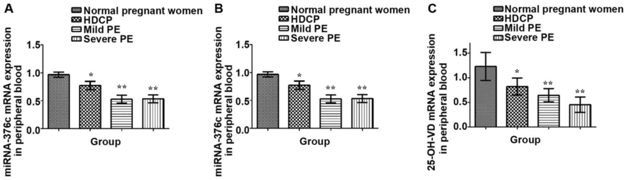
Expression of miRNA-376c and 25-OH-VD
mRNA in peripheral blood of pregnant women
miRNA-376c and 25-OH-VD in peripheral blood and
placenta of patients in 4 groups were detected via RT-qPCR. The
detection results of expression of miRNA-376c in the peripheral
blood and placenta of pregnant women with severe PE, mild PE and
HDCP and normal pregnant women showed that the expression levels in
mild PE, HDCP and severe PE groups were decreased, and there were
statistically significant differences compared with that in the
normal group (P<0.05). With the progression of the disease, the
expression of miRNA-376c in the peripheral blood and placental
tissues was decreased. The expression of 25-OH-VD mRNA showed a
decreasing trend with the aggravation of the disease, and there
were statistically significant differences in the mild PE, HDCP and
severe PE groups compared with the normal group (P<0.05)
(Fig. 1).
Correlation analyses of expression of
miRNA-376c and 25-OH-VD mRNA in peripheral blood and placental
tissues with clinical data
In this study, the correlations of the expression of
miRNA-376c and 25-OH-VD mRNA with maternal age, urine protein,
fetal weight and blood pressure in mild PE, HDCP and severe PE
groups were analyzed. Results showed that the decreased expression
of miRNA-376c in peripheral blood and placental tissues was
positively correlated with maternal age and fetal weight
(P<0.01), but negatively correlated with blood pressure and
urine protein level in patients (P<0.01). The expression of
25-OH-VD mRNA in placental tissues was positively correlated with
maternal age and fetal weight (P<0.01), but negatively
correlated with blood pressure and urine protein level in patients
(P<0.01) (Table III).
| Table III.Correlation analyses of expressions of
miRNA-376c and 25-OH-VD mRNA with clinical data. |
Table III.
Correlation analyses of expressions of
miRNA-376c and 25-OH-VD mRNA with clinical data.
|
|
| miRNA-376c in
peripheral blood | miRNA-376c in
placental tissues | 25-OH-VD in
placental tissues |
|---|
|
|
|
|
|
|
|---|
| Variables | n | rs | P-value | rs | P-value | rs | P-value |
|---|
| Age | 199 | 0.731 | <0.01 | 0.713 | <0.01 | −0.614 | <0.01 |
| Fetal weight | 199 | 0.647 | <0.01 | 0.434 | <0.01 | −0.517 | <0.01 |
| Blood pressure | 199 | −0.541 | <0.01 | −0.461 | <0.01 | 0.521 | <0.01 |
| Urine protein | 132 | −0.614 | <0.01 | −0.467 | <0.01 | 0.519 | <0.01 |
Changes in expression of miRNA-376c in
peripheral blood and placental tissues and expression of 25-OH-VD
mRNA in placental tissues
Changes in the expression of miRNA-376c in the
peripheral blood and placental tissues and the expression of
25-OH-VD mRNA in the placental tissues of pregnant women in the 4
groups were studied. Results revealed that the expression of
miRNA-376c and 25-OH-VD mRNA were decreased. There were
significantly positive correlations between miRNA-376c in
peripheral blood and 25-OH-VD mRNA in placental tissues, and
between miRNA-376c in placental tissues and 25-OH-VD mRNA
(P<0.01), suggesting that with the decrease of miRNA-376c
expression in the peripheral blood or placental tissues and the
expression of 25-OH-VD mRNA in the placental tissues is also
decreased. Therefore, miRNA-376c may affect the expression of
25-OH-VD gene and play an important role in the occurrence and
development of hypertension. Besides, the expression of miRNA-376c
in peripheral blood and placental tissues was consistent and showed
a positive correlation (P<0.01), indicating that miRNA-376c in
peripheral blood may reflect the expression level of miRNA-376c in
placental tissues.
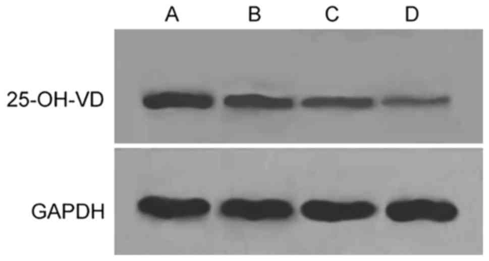
miRNA-376c and 25-OH-VD mRNA and
protein expression in placental tissues and their correlations
Finally, 10 patients were randomly selected from
each group to analyze the miRNA-376c and 25-OH-VD mRNA and protein
expression levels in placental tissues. Results revealed that the
25-OH-VD protein expression were significantly downregulated in the
4 groups (Fig. 2). The expression of
miRNA-376c in placental tissues was positively correlated with the
expression of 25-OH-VD protein, but negatively correlated with the
expression of 25-OH-VD mRNA, suggesting that miRNA-376c promotes
the production of 25-OH-VD protein through regulating 25-OH-VD mRNA
in tissues.
Discussion
As a kind of non-coding RNA molecule with 19–23
nucleotides in length, miRNA regulates the different expression of
molecules at the post-transcriptional level, thus playing an
important regulatory role in the body (18). miRNA is widely distributed in animals
and plants. The regulation of miRNA can affect the cell
proliferation, apoptosis, differentiation and other processes, and
miRNA is also involved in the occurrence and development of tumors
(19). At present, the relationship
between miRNA and human diseases has gradually gained people's
attention. As a member of miRNA-379 and miR-656 clusters,
miRNA-376c is located on a large miRNA cluster in the DIO3 region
of human chromosome 14.33. In this cluster, most miRNAs are
differentially expressed in the placenta, but its mechanism remains
unclear yet at present (20).
PE, as one of HDCP, is closely related to the health
and safety of pregnant women. It has been reported that the damage
and dysfunction of vascular endothelial cells (VEC) may be the
central part of the pathogenesis, eventually leading to eclampsia.
After VEC injury, tissue hypoxia, hemoconcentration, enhanced
permeability, increased expression of coagulation factors and
decreased expression of anticoagulant protein fibrinolysis factor
will occur, leading to imbalance of the coagulation and
fibrinolytic systems, ultimately resulting in thrombosis (21). Under the prethrombotic state,
atherosclerosis in placental vasculature, placental ischemia and
hypoxia as well as dysfunction may happen. As a result, the
maternal body will secrete a large number of plasma cytotoxic
substances, damaging the VEC function of maternal body and
resulting in PE. It has been found in recent studies that miRNA has
some differences in the expression in PE placental and normal
placental tissues, which may indicate that miRNA is involved in the
occurrence and development of PE, helping us acquire a better
understanding of the pathogenesis of PE (22).
This study was performed strictly in accordance with
the inclusion and exclusion criteria. The expression of miRNA-376c
and 25-OH-VD mRNA in peripheral blood and placental tissues in the
normal pregnant women and those with HDCP, mild PE and severe PE
were detected, and it was found that the expression level of
miRNA-376c was significantly decreased with the gradual aggravation
of the disease, and it was positively correlated with the
expression of 25-OH-VD mRNA and protein, suggesting that miRNA-376c
may participate in the occurrence and development of PE through
regulating the expression of 25-OH-VD gene in tissues. Fu et
al (23) said in his article
that the miRNA-376c expression levels in placental tissues of
Canadians and Chinese with severe PE are significantly decreased
compared with that in the normal group in the same period, and the
expression level in patients with advanced PE also shows a
decreasing trend compared with that in normal subjects, which is
basically consistent with the results obtained in this study,
further proving the experimental results. The correlations between
miRNA-376c expression and clinical data were analyzed, and it was
found that the miRNA-376c expression was positively correlated with
maternal age and fetal weight, but negatively correlated with blood
pressure and urine protein of pregnant women. With the aggravation
of the disease, the expression of miRNA-376c in patients with
severe PE was significantly decreased compared with those in other
groups, the fetal weight was small and most fetuses were premature.
It has been reported in the literature (24) that the risks of neonatal defect
disease and premature delivery are higher in PE patients.
Finally, the correlations of miRNA-376c expression
in placental tissues and peripheral blood of pregnant women in the
4 groups were compared, and results showed that there was a
positive correlation between them, suggesting that significant
changes in miRNA expression can be observed in both peripheral
blood and placental tissues. Recently, some studies have shown
(25) that miRNA can be detected in
serum and plasma and it can be used as a diagnostic tool. According
to the results of this study, it was found that the expression of
miRNA-376c in peripheral blood was similar to that in placental
tissues, and the collection of peripheral blood was characterized
by convenience and small trauma compared with that of peripheral
tissues. Therefore, it is suitable for pregnant women.
At the same time, this study had some deficiencies.
Only the miRNA-376c expression in placental tissues and peripheral
blood were detected and investigated preliminarily, but its
occurrence mechanism and process were not studied in depth.
Besides, our study was not representative due to the insufficient
samples, and whether the race and geographical differences may lead
to biased results, remains unknown. In future studies, the sample
size should be increased to better prove our views, laying a
foundation for clinical diagnosis.
In conclusion, the expression of miRNA-376c in
placental tissues and peripheral blood in varying degrees of HDCP
were detected in this study. Results showed that the miRNA-376c
expression was significantly decreased with the progression of the
disease. The expression of 25-OH-VD mRNA and protein in placental
tissues were also obviously decreased and they were positively
correlated. Finally, changes in miRNA-376c and 25-OH-VD were
significantly correlated with blood pressure and urine protein in
pregnant women, indicating that miRNA-376c and 25-OH-VD interact
with each other and participate in the occurrence and development
of HDCP.
Acknowledgements
Not applicable.
Funding
No funding was received.
Availability of data and materials
All data generated or analyzed during this study are
included in this published article.
Authors contributions
JL and JD designed the study and performed the
experiments. CW, ZW and JB collected the patient data. JL and SZ
analyzed the patient data. All authors have read and approved the
final manuscript.
Ethics approval and consent to
participate
This study was approved by the Medical Ethics
Committee of Jining First People's Hospital (Jining, China). All
patients and their families signed the informed consent.
Patient consent for publication
Not applicable.
Competing interests
The authors declare that they have no competing
interests.
References
|
1
|
American College of Obstetricians and
Gynecologists, . Task Force on Hypertension in Pregnancy:
Hypertension in pregnancy. Report of the American College of
Obstetricians and Gynecologists' task force on hypertension in
pregnancy. Obstet Gynecol. 122:1122–1131. 2013.PubMed/NCBI
|
|
2
|
Taylor RN, Roberts JM, Cunningham FG and
Lindheimer MD: Chesley's Hypertensive Disorders in Pregnancy. 4th
edition. Elsevier BV; Amsterdam: 2014
|
|
3
|
Creanga AA, Berg CJ, Syverson C, Seed K,
Bruce FC and Callaghan WM: Pregnancy-related mortality in the
United States, 2006–2010. Obstet Gynecol. 125:5–12. 2015.
View Article : Google Scholar : PubMed/NCBI
|
|
4
|
Li XL, Chen TT, Dong X, Gou WL, Lau S,
Stone P and Chen Q: Early onset preeclampsia in subsequent
pregnancies correlates with early onset preeclampsia in first
pregnancy. Eur J Obstet Gynecol Reprod Biol. 177:94–99. 2014.
View Article : Google Scholar : PubMed/NCBI
|
|
5
|
Henderson JT, Whitlock EP, O'Connor E,
Senger CA, Thompson JH and Rowland MG: Low-dose aspirin for
prevention of morbidity and mortality from preeclampsia: A
systematic evidence review for the U.S. Preventive Services Task
Force. Ann Intern Med. 160:695–703. 2014. View Article : Google Scholar : PubMed/NCBI
|
|
6
|
Abalos E, Cuesta C, Grosso AL, Chou D and
Say L: Global and regional estimates of preeclampsia and eclampsia:
A systematic review. Eur J Obstet Gynecol Reprod Biol. 170:1–7.
2013. View Article : Google Scholar : PubMed/NCBI
|
|
7
|
Abalos E, Cuesta C, Carroli G, Qureshi Z,
Widmer M, Vogel JP and Souza JP: WHO Multicountry Survey on
Maternal and Newborn Health Research Network: Pre-eclampsia,
eclampsia and adverse maternal and perinatal outcomes: a secondary
analysis of the World Health Organization Multicountry Survey on
Maternal and Newborn Health. Int BJOG. 35:14–24. 2014. View Article : Google Scholar
|
|
8
|
Ghulmiyyah L and Sibai B: Maternal
mortality from preeclampsia/eclampsia. Semin Perinatol. 36:56–59.
2012. View Article : Google Scholar : PubMed/NCBI
|
|
9
|
Vitsios DM, Davis MP, van Dongen S and
Enright AJ: Large-scale analysis of microRNA expression,
epi-transcriptomic features and biogenesis. Nucleic Acids Res.
45:1079–1090. 2017. View Article : Google Scholar : PubMed/NCBI
|
|
10
|
Lin S and Gregory RI: MicroRNA biogenesis
pathways in cancer. Nat Rev Cancer. 15:321–333. 2015. View Article : Google Scholar : PubMed/NCBI
|
|
11
|
Ameres SL and Zamore PD: Diversifying
microRNA sequence and function. Nat Rev Mol Cell Biol. 14:475–488.
2013. View
Article : Google Scholar : PubMed/NCBI
|
|
12
|
Ouyang Y, Mouillet JF, Coyne CB and
Sadovsky Y: Review: Placenta-specific microRNAs in exosomes - good
things come in nano-packages. Placenta. 35 Suppl:S69–S73. 2014.
View Article : Google Scholar : PubMed/NCBI
|
|
13
|
Mayor-Lynn K, Toloubeydokhti T, Cruz AC
and Chegini N: Expression profile of microRNAs and mRNAs in human
placentas from pregnancies complicated by preeclampsia and preterm
labor. Reprod Sci. 18:46–56. 2011. View Article : Google Scholar : PubMed/NCBI
|
|
14
|
Principi N, Bianchini S, Baggi E and
Esposito S: Implications of maternal vitamin D deficiency for the
fetus, the neonate and the young infant. Eur J Nutr. 52:859–867.
2013. View Article : Google Scholar : PubMed/NCBI
|
|
15
|
Wei SQ, Qi HP, Luo ZC and Fraser WD:
Maternal vitamin D status and adverse pregnancy outcomes: A
systematic review and meta-analysis. J Matern Fetal Neonatal Med.
26:889–899. 2013. View Article : Google Scholar : PubMed/NCBI
|
|
16
|
Baker AH and Delles C: Is microRNA-376c a
biomarker or mediator of preeclampsia? Hypertension. 61:767–769.
2013. View Article : Google Scholar : PubMed/NCBI
|
|
17
|
Livak KJ and Schmittgen TD: Analysis of
relative gene expression data using real-time quantitative PCR and
the 2(-Delta Delta C(T)) Method. METHODS. 25:402–408. 2001.
View Article : Google Scholar : PubMed/NCBI
|
|
18
|
Liang Y, Ridzon D, Wong L and Chen C:
Characterization of microRNA expression profiles in normal human
tissues. BMC Genomics. 8:1662007. View Article : Google Scholar : PubMed/NCBI
|
|
19
|
Agarwal V, Bell GW, Nam JW and Bartel DP:
Predicting effective microRNA target sites in mammalian mRNAs.
eLife. 4:e050052015. View Article : Google Scholar
|
|
20
|
Liu J, Wang L, Su Z, Wu W, Cai X, Li D,
Hou J, Pei D and Pan G: A reciprocal antagonism between miR-376c
and TGF-β signaling regulates neural differentiation of human
pluripotent stem cells. FASEB J. 28:4642–4656. 2014. View Article : Google Scholar : PubMed/NCBI
|
|
21
|
Chen Q, Sousa JD, Snowise S, Chamley L and
Stone P: Reduction in the severity of early onset severe
preeclampsia during gestation may be associated with changes in
endothelial cell activation: A pathological case report. Hypertens
Pregnancy. 35:32–41. 2016. View Article : Google Scholar : PubMed/NCBI
|
|
22
|
Korkes HA, de Oliveira LG, Sass N,
Karumanchi SA and Rajakumar A: 79 sFlt-1 and miRNA-210 expressions
have strong correlation in preeclamptic placentas: Biomarkers,
prediction of preeclampsia. Pregnancy Hypertens: An Int J Womens
Cardiovasc Health. 6:2162016. View Article : Google Scholar
|
|
23
|
Fu G, Ye G, Nadeem L, Ji L, Manchanda T,
Wang Y, Zhao Y, Qiao J, Wang YL, Lye S, et al: MicroRNA-376c
impairs transforming growth factor-β and nodal signaling to promote
trophoblast cell proliferation and invasion. Hypertension.
61:864–872. 2013. View Article : Google Scholar : PubMed/NCBI
|
|
24
|
Brodwall K, Leirgul E, Greve G, Vollset
SE, Holmstrøm H, Tell GS and Øyen N: Possible common aetiology
behind maternal preeclampsia and congenital heart defects in the
child: A Cardiovascular Diseases in Norway Project Study. Paediatr
Perinat Epidemiol. 30:76–85. 2016. View Article : Google Scholar : PubMed/NCBI
|
|
25
|
Chim SS, Shing TK, Hung EC, Leung TY, Lau
TK, Chiu RW and Lo YM: Detection and characterization of placental
microRNAs in maternal plasma. Clin Chem. 54:482–490. 2008.
View Article : Google Scholar : PubMed/NCBI
|