Introduction
Osteoporosis (OP) is a common but serious skeletal
disorder where increased bone weakness increases the risk of broken
bone, especially for the older patients. OP is a major public
health problem, and a serious economical burden for the patients
(1). Although little is known about
how OP develops, numerous factors (2), including sex, genetics, age, hormones
and diet, have been identified as being involved in the development
of OP or identified to be able to increase the likelihood of
OP.
OP has been considered to be a disease associated
with abnormal calcium metabolism. Several lines of evidence
supported the hypothesis (3,4). Nonetheless, a growing number of
clinical observations strongly suggest the association of iron
overload with bone diseases, particularly with osteoporosis
(5,6). Therefore, elevated iron level has been
suggested to be implicated in and has been linked to the risk
factor of OP. Actually, the first clinical observation was made by
Sinigaglia and colleagues (7) in
patients with OP in the setting of genetic hemochromatosis.
Although the observation was obviously somewhat compounded by the
linkage between iron concentration and hemochromatosis in OP, it
was supported by further clinical investigations reporting the
association between iron overload and bone disorders, including
osteomalacia (8), osteoporosis
(9), altered microarchitecture
(10) and bone fractures (11). Despite this, no direct clinical
evidence has been established until now regarding the concentration
of hepcidin, a key regulator in homeostasis of iron, as well as the
iron concentration in the peripheral sera of patients with OP.
Hepcidin, also LEAP-1 (12), HEPC (13) or HAMP (14), was originally discovered to be
involved in the maintenance of iron homeostasis, which is necessary
for the regulation of iron storage and iron absorption (15). Any mutation in this gene was found to
be able to cause hemochromatosis (16). Hepcidin has been suggested to have
anti-osteoporosis effects by preventing iron overload (17), suggesting that the level of hepcidin
seems to be inversed with level of iron accumulation. Sun et
al (18) confirmed the in
vivo use of a transgenic mouse model. In addition, the relevant
studies performed concerning hepcidin in osteoporosis were either
from in vitro cell culture system or in vivo animal
models. Clinical status of hepcidin remains to be investigated in
patients with OP.
In order to understand the status of hepcidin,
soluble TFR2 as well as the iron concentration in the sera of
patients with OP, we detected the concentration of hepcidin,
soluble TFR2 and iron in sera based on biochemical autoanalyzer
(Cobas 8000; Roche Diagnostics, Indianapolis, IN, USA). It was
demonstrated that iron concentration and soluble TFR2 were
significantly higher in the sera of patients with OP than that of
healthy control and that concentration of hepcidin was pronouncedly
lower in OP than that in control. To further explore the
biochemical role of hepcidin, we showed that hepcidin can interact
with TFR2 using immunoprecipitation method.
The present study is the first to establish the
direct clinical evidence for hepcidin and iron overload in OP,
suggesting that upregulation of hepcidin could be used as an
alternative therapeutic strategy in the management of OP.
Materials and methods
Clinical serum samples
The present study was approved by the Medical Ethics
Committee of Qilu Hospital of Shandong University (Qingdao, China).
Written informed consent was obtained from each participant
involved in the study. Forty serum samples from patients with OP
were collected from the Department of Nuclear Medicine, The
Affiliated Hospital of Qingdao University. As healthy control, 40
serum samples were collected from the Department of Laboratory
Medicine, Qilu Hospital of Shandong University (Qingdao).
Biochemical analysis of hepcidin and
iron in sera
The concentration of iron in sera from OP group and
healthy control group was measured by Cobas 8000 model Roche
autoanalyser (Cobas 8000; Roche Diagnostics). Given that the
preproprotein of hepcidin was post-translationally cleaved into
mature peptides of 20, 22 and 25 amino acids, here we focused only
on hepcidin 25. Serum hepcidin 25 (bioactive) was measured by
enzyme-linked immunosorbent assay (ELISA) (cat. no. EIA-5782; DRG
International, Inc., Springfield Township, NJ, USA). In a similar
way, soluble TFR2 was also measured using ELISA kit (cat. no.
E-EL-H2346; Biofavor Biotech, Inc., Wuhan, China).
293T cell culture and transfection
constructs
293T cells were maintained at 37°C in Dulbeccos
modified Eagles medium (DMEM) supplemented with 10% fetal bovine
serum (FBS), 100 U/ml penicillin and 100 µg/ml streptomycin (Gibco,
Carlsbad, CA, USA). Cells were transiently transfected with plasmid
DNA by Lipofectamine 2000 (Invitrogen), unless otherwise specified.
For Lipofectamine transfection, cells were transfected with 2–8 µg
plasmid DNA in Opti-MEM I reduced serum media (Invitrogen,
Carlsbad, CA, USA) containing Lipofectamine 2000 at a ratio of 1 µg
DNA to 2 µl Lipofectamine for 5 h before replacing the media with
DMEM. Cells were allowed to incubate for ~24 h before being
harvested. The expression plasmids used are described below:
Eukaryotic expression vector harboring human transferrin receptor 2
(TFR2) gene tagged with Myc-DDK (cat. no. RC220060; and the vector
harboring human hepcidin gene tagged with GFP (cat. no. RG204620
were commercially available from Origene Technologies, Inc.,
Rockville, MD, USA).
Immunoprecipitation (IP)
The two vectors were co-transfected into HEK293T
cells, followed by collection with SDS protein lysis buffer. The
lysates were centrifuged at 12,000 × g for 10 min at 4°C and the
resulting supernatant diluted 5-fold with IP buffer (50 nM
Tris-HCI, pH 7.5, 150 nM NaCl, 2 mM EDTA, 1% NP-40 and 10 mM
N-ethylmaleimide). The lysates were pre-cleared by incubating with
rabbit sera for 30 min followed by incubation with protein
A-Sepharose beads (GE Healthcare) for 1 h. The lysates were
recovered by centrifugation at 12,000 × g for 10 min at 4°C after
which the supernatant was moved to a fresh tube. The supernatant
was incubated with 10 µl of rabbit monoantibody to hepcidin for 1.5
h followed by protein A-Sepharose beads for another 1.5 h. The
incubation was performed at 4°C with gentle rotation. The beads
were recovered by centrifugation and washed 4 times with IP buffer.
The proteins were eluted from the beads by incubation with sample
loading buffer at 37°C for 10 min. Equal volumes of supernatant
were separated on SDS-PAGE and the precipitated proteins detected
by immunoblotting (IB). Rabbit mono-antibodies to human hepcidin
(cat. no. ab187778), TFR2 (cat. no. ab185550) and IgG (cat. no.
ab218427) were all obtained commercially from Abcam (Cambridge, MA,
USA).
Statistical analysis
Statistical analyses were carried out using SPSS
17.0 version (SPSS, Inc., Chicago, IL, USA). Continuous variables
are expressed as the mean ± standard error of mean (SEM). Normality
of the distribution was assessed with the Kolmogorov-Smirnov test.
The differences between groups in terms of numerical variables were
examined using the independent sample t-test or the Mann-Whitney U
test, according to the provided condition of parametric or
non-parametric distribution. Pearson's correlation analysis was
performed to evaluate the relationship between numeric variables.
P<0.05 was considered to be statistically significant.
Results
Baseline characteristics
Baseline characteristics of the patient and control
groups are shown in Table I. There
were no differences in age, sex and BMI between the two groups.
Nevertheless, there was a significant difference of iron, hepcidin
and TFR2 concentration between the two groups. Iron concentration
was presented to be markedly higher in the sera of OP group
(18.57±3.36 µM/ml) than that of healthy control group (9.34±3.15
µM/ml), as exemplified by Roche biochemical autoanalyzer.
Similarly, the concentration of soluble TFR2 was also significantly
elevated in OP (19.31±3.41 ng/ml) than that (8.70±3.26 ng/ml) in
healthy control. By contrast, the concentration of hepcidin was
shown to be remarkably lower in sera of OP (11.48±4.12 ng/ml) than
that (29.88±4.23 ng/ml) of healthy control. To observe whether
there was a significant correlation between soluble TRF2 and
hepcidin, Pearson's correlation analysis was performed. It
indicated that there was a significant negative correlation between
soluble TRF2 vs. hepcidin (Table
II).
 | Table I.Baseline characteristics of the
patient with OP and healthy controls. |
Table I.
Baseline characteristics of the
patient with OP and healthy controls.
| Variable | OP group (n=40) | Healthy control
(n=40) | P-value |
|---|
| Age (years) | 51.22±7.38 | 50.45±10.02 | 0.538 |
| Sex (F/M) | 29/11 | 26/14 | 0.782 |
| BMI
(kg/m2) | 32.16±6.63 | 31.06±3.87 | 0.314 |
| Iron (µm/ml) | 18.57±3.36 | 9.34±3.15 | 0.012 |
| Hepcidin (ng/ml) | 11.48±4.12 | 29.88±4.23 | 0.019 |
| TFR2 (ng/ml) | 19.31±3.41 | 8.70±3.26 | 0.022 |
| Table II.The correlation between hepcidin and
TRF2. |
Table II.
The correlation between hepcidin and
TRF2.
|
| Hepcidin (ng/ml) |
|---|
|
|
|
|---|
| Variable | r | P-value |
|---|
| Age (years) | 0.525 | 0.056 |
| BMI
(kg/m2) | 0.071 | 0.612 |
| Iron (µM/ml) | −0.031 | 0.021 |
| Hepcidin (ng/ml) | – | – |
| TFR2 (ng/ml) | −0.258 | 0.038 |
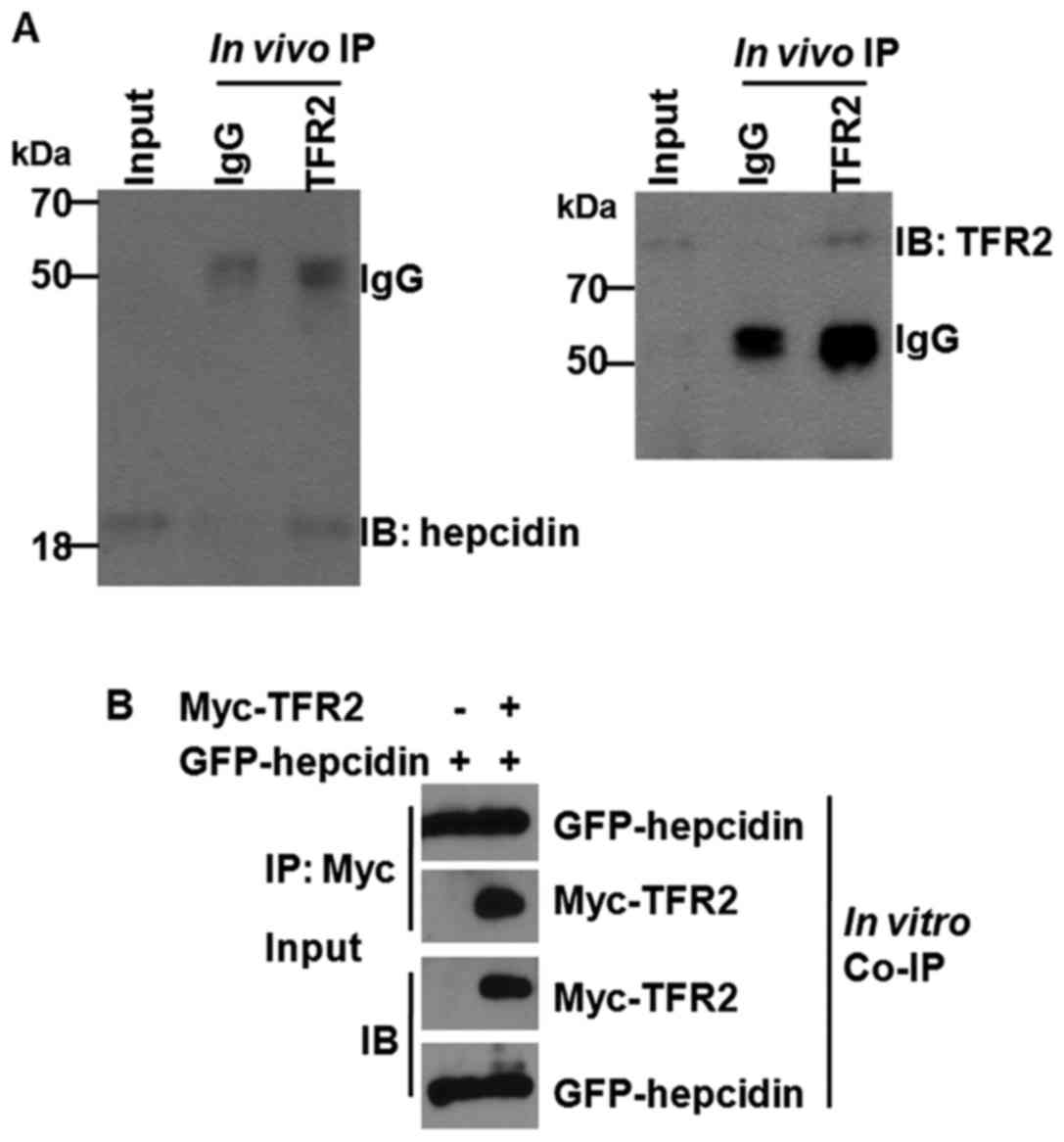
Hepcidin can interplay with TFR2
Having identified a significant correlation between
soluble TRF2 and hepcidin, we determined whether there is a direct
interaction between hepcidin and TFR2. To approach the problem, we
conducted the IP in vivo. To ensure that our IP contained
only hepcidin and TFR2 proteins and were devoid of its associated
proteins, the IP was carried out in the presence of SDS to break up
any potential protein complex. As shown in Fig. 1A, the specific monoantibody to TFR2
immunoprecipitated the TFR2 and hepcidin proteins successfully. The
specificity of the IP was evident from the failure of the
pre-immune IgG to immunnoprecipitate the hepcidin protein. The
signal of the bands was actually faint, at least not as significant
as the brightness of IgG, which was used as control. For further
verification, the Co-IP was performed in vitro using
co-transfection of eukaryotic expression vectors of hepcidin and
TFR2 into 293T cells (Fig. 1B). As
expected, in vitro Co-IP result confirmed that hepcidin can
interplay with TFR2, with the signal of band being strong. The
results demonstrated that hepcidin can interplay with TFR2.
Discussion
To the best of our knowledge, this is the first
study on hepcidin and TFR2 expression in sera of OP. Hepcidin was
significantly reduced in the sera of patients with OP compared with
healthy controls, whereas the iron concentration and soluble TFR2
was pronouncedly elevated in the sera of osteoporosis relative to
healthy control. We also report that hepcidin can interplay with
TFR2 in a protein-protein interaction fashion. These results
present the biochemical profiles of hepcidin and TFR2 expression in
sera of OP, which may account for the clinical phenotype that
elevated iron concentration was observed in the sera of patients
with OP.
Hepcidin is a protein that in humans is encoded by
the HAMP gene, which is a key regulator of the entry of iron
into the circulation in mammals (15). Hepcidin inhibits iron transport by
binding to the iron export channel ferroportin that is located on
the basolateral surface of gut enterocytes and the plasma membrane
of reticuloendothelial cells (19,20). The
first direct experimental evidence establishing the linkage between
hepcidin and OP came from the study performed by Xu and colleagues
(17) reporting that hepcidin was
shown to be significantly capable of increasing intracellular
calcium concentration in osteoblast cell line hFOB1.19. The study,
albeit carried out in an in vitro cell culture system, sheds
new light into the crucial role of iron homeostasis in bone
metabolism (5). Abnormal calcium
metabolism has been traditionally taken to be associated with the
development of OP (5). Nevertheless,
only abnormal calcium metabolism itself was obviously not
sufficient to account for the pathogenesis of OP. Given this,
imbalance of iron may be an alternative hypothesis explaining the
etiology of OP. The present study, therefore, was important in
disclosing the imbalance of iron in OP. We demonstrated using ELISA
that hepcidin was markedly lower in the sera of OP than that of
healthy control, which was basically in line with observations made
by Sun et al (18). Using a
murine model where the hepcidin gene was double knocked out the
study by Sun et al (18)
identified that, hepcidin deficiency resulted in a marked reduction
of bone load-bearing ability, indicating a link between hepcidin
deficiency and bone loss. Considering the biochemical role of
hepcidin (20), reduced level of
hepcidin can lead to the accumulation of iron, as we observed by
Roche biochemical autoanalyzer, which was supported by earlier
several lines of evidence (9,21).
However, the manner of hepcidin down-regulation in OP remains
unknown and deserves further investigation.
It has been well-established that hepcidin inhibited
iron transport through binding to ferroportin (19,20).
However, the interaction between hepcidin and transferrin receptor
2 (TFR2) remains to be elucidated as no direct evidence has been
established concerning the interaction between hepcidin and TFR2,
although association studies have suggested that there was a
negative correlation between them in the context of anemia
(22) and in children with
vegetarian diet (23). In the
present study, we showed using ELISA approach that soluble TFR2 was
also significantly higher in the sera of OP than that of healthy
controls. In the case of concentration trend of hepcidin and TFR2
in sera, our observation was in total agreement with previous
reports performed in the setting of anemia (22) and vegetarian (23). In addition, we have shown for the
first time, to the best of our knowledge, that hepcidin can
directly interact with TFR2, which could explain the reason for
hepcidin being detected to be lower in sera of OP relative to
healthy control.
There were some limitations that deserve to be
noted. Firstly, our conclusion was established on the limited
number of sample that requires to be further confirmed in larger
sample size. Secondly, direct extrapolation of our conclusion from
the study should be approached with caution in that, a higher
concentration of iron detected in serum relative to normal range or
control does not necessarily mean the onset of OP. It may also be
associated with anemia and hemochromatosis, therefore a high
concentration of iron in sera seems to be sufficiently necessary
but insufficient condition to the judgment of onset of OP. Thirdly,
hepcidin has been shown to be able to increase the intracellular
calcium (17), the calcium
concentration should have been detected.
In conclusion, the present study supports the role
of hepcidin in the development of OP, suggesting that upregulation
of hepcidin could be used as a novel alternative therapeutic
strategy in the management of OP.
Acknowledgements
The present study was supported by the Department of
Laboratory Medicine, Qilu Hospital of Shandong University (Qingdao,
China).
Funding
This research did not receive any specific grant
from funding agencies in the public, commercial, or not-for-profit
sectors.
Availability of data and material
The datasets used and/or analyzed during the present
study are available from the corresponding author on reasonable
request.
Authors' contributions
BL and CL contributed to the conception of the
study. WZ contributed significantly to the data analysis and study
preparation. MS and SD performed the data analyses and wrote the
study. JS helped perform the data analysis with constructive
discussions. All authors have read and approved the final
study.
Ethics approval and consent to
participate
The present study was approved by the Medical Ethics
Committee of Qilu Hospital of Shandong University (Qingdao, China).
Written informed consent was obtained from each participant
involved in the study.
Patient consent for publication
Not applicable.
Competing interests
The authors declare that they have no competing
interests.
References
|
1
|
Qu B, Ma Y, Yan M, Wu HH, Fan L, Liao DF,
Pan XM and Hong Z: The economic burden of fracture patients with
osteoporosis in western China. Osteoporos Int. 25:1853–1860. 2014.
View Article : Google Scholar : PubMed/NCBI
|
|
2
|
Hendrickx G, Boudin E and Van Hul W: A
look behind the scenes: The risk and pathogenesis of primary
osteoporosis. Nat Rev Rheumatol. 11:462–474. 2015. View Article : Google Scholar : PubMed/NCBI
|
|
3
|
Rull Ochoa-Hortal MA, Cano-García MC,
Arrabal-Martín M and Arrabal-Polo MA: Lithogenic factors in
postmenopausal women with osteoporotic fracture. Minerva
Endocrinol. 42:41–45. 2017.PubMed/NCBI
|
|
4
|
Rull MA, Cano-García MC, Arrabal-Martín M
and Arrabal-Polo MA: The importance of urinary calcium in
postmenopausal women with osteoporotic fracture. Can Urol Assoc J.
9:E183–E186. 2015. View Article : Google Scholar : PubMed/NCBI
|
|
5
|
Li GF, Pan YZ, Sirois P, Li K and Xu YJ:
Iron homeostasis in osteoporosis and its clinical implications.
Osteoporos Int. 23:2403–2408. 2012. View Article : Google Scholar : PubMed/NCBI
|
|
6
|
Tian Q, Wu S, Dai Z, Yang J, Zheng J,
Zheng Q and Liu Y: Iron overload induced death of osteoblasts in
vitro: Involvement of the mitochondrial apoptotic pathway. PeerJ.
4:e26112016. View Article : Google Scholar : PubMed/NCBI
|
|
7
|
Sinigaglia L, Fargion S, Fracanzani AL,
Binelli L, Battafarano N, Varenna M, Piperno A and Fiorelli G: Bone
and joint involvement in genetic hemochromatosis: Role of cirrhosis
and iron overload. J Rheumatol. 24:1809–1813. 1997.PubMed/NCBI
|
|
8
|
Mahachoklertwattana P, Sirikulchayanonta
V, Chuansumrit A, Karnsombat P, Choubtum L, Sriphrapradang A,
Domrongkitchaiporn S, Sirisriro R and Rajatanavin R: Bone
histomorphometry in children and adolescents with beta-thalassemia
disease: Iron-associated focal osteomalacia. J Clin Endocrinol
Metab. 88:3966–3972. 2003. View Article : Google Scholar : PubMed/NCBI
|
|
9
|
Valenti L, Varenna M, Fracanzani AL, Rossi
V, Fargion S and Sinigaglia L: Association between iron overload
and osteoporosis in patients with hereditary hemochromatosis.
Osteoporos Int. 20:549–555. 2009. View Article : Google Scholar : PubMed/NCBI
|
|
10
|
Tsay J, Yang Z, Ross FP,
Cunningham-Rundles S, Lin H, Coleman R, Mayer-Kuckuk P, Doty SB,
Grady RW, Giardina PJ, et al: Bone loss caused by iron overload in
a murine model: Importance of oxidative stress. Blood.
116:2582–2589. 2010. View Article : Google Scholar : PubMed/NCBI
|
|
11
|
Chen B, Li GF, Shen Y, Huang XI and Xu YJ:
Reducing iron accumulation: A potential approach for the prevention
and treatment of postmenopausal osteoporosis. Exp Ther Med.
10:7–11. 2015. View Article : Google Scholar : PubMed/NCBI
|
|
12
|
Krause A, Neitz S, Mägert HJ, Schulz A,
Forssmann WG, Schulz-Knappe P and Adermann K: LEAP-1, a novel
highly disulfide-bonded human peptide, exhibits antimicrobial
activity. FEBS Lett. 480:147–150. 2000. View Article : Google Scholar : PubMed/NCBI
|
|
13
|
Pigeon C, Ilyin G, Courselaud B, Leroyer
P, Turlin B, Brissot P and Loréal O: A new mouse liver-specific
gene, encoding a protein homologous to human antimicrobial peptide
hepcidin, is overexpressed during iron overload. J Biol Chem.
276:7811–7819. 2001. View Article : Google Scholar : PubMed/NCBI
|
|
14
|
Truksa J, Gelbart T, Peng H, Beutler E,
Beutler B and Lee P: Suppression of the hepcidin-encoding gene Hamp
permits iron overload in mice lacking both hemojuvelin and
matriptase-2/TMPRSS6. Br J Haematol. 147:571–581. 2009. View Article : Google Scholar : PubMed/NCBI
|
|
15
|
Ganz T: Hepcidin, a key regulator of iron
metabolism and mediator of anemia of inflammation. Blood.
102:783–788. 2003. View Article : Google Scholar : PubMed/NCBI
|
|
16
|
Beaumont-Epinette MP, Delobel JB, Ropert
M, Deugnier Y, Loréal O, Jouanolle AM, Brissot P and Bardou-Jacquet
E: Hereditary hypotransferrinemia can lead to elevated transferrin
saturation and, when associated to HFE or HAMP mutations, to iron
overload. Blood Cells Mol Dis. 54:151–154. 2015. View Article : Google Scholar : PubMed/NCBI
|
|
17
|
Xu Y, Li G, Du B, Zhang P, Xiao L, Sirois
P and Li K: Hepcidin increases intracellular Ca2+ of
osteoblast hFOB1.19 through L-type Ca2+ channels. Regul
Pept. 172:58–61. 2011. View Article : Google Scholar : PubMed/NCBI
|
|
18
|
Sun L, Guo W, Yin C, Zhang S, Qu G, Hou Y,
Rong H, Ji H and Liu S: Hepcidin deficiency undermines bone
load-bearing capacity through inducing iron overload. Gene.
543:161–165. 2014. View Article : Google Scholar : PubMed/NCBI
|
|
19
|
Gulec S, Anderson GJ and Collins JF:
Mechanistic and regulatory aspects of intestinal iron absorption.
Am J Physiol Gastrointest Liver Physiol. 307:G397–G409. 2014.
View Article : Google Scholar : PubMed/NCBI
|
|
20
|
Rossi E: Hepcidin - the iron regulatory
hormone. Clin Biochem Rev. 26:47–49. 2005.PubMed/NCBI
|
|
21
|
Rossi F, Perrotta S, Bellini G, Luongo L,
Tortora C, Siniscalco D, Francese M, Torella M, Nobili B, Di Marzo
V, et al: Iron overload causes osteoporosis in thalassemia major
patients through interaction with transient receptor potential
vanilloid type 1 (TRPV1) channels. Haematologica. 99:1876–1884.
2014. View Article : Google Scholar : PubMed/NCBI
|
|
22
|
Choi HS, Song SH, Lee JH, Kim HJ and Yang
HR: Serum hepcidin levels and iron parameters in children with iron
deficiency. Korean J Hematol. 47:286–292. 2012. View Article : Google Scholar : PubMed/NCBI
|
|
23
|
Ambroszkiewicz J, Klemarczyk W, Mazur J,
Gajewska J, Rowicka G, Strucińska M and Chełchowska M: Serum
hepcidin and soluble transferrin receptor in the assessment of iron
metabolism in children on a vegetarian diet. Biol Trace Elem Res.
180:182–190. 2017. View Article : Google Scholar : PubMed/NCBI
|