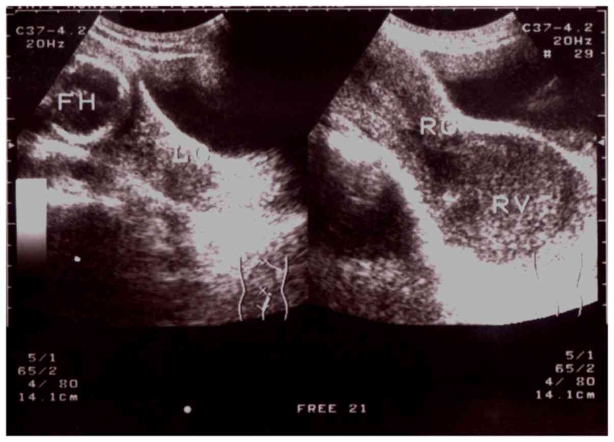
Ultrasonic evaluation of congenital vaginal oblique septum syndrome: A study of 21 cases

  • Authors:
    • Yong‑Hao Gai
    • Hui‑Li Fan
    • Ye Yan
    • Shi‑Feng Cai
    • Yue‑Zhong Zhang
    • Ru‑Xin Song
    • Shu‑Liang Song
  • View Affiliations

  • Published online on: July 6, 2018     https://doi.org/10.3892/etm.2018.6411
  • Pages: 2066-2070
Metrics: Total Views: 0 (Spandidos Publications: | PMC Statistics: )
Total PDF Downloads: 0 (Spandidos Publications: | PMC Statistics: )


Abstract

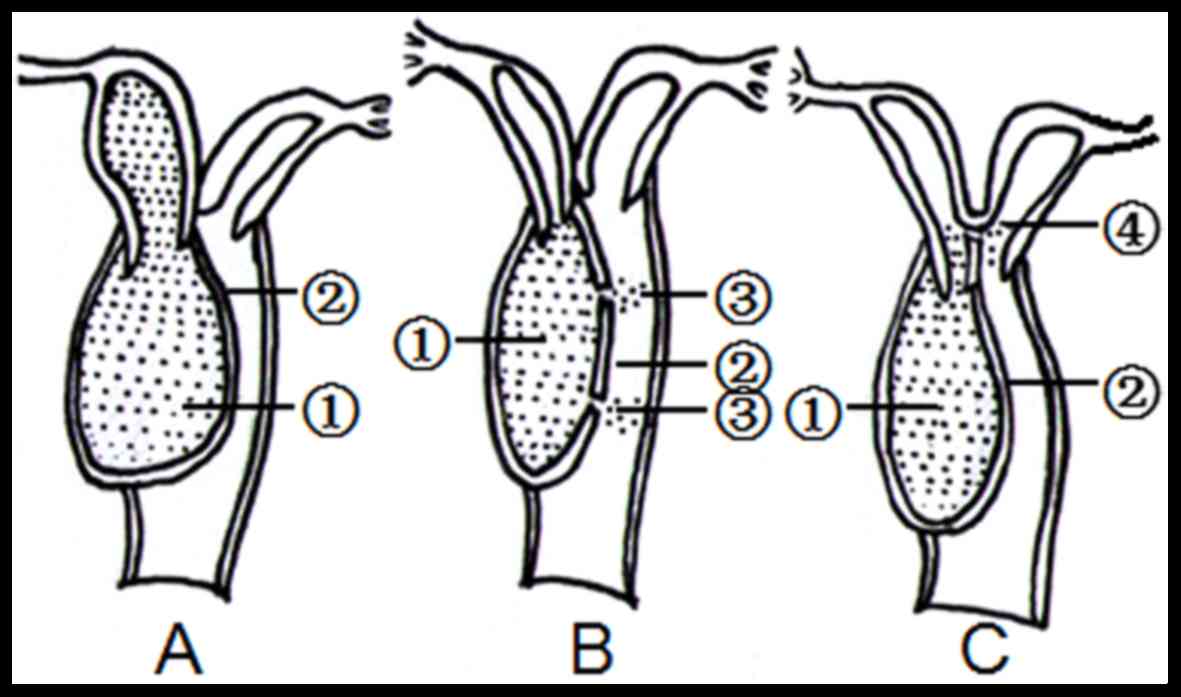
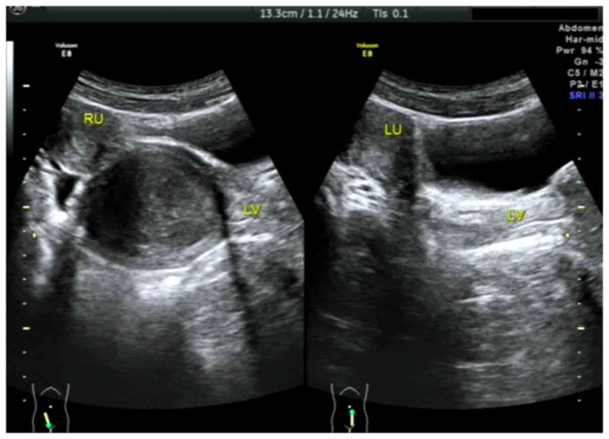
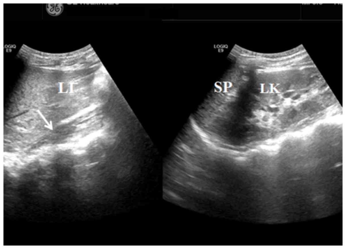
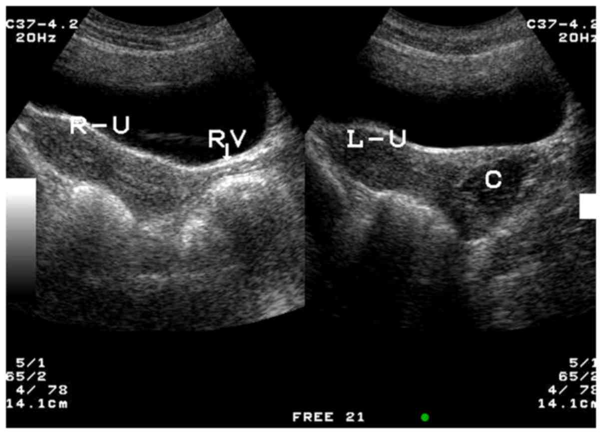
The aim of the present study was to investigate the ultrasound features and classify the lesion types of congenital vaginal oblique septum syndrome (CVOS) in 21 patients prior to surgery. Grey‑scale pelvic ultrasound was performed to evaluate the uterus, vagina and kidneys in 21 patients with suspected CVOS. Ultrasound features, including the presence of a double uterus, hematocolpos masses and renal absence, in CVOS types I, II and III were studied and compared with intra‑operative results and the results of surgery. Ultrasound identified the presence of double uteruses and cervices with ipsilateral renal agenesis on the oblique septum side in all 21 patients. There were 14 hematocolpos lesions on the right and 7 on the left of the vagina. Type I CVOS was diagnosed in 15 patients with a large hematocolpos mass (volume, 64‑268 ml) and these diagnoses were confirmed by surgery. Furthermore, there were 4 patients with type II and 2 patients with type III CVOS exhibiting small hematocolpos lesions (volume, 5‑36 ml) identified by ultrasound, which were all confirmed by surgery. Therefore, ultrasound imaging is useful tool to evaluate the abnormal features of CVOS and determine the type of CVOS in patients prior to surgical intervention.
View Figures
View References

Related Articles

Journal Cover

September-2018
Volume 16 Issue 3

Print ISSN: 1792-0981
Online ISSN:1792-1015

Sign up for eToc alerts

Recommend to Library

Copy and paste a formatted citation
x
Spandidos Publications style
Gai YH, Fan HL, Yan Y, Cai SF, Zhang YZ, Song RX and Song SL: Ultrasonic evaluation of congenital vaginal oblique septum syndrome: A study of 21 cases. Exp Ther Med 16: 2066-2070, 2018
APA
Gai, Y., Fan, H., Yan, Y., Cai, S., Zhang, Y., Song, R., & Song, S. (2018). Ultrasonic evaluation of congenital vaginal oblique septum syndrome: A study of 21 cases. Experimental and Therapeutic Medicine, 16, 2066-2070. https://doi.org/10.3892/etm.2018.6411
MLA
Gai, Y., Fan, H., Yan, Y., Cai, S., Zhang, Y., Song, R., Song, S."Ultrasonic evaluation of congenital vaginal oblique septum syndrome: A study of 21 cases". Experimental and Therapeutic Medicine 16.3 (2018): 2066-2070.
Chicago
Gai, Y., Fan, H., Yan, Y., Cai, S., Zhang, Y., Song, R., Song, S."Ultrasonic evaluation of congenital vaginal oblique septum syndrome: A study of 21 cases". Experimental and Therapeutic Medicine 16, no. 3 (2018): 2066-2070. https://doi.org/10.3892/etm.2018.6411