Introduction
Circulating tumor cells (CTCs) are cells that detach
from primary or metastatic solid tumors into the vasculature, where
they can be sampled from the circulating blood stream (1). CTCs are commonly identified in the
peripheral blood supply of diverse solid tumors, including breast
cancer (2,3), colorectal cancer (4), prostate cancer (5) etc. The migration of CTCs seems to be an
early event in human carcinogenesis, even before the cancer is
visible in clinical imaging. Experimental data in animal models has
shown that tumors measuring less than 1 mm could be associated with
the presence of CTCs in the bloodstream (6).
Zhangjiang Center for Translational Medicine
published a series of studies of CTCs in breast cancer (7), pancreatic cancer (8) and colorectal cancer (9) using combined subtraction enrichment and
immunostaining-fluorescence in situ hybridization
(SE-iFISH), cooperated with other research teams. These studies
demonstrated SE-iFISH CTC detection 91% positive rate for breast
cancer patients (7), a sensitivity
of 88% and specificity of 90% in pancreatic cancer and healthy
individuals at the cutoff value of 2 cells/7.5 ml (8), and a sensitivity of 90.9% and
specificity of 82.4% in colorectal cancer and healthy individuals
at the cutoff value of one cell/7.5 ml (9). From these studies, CTC detection by
SE-iFISH showed high sensitivity and specificity for distinguishing
breast cancer, pancreatic cancer and colorectal cancer from healthy
people. CTCs detection, up to date, provided potential biomarkers
for screening of cancer or precancerosis.
SE-iFISH is a novel strategy to detect CTCs in blood
(10), which enrich CTCs through the
removal of WBCs using anti-CD45 antibody conjugated immunomagnetic
particles, independent of EpCAM expression and tumor cell size.
Centromere Probe 8 (CEP8), cytokeratin (CK), CD45,
4′,6-diamidino-2-phenylindole (DAPI) were combined to identify
CTCs. Since aneuploidy is a typical common cytogenetic abnormality
in tumor cells, this feature could be exploited for CTC detection.
FISH was performed on CEP8 to identify aneuploidy cells. CTCs were
confirmed to be negative for CD45, positive for DAPI and either
positive for PanCK staining or aneuploidy chromosome 8. Cells with
characteristics of CK-/CD45+/DAPI+/CEP8=2 were WBCs.
The present case report describes a healthy female
who accepted a CTC test (by SE-iFISH platform) in Quanzhou No. 1
Hospital, and the result was 8 CTCs/7.5 ml, which indicated a high
risk of cancer. WES of these CTCs was performed to analyze their
mutation profiles to track the lesion.
Materials and methods
Subtraction enrichment of CTCs
7.5 ml peripheral blood was collected by ACD
anticoagulant tubes (BD Biosciences, Franklin Lakes, NJ, USA).
Reagents for subtraction enrichment are Cytelligen CTC enrichment
kit (Cytelligen, Inc., San Diego, CA, USA). In brief, peripheral
blood (7.5 ml) was centrifuged at 800 × g for 8 min at room
temperature, then supernatant was discarded. The left sample was
transferred to a centrifuge tube containing 3 ml hCTC Separation
Matrix. After centrifuging for 8 min at 450 g, the cell suspension
was collected from the buffy-coat layer. 150 ul immunomagnetic
particles conjugated anti-CD45 antibody was added into the cell
suspension, which was inoculated at room temperature for 10 min and
then placed on a magnetic stand (Promega Corporation, Madison, WI,
USA) till the liquid became clear. The supernatant was pipetted off
the magnetic field (non-magnetic bead-binding cell suspension) to
remove leukocytes by centrifuging at 500 rpm for 2 min. Sedimented
cells were thoroughly mixed with cell fixative and applied onto the
coated CTC slides for subsequent identification.
Identification of CTCs
Reagents for CTC identification were provided by the
Human Tumor Cell Identification kit (Cytelligen, Inc.). To identify
aneuploidy CTCs, fluorescence in situ hybridization (FISH)
and immunocytochemistry are used in combination. After a series of
pre-treatment containing drying, washing and dehydration, 10 µl of
probe solution containing fluorescence-labeled alpha-satellite
probes for the centromeres of the chromosome (CEP8) (2 µg/ml) was
added and then covered with a coverslip and sealed with neutral
resin. The hybridization procedure was as follows: degeneration at
75°C for 5 min, followed by hybridization at 37°C overnight. Then
the slide was rinsed with FR3 and added with monoclonal antibody
anti-CD45 conjugated to Alexa Fluor 594 (Invitrogen; Thermo Fisher
Scientific, Inc., Waltham, MA, USA) and anti-PanCK (CK4, 5, 6, 8,
10, 13 and 18) (Invitrogen; Thermo Fisher Scientific, Inc.) before
inoculation at room temperature for 2 h. After rinsing with PBS,
the slides were mounted with mounting medium containing DAPI and
photographed with a fluorescence microscope (Nikon Corporation,
Tokyo, Japan). CTCs were confirmed to be negative for CD45 and
either positive for PanCK staining or aneuploidy chromosome 8.
Laser capture microdissection and
whole genome amplification of CTCs
The CTC fixed slide was put in ZEISS Palm MicroBeam
Laser Micro Dissection System (Zeiss AG, Oberkochen, Germany). We
found CTCs according to the coordinate recorded in process of CTC
identification, and collected CTCs by laser micro dissection.
DNA amplification experiment of CTCs was according
to the kit instruction of MalBac single cell genome amplification
(YK001A/B; Yikon Genomics, Jiangsu, China). The product was
quantified using DNA electrophoresis.
Whole exome sequencing
Whole exome sequencing was performed on DNA of
blood, intestinal polyp and CTCs. For library construction, whole
exome DNA capture was performed using Agilent SureSelect Human All
ExonV5 kits following the manufacturer's instructions (Agilent
Technologies, Inc., Santa Clara, CA, USA). Subsequent to the
quality test, the qualified library was sequenced as 125 bp
paired-end reads on an Illumina Hiseq 2500 platform (Illumina,
Inc., San Diego, CA, USA).
Data analysis of whole exome
sequencing
For whole exome sequencing, clean data was obtained
after filtering adapter, low quality reads and reads with
proportion of N>10%. Reads were aligned to the reference human
genome (UCSC hg19) through Burrows-Wheeler Aligner. Next, the
Picard and Genome Analysis Tool kit (GATK) methods were adopted for
duplicate removal, local realignment and base quality
recalibration. Finally, the GATK Unified Genotyper was used for
single nucleotide variation (SNV)/inDel annotation.
Somatic SNP/InDel detection was performed with
Varscan2 software. Variants were annotated using the ANNOVAR
software tool. Mutations of CTCs were aligned to COSMIC database
(http://cancer.sanger.ac.uk/cosmic/).
The data in COSMIC is curated from number of high-quality sources
and combined into a single resource. The sources include:
Peer-reviewed journal articles, CGP laboratories at the Sanger
Institute, TCGA data portal, the ICGC data portal, IARC p53
database. We selected cancer-related mutations and annotated
relevant primary organ.
Droplet digital PCR
Each PCR (Bio-Rad Laboratories, Inc., Hercules, CA,
USA) reaction system (in a total reaction volume of 20 µl)
contained: 10 µl Bio-Rad 2×ddPCR supermix, 1 µl primer and probe, 2
µl DNA template (100 ng) and 7 µl H2O. Droplets were
generated and analyzed using the QX100 system (Bio-Rad
Laboratories). Amplifications were performed using the following
conditions: 1 cycle of 95°C for 10 min, 45 cycles of 94°C for 10
sec and 60°C for 45 sec, 1 cycle of 98°C for 10 min, and 1 cycle of
25°C for 10 sec. QuantaSoft analysis software (Bio-Rad
Laboratories) enabled abundance to be calculated for each
sample.
Case report
A female underwent circulating tumor cell detection
using SE-iFISH platform in the surgical oncology department,
Quanzhou No.1 hospital, Fujian, China, in Dec 22, 2014. She was
56-year-old, during menopause, and has no family history of cancer.
The results were 8 CTCs/7.5 ml which indicated a high risk of
cancer (Fig. 1). However, the levels
of AFP, cancer antigen (CA)-125, CA19-9, carcinoembryonic antigen
(CEA) and CA15-3 were in normal range. This patient also accepted
an HPV typing (HPV16, 18, 31, 33, 35,39, 45, 51, 52, 56, 58, 59,
68, 6, 11, 42, 43, 44, 53, 66 and CP8304) test, and the results
were negative. The thinprep cytology test showed no intraepithelial
neoplasia. Written informed consent was obtained from this person
for the present study.
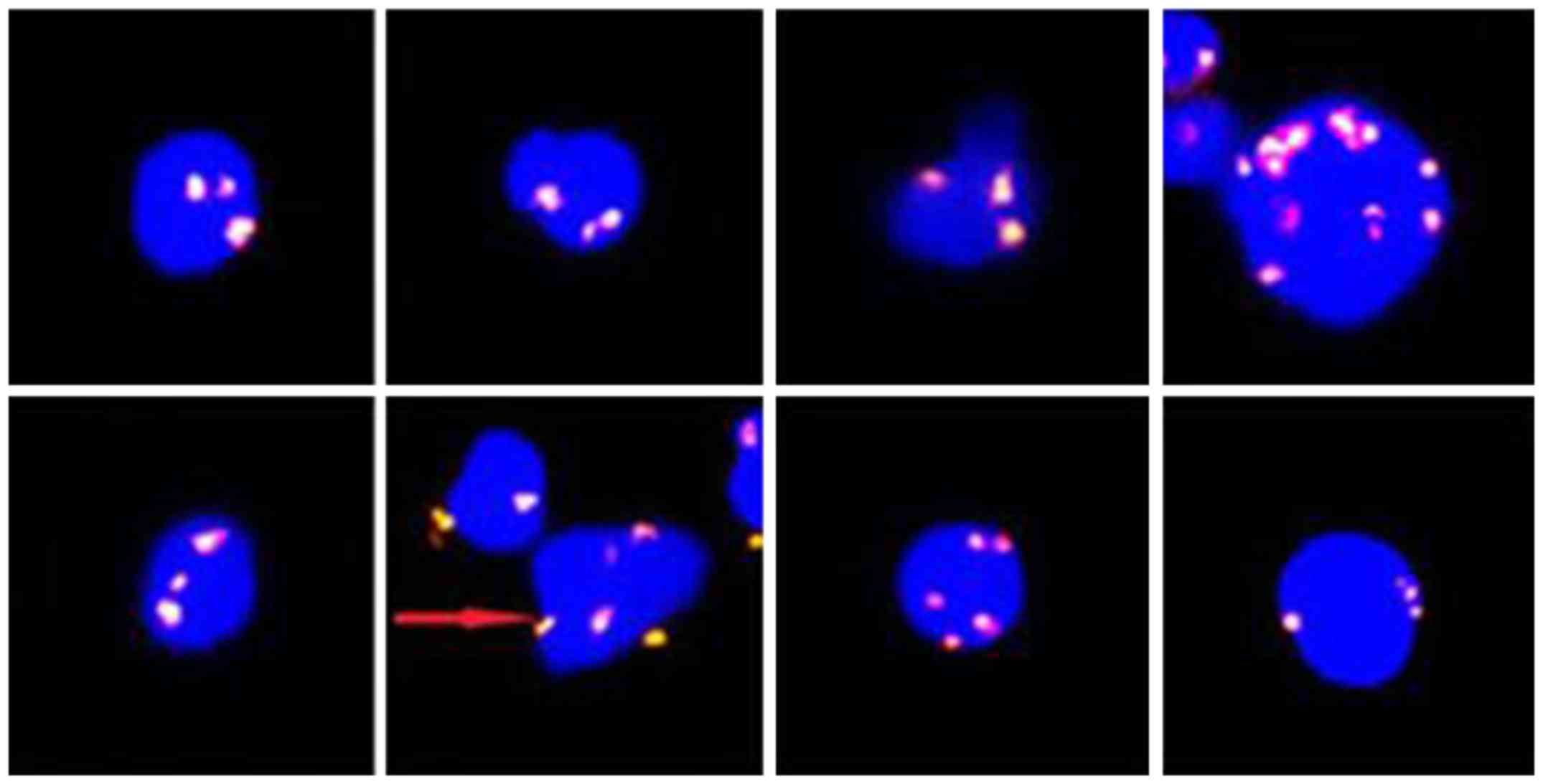 | Figure 1.Eight polyploid CTCs
(CK-/CD45-/DAPI+/CEP8+) identified by the SE-iFISH platform in one
female. Magnification, ×400. DAPI, blue; CEP8, orange; CK, green;
CD45, red. Red arrow, CTC in this image is this polyploidy pointed
by red arrow, rather than another diploid. CTCs, circulating tumor
cells; CEP8, Centromere Probe 8; CK, cytokeratin; DAPI,
4′,6-diamidino-2-phenylindole; SE-iFISH,
immunostaining-fluorescence in situ hybridization. |
To define the mutation spectrum of CTCs, we
performed laser capture microdissection to isolate the CTCs, and
whole genome amplification and whole exome capture DNA sequencing
(WES) on the 8 CTCs from this female. Sequencing achieved 81.5×
mean coverage on targeted exons. 34215 SNVs and 6807 InDels were
defined in the CTCs. Mapping these variations in COSMIC (http://cancer.sanger.ac.uk/cosmic/), 42
variations and InDels were correlated significantly with cancer
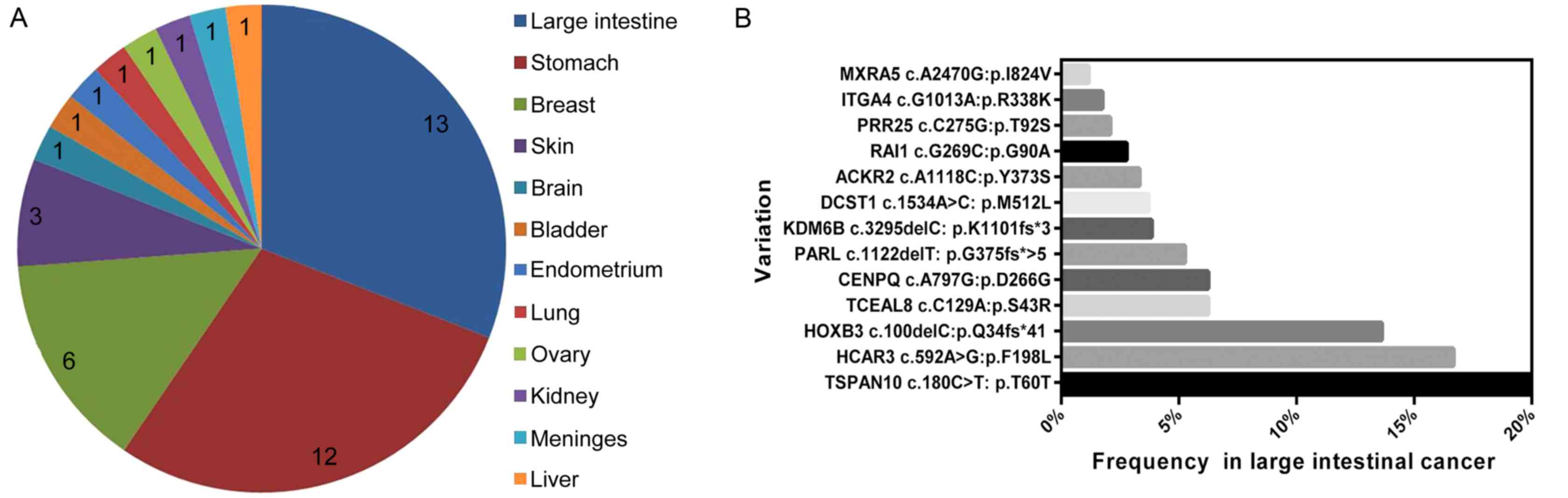
(Table I). Notably, 13, 12 and 6
mutations were related to large intestinal cancer, gastric cancer
and breast cancer, respectively (Fig.
2A). Fig. 2B shows 13 gene
variations and their mutational frequency in large intestinal
cancer, according to a TCGA study.
 | Table I.Cancer-related variations annotation
in COSMIC database. |
Table I.
Cancer-related variations annotation
in COSMIC database.
| Gene | Loci | Ref | Alt | Histology | Organ |
|---|
| HOXB3 | 46629737 | TG/TG | TG/T | Adenocarcinoma | Colon |
| CENPQ | 49459978 | AA | AG | Adenocarcinoma | Colon |
| TSPAN10 | 79612161 | CC | TT | Adenocarcinoma | Colon |
| TCEAL8 | 102508779 | GG | GT | Adenocarcinoma | Rectum |
| MXRA5 | 3241256 | TT | CC | Adenocarcinoma | Colon |
| PRR25 | 855717 | CC | GG | Adenocarcinoma | Rectum |
| HCAR3 | 123200693 | AA | GG | Adenocarcinoma | Colon |
| PARL | 183547404 | CT/CT | CT/C | Adenocarcinoma | Colon |
| KDM6B | 7752901 | GC/GC | G/G | Adenocarcinoma | Colon |
| DCST1 | 155019710 | AA | CC | Adenocarcinoma | Rectum |
| RAI1 | 17696531 | GG | CC | Adenocarcinoma | Colon |
| ITGA4 | 182347350 | GG | GA | Adenocarcinoma | Cecum |
| ACKR2 | 42907112 | AA | AC | Adenocarcinoma | Cecum |
| FCRLA | 161683136 | GG | AA | Adenocarcinoma | Stomach |
| TNRC18 | 5396715 | TT | CC | Adenocarcinoma | Stomach |
| ASAP1 | 131124559 | TT | CC | Adenocarcinoma | Stomach |
| CCDC153 | 119063908 | CC | TT | Adenocarcinoma | Stomach |
| OR10G7 | 123909627 | TT | CC | Adenocarcinoma | Stomach |
| CLEC1B | 10149406 | TT | CC | Adenocarcinoma | Stomach |
| BRCA1 | 41244000 | TT | CC | Adenocarcinoma | Stomach |
| LGALS14 | 40199914 | CC | GG | Adenocarcinoma | Stomach |
| CLC | 40225646 | GG | AA | Adenocarcinoma | Stomach |
| CHGB | 5904040 | GG | AA | Adenocarcinoma | Stomach |
| IGLL1 | 23915652 | GG | GA | Adenocarcinoma | Stomach |
| GCNT2 | 10557242 | GA/GA | GA/G | Adenocarcinoma | Stomach |
| MINA | 97664725 | CC | TT | Cancer | Breast |
| LINC01168,
LOC100128127 | 134886618 | GG | CC | Cancer | Breast |
| WDR90 | 701656 | CC | TT | Cancer | Breast |
| CHTF18 | 840378 | AA | GG | Cancer | Breast |
| ZNF286A | 15611495 | TT | CC | Cancer | Breast |
| BPIFB3 | 31656632 | CC | CG | Cancer | Breast |
| TNN | 175067689 | GG | AA | Squamous cell
carcinoma | Skin, face |
| MAN2B2 | 6602344 | GG | GT | Malignant
melanoma | Skin, arm |
| SMOC1 | 70420202 | GG | GA | Malignant
melanoma | Skin |
| SRSF4 | 29481412 | CC | AA | Hepatocellular
carcinoma | Liver |
| COL15A1 | 101778265 | GG | TT | Astrocytoma | Brain |
| IDI2 | 1065491 | GG | TT | Renal clear cell
carcinoma | Kidney |
| KRTAP4-7 | 39240627 | TT | TC | Transitional cell
carcinoma | Bladder |
| LTBP4 | 41118056 | AA | GG | Meningioma | Meninges |
| TGM3 | 2290333 | CC | AA | Adenocarcinoma | Lung |
| UMODL1 | 43546494 | GG | GA | Endometrioid
carcinoma | Endometrium |
| TMEM37 | 120194651 | A/A |
AGTGTGC/AGTGTGC | Serous
carcinoma | Ovary |
SNV annotation in COSMIC showed a high risk of large
intestinal cancer, followed by gastric cancer and breast cancer. To
check for lesions, the patient underwent a series of imaging
examinations, including colonoscopy, gastroscopy and color Doppler
ultrasound (in the thyroid gland, cervical lymph node, mammary
gland and draining lymph nodes, liver, spleen, gallbladder,
pancreas, uterus and adnexa, bladder and adjacent tissue).
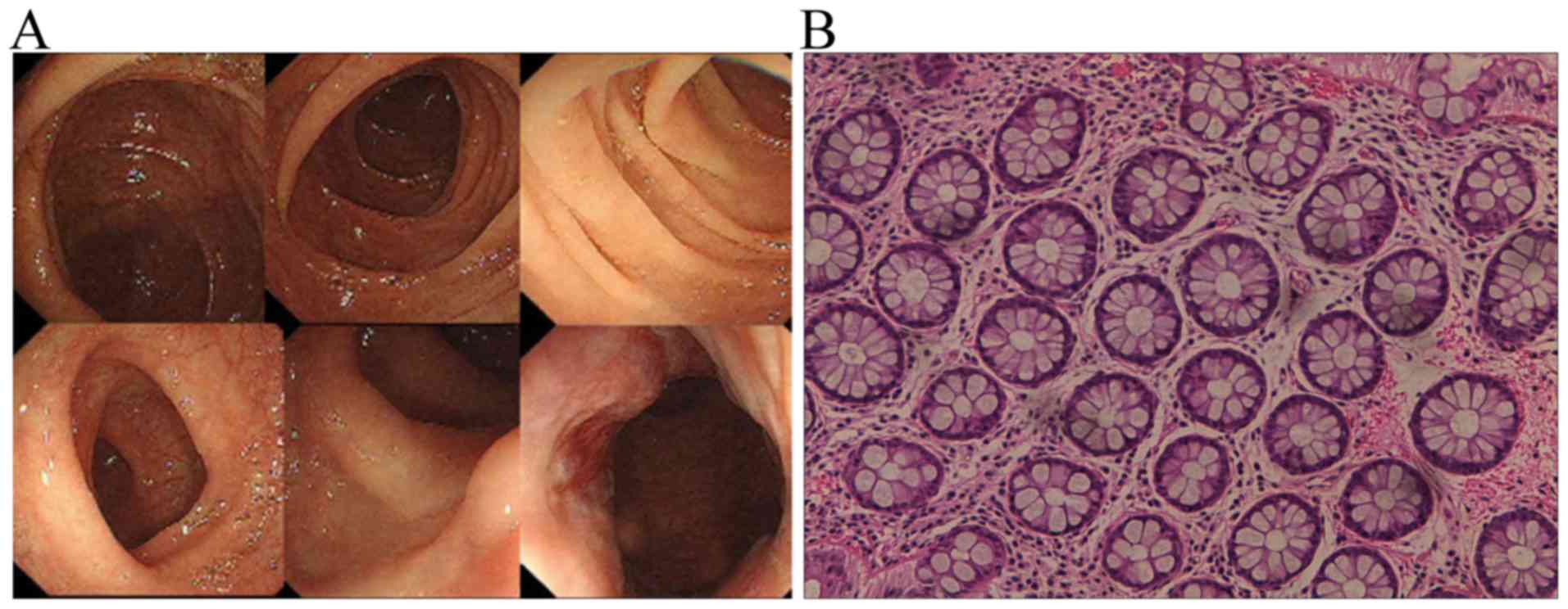
A hemispherical polyp (0.4 cm) was observed in the
sigmoid 18 cm away from the anus (Fig.
3A), which was then resected via endoscopic therapy.
Pathological diagnosis showed that it was a hyperplastic polyp
(Fig. 3B). No other lesions were
detected in the stomach or breast by both gastroscopy and color
Doppler ultrasound.
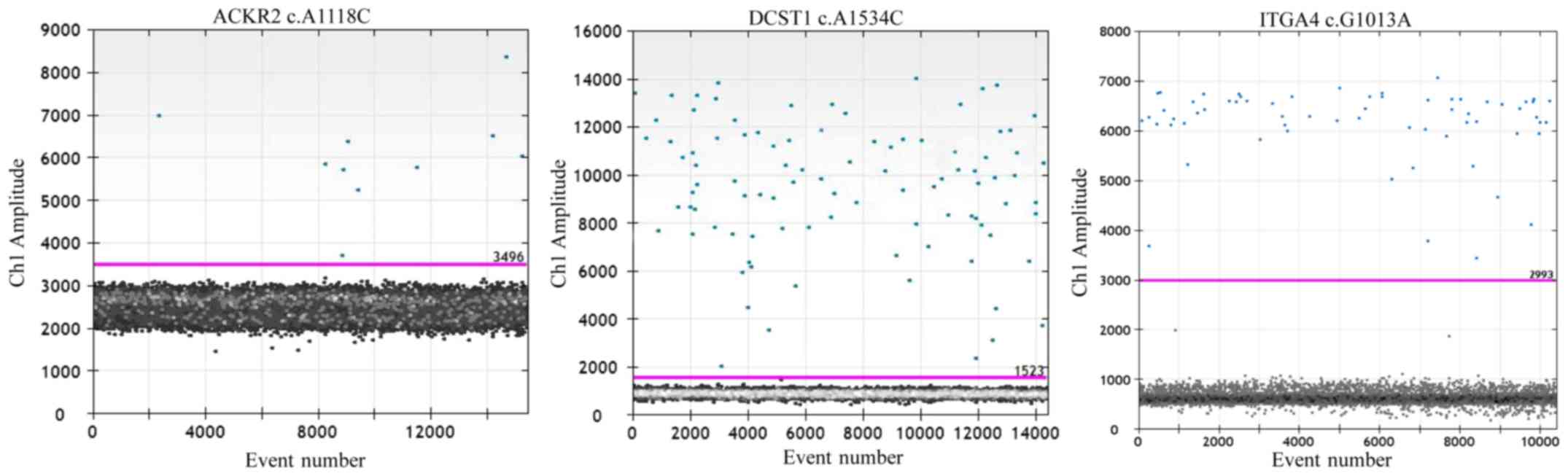
WES was performed on DNA of resected intestinal
polyp, and achieved 107.71× mean coverage on targeted exons. But
none of 13 SNVs in CTCs was found in polyp, which were related with
large intestine cancer in COSMIC database. For higher sequencing
depth, we analyzed these 13 SNVs in polyp's DNA using droplet
digital PCR (ddPCR). ACKR2 c.A1118C, DCST1 c.A1534C, ITGA4 c.G1013A
were positive in intestinal polyp (Fig.
4), other 10 SNVs were not detected.
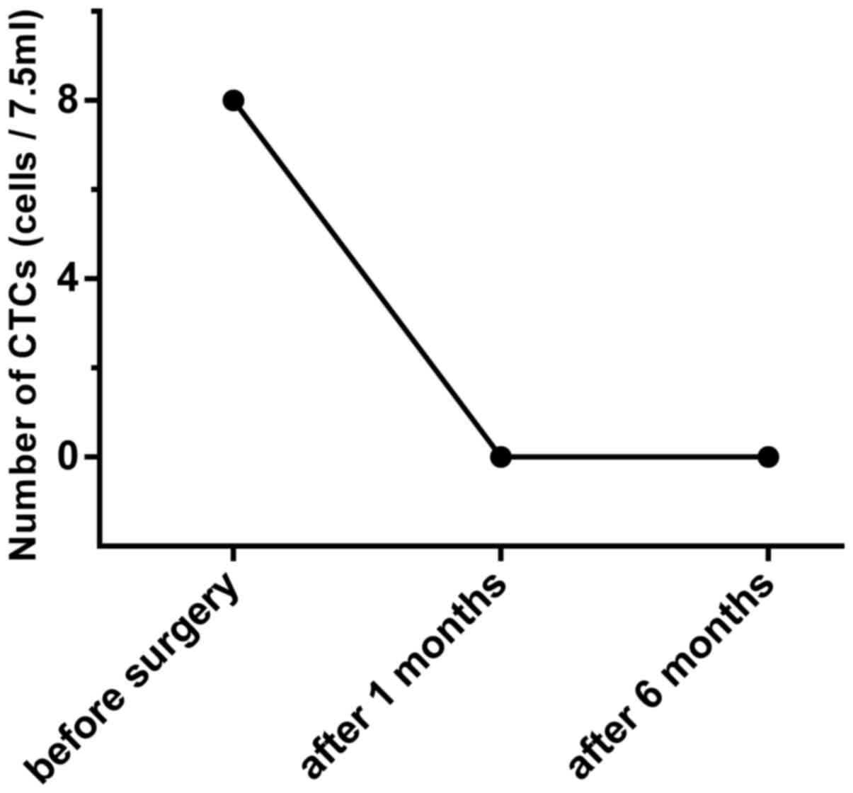
CTCs in the peripheral blood of this patient were
monitored after surgery, at 1 and 6 months during the follow-up.
The number of CTCs reduced to 0 CTCs/7.5 ml, indicating a low risk
of residual lesions (Fig. 5). And
level of AFP, CA-125, CA19-9, CEA and CA15-3 in plasma were in
normal range at 1 and 6 months during follow-up.
Discussion
CTCs in the blood have been suggested to be
potential surrogate markers for minimal residual disease, and the
precursor of metastatic disease (1).
Their presence represents an independent prognostic factor for
reduced disease-free and overall survival (3,4). The
migration of CTCs seems to be an early event in human
carcinogenesis, even before the cancer is visible in clinical
imaging. CTCs have been detected in the blood of model animals when
the tumor size is <1 mm (6). The
study of Ilie et al (11)
showed that CTC+ patients with chronic obstructive
pulmonary disease (COPD) were all diagnosed with lung cancer during
a 1–4 years follow up period. The search for CTCs, at present, may
facilitate an early diagnosis of cancer or precancerosis.
Many studies have proven that the SE-iFISH platform
(identified by CEP8, CK and CD45 (12)) has a higher CTC detection rate than
the Cellsearch system (7,8,10,13). The
high detection rate of CTCs by the SE-iFISH platform was attributed
to the following reasons, as previously reported (7). Firstly, the subtraction enrichment of
the SE-iFISH platform uses immunomagnetic particles conjugated with
anti-CD45 antibody to wipe off the WBCs, which doesn't depend on
the EpCAM expression of CTCs; the expression of EpCAM on CTCs may
decrease during epithelial-mesenchymal transition (EMT). Secondly,
the SE-iFISH system not only identifies CK+ CTCs by
immunostaining, but also aneuploid CTCs by CEP8-fluorescence in
situ hybridization; aneuploidy is a typical common cytogenetic
abnormality malignant cells (14).
In this case, 8 CTCs/7.5 ml were detected in one
healthy female by CK-/CD45-/DAPI+/CEP>2, indicating a high risk
of cancer. Via mapping of the CTC mutation spectrum in the COSMIC
database, we proposed that the cells may have derived from large
intestinal cancer, gastric cancer or breast cancer, or their
precancerous lesions. A 0.4 cm hemispherical polyp was observed in
the sigmoid, 18 cm away from the anus (Fig. 3A), which was diagnosed as a
hyperplastic polyp by pathological determination (Fig. 3B). However, no lesions were found in
the stomach or breast by gastroscopy and color Doppler ultrasound.
There are small subsets of hyperplastic polyps with risk for
development of colorectal cancer, although vast majority of
hyperplastic polyps are innocent (15). There exists a degree of genetic and
perhaps morphologic heterogeneity amongst hyperplastic polyps
(16). It has been proposed that
hyperplastic polyps may serve as the initial lesion in a serrated
neoplasia pathway that results in the 15% sporadic colorectal
adenocarcinomas that are microsatellite unstable (17–19).
A total of 13 SNVs above (Fig. 2B) weren't detected in resected
intestinal polyp using WES, while three SNVs including ACKR2
c.A1118C, DCST1 c.A1534C, ITGA4 c.G1013A were detected by ddPCR
(Fig. 4). That may be attributed to
following reasons. Tumor evolve from benign to malignant lesions by
acquiring a series of mutations over a long time. Genetic
variations emerged in a minor fraction of a cell population before
histology change during tumorigenesis (20). The cells with gene mutations may
persist, but the cell numbers are very small compared with wild
type cells. Sample for DNA extraction couldn't represent overall
perspective of polyp because of heterogeneity. WES of hyperplastic
polyp in this case achieved limited sequencing depth about 100×,
which was hard to detect these rare mutations. While, ddPCR have a
high degree of sensitivity, which is available for detecting 0.001%
mutant fractions.
The CTC detection results were 0 CTC/7.5 ml in this
patient's blood at 1 and 6 months after removal of the intestinal
polyp. The study by Wu et al (9) reported that a decline of the CTC count
after surgery indicated better prognosis, while an increase
indicated fast recurrence for colorectal cancer patients. From the
results above, we can deduce that the intestinal polyp could have
been the main source of the 8 CTCs in this patient. And this
intestinal polyp had risk for development of early colorectal
cancer.
Sporadic CRC is a somatic genetic disease that may
be influenced by the local colonic environment and the individual's
background genetic makeup (21).
Patients often present after 60 years of age, with most cancers
originating from precursor initiating adenomas that, over 1–2
decades, transform into cancer. Because tumor cell dissemination
appears to be an early event in tumor progression, CTCs may appear
at very early stages of tumor development. Genomic alterations
occur during the initiation and progression of a normal colonic
cell into a neoplastic and malignant cell. Changes in the nucleic
acids of the cancer cell might be detected from blood circulation,
such as CTCs (22). Early screening
of this population at age 50 years or older is effective and
sustainable, reducing mortality from CRC and decreasing the
incidence of CRC. Preventive surgery for adenoma, polyps or cancer
is a vital approach towards a cure (23).
There is few study focused on CTCs' organ derivation
for person who isn't previously diagnosed with cancer using WES,
although some studies performed whole-exome sequencing of CTCs on
patients with cancer (24,25). In summary, SE-iFISH CTC detection
represents a potential tool for early stage cancer screening, and
next generation sequencing of CTCs provides a window into the
source of the CTCs and the properties of the solid tumor.
Acknowledgements
Not applicable.
Funding
The present study was supported by grants from
Fujian Natural Science Foundation (grant nos. 2016J01612 and
2018J01198), and Fujian Medical Innovation Project (grant no.
2017-CX-47).
Availability of data and materials
The datasets generated during the current study are
available from the corresponding author on reasonable request.
Authors' contributions
QXP and ZJS contributed to the study design; ZJS and
JMZ were major contributors to writing the manuscript; ZJS
interpreted the clinical data of patient; SYK, JZ and XYL collected
samples and performed experiments; YW and JMZ analyzed the
whole-exome sequencing data. QHS contributed to data analysis and
manuscript revision.
Ethics approval and consent to
participate
Ethical approval for the recruitment of human
subjects was obtained from the Ethics Committee of First Hospital
of Quanzhou Affiliated to Fujian Medical University and was
consistent with ethical guidelines provided by the Declaration of
Helsinki (1975). Written informed consent was obtained from each
patient.
Patient consent for publication
All individuals whose data were used provided
informed consent for publication.
Competing interests
The authors declare that they have no competing
interests.
References
|
1
|
Yap TA, Lorente D, Omlin A, Olmos D and de
Bono JS: Circulating tumor cells: A multifunctional biomarker. Clin
Cancer Res. 20:2553–2568. 2014. View Article : Google Scholar : PubMed/NCBI
|
|
2
|
Magbanua MJ, Carey LA, DeLuca A, Hwang J,
Scott JH, Rimawi MF, Mayer EL, Marcom PK, Liu MC, Esteva FJ, et al:
Circulating tumor cell analysis in metastatic triple-negative
breast cancers. Clin Cancer Res. 21:1098–1105. 2015. View Article : Google Scholar : PubMed/NCBI
|
|
3
|
Cristofanilli M, Budd GT, Ellis MJ,
Stopeck A, Matera J, Miller MC, Reuben JM, Doyle GV, Allard WJ,
Terstappen LW and Hayes DF: Circulating tumor cells, disease
progression, and survival in metastatic breast cancer. N Engl J
Med. 351:781–791. 2004. View Article : Google Scholar : PubMed/NCBI
|
|
4
|
Cohen SJ, Punt CJ, Iannotti N, Saidman BH,
Sabbath KD, Gabrail NY, Picus J, Morse M, Mitchell E, Miller MC, et
al: Relationship of circulating tumor cells to tumor response,
progression-free survival, and overall survival in patients with
metastatic colorectal cancer. J Clin Oncol. 26:3213–3221. 2008.
View Article : Google Scholar : PubMed/NCBI
|
|
5
|
Scher HI, Heller G, Molina A, Attard G,
Danila DC, Jia X, Peng W, Sandhu SK, Olmos D, Riisnaes R, et al:
Circulating tumor cell biomarker panel as an individual-level
surrogate for survival in metastatic castration-resistant prostate
cancer. J Clin Oncol. 33:1348–1355. 2015. View Article : Google Scholar : PubMed/NCBI
|
|
6
|
Rhim AD, Mirek ET, Aiello NM, Maitra A,
Bailey JM, McAllister F, Reichert M, Beatty GL, Rustgi AK,
Vonderheide RH, et al: EMT and dissemination precede pancreatic
tumor formation. Cell. 148:349–361. 2012. View Article : Google Scholar : PubMed/NCBI
|
|
7
|
Sheng Y, Wang T, Li H, Zhang Z, Chen J, He
C, Li Y, Lv Y, Zhang J, Xu C, et al: Comparison of analytic
performances of Cellsearch and iFISH approach in detecting
circulating tumor cells. Oncotarget. 8:8801–8806. 2017. View Article : Google Scholar : PubMed/NCBI
|
|
8
|
Gao Y, Zhu Y, Zhang Z, Zhang C, Huang X
and Yuan Z: Clinical significance of pancreatic circulating tumor
cells using combined negative enrichment and
immunostaining-fluorescence in situ hybridization. J Exp Clin
Cancer Res. 35:662016. View Article : Google Scholar : PubMed/NCBI
|
|
9
|
Wu W, Zhang Z, Gao XH, Shen Z, Jing Y, Lu
H, Li H, Yang X, Cui X, Li Y, et al: Clinical significance of
detecting circulating tumor cells in colorectal cancer using
subtraction enrichment and immunostaining-fluorescence in situ
hybridization (SE-iFISH). Oncotarget. 8:21639–21649.
2017.PubMed/NCBI
|
|
10
|
Li Y, Zhang X, Ge S, Gao J, Gong J, Lu M,
Zhang Q, Cao Y, Wang DD, Lin PP and Shen L: Clinical significance
of phenotyping and karyotyping of circulating tumor cells in
patients with advanced gastric cancer. Oncotarget. 5:6594–6602.
2014. View Article : Google Scholar : PubMed/NCBI
|
|
11
|
Ilie M, Hofman V, Long-Mira E, Selva E,
Vignaud JM, Padovani B, Mouroux J, Marquette CH and Hofman P:
‘Sentinel’ circulating tumor cells allow early diagnosis of lung
cancer in patients with chronic obstructive pulmonary disease. PLoS
One. 9:e1115972014. View Article : Google Scholar : PubMed/NCBI
|
|
12
|
Ge F, Zhang H, Wang DD, Li L and Lin PP:
Enhanced detection and comprehensive in situ phenotypic
characterization of circulating and disseminated heteroploid
epithelial and glioma tumor cells. Oncotarget. 6:27049–27064. 2015.
View Article : Google Scholar : PubMed/NCBI
|
|
13
|
Zhang Y, Wang F, Ning N, Chen Q, Yang Z,
Guo Y, Xu D, Zhang D, Zhan T and Cui W: Patterns of circulating
tumor cells identified by CEP8, CK and CD45 in pancreatic cancer.
Int J Cancer. 136:1228–1233. 2015. View Article : Google Scholar : PubMed/NCBI
|
|
14
|
Zhang J, Li S, Liu F, Zhou L, Shao N and
Zhao X: SELEX aptamer used as a probe to detect circulating tumor
cells in peripheral blood of pancreatic cancer patients. PLoS One.
10:e01219202015. View Article : Google Scholar : PubMed/NCBI
|
|
15
|
Hyman NH, Anderson P and Blasyk H:
Hyperplastic polyposis and the risk of colorectal cancer. Dis Colon
Rectum. 47:2101–2104. 2004. View Article : Google Scholar : PubMed/NCBI
|
|
16
|
Goldstein NS, Bhanot P, Odish E and Hunter
S: Hyperplastic-like colon polyps that preceded
microsatellite-unstable adenocarcinomas. Am J Clin Pathol.
119:778–796. 2003. View Article : Google Scholar : PubMed/NCBI
|
|
17
|
Jass JR, Young J and Leggett BA:
Hyperplastic polyps and DNA microsatellite unstable cancers of the
colorectum. Histopathology. 37:295–301. 2000. View Article : Google Scholar : PubMed/NCBI
|
|
18
|
Jass JR: Serrated route to colorectal
cancer: Back street or super highway? J Pathol. 193:283–285. 2001.
View Article : Google Scholar : PubMed/NCBI
|
|
19
|
Iino H, Jass JR, Simms LA, Young J,
Leggett B, Ajioka Y and Watanabe H: DNA microsatellite instability
in hyperplastic polyps, serrated adenomas, and mixed polyps: A mild
mutator pathway for colorectal cancer? J Clin Pathol. 52:5–9. 1999.
View Article : Google Scholar : PubMed/NCBI
|
|
20
|
Vogelstein B, Papadopoulos N, Velculescu
VE, Zhou S, Diaz LA Jr and Kinzler KW: Cancer genome landscapes.
Science. 339:1546–1558. 2013. View Article : Google Scholar : PubMed/NCBI
|
|
21
|
Carethers JM and Jung BH: Genetics and
genetic biomarkers in sporadic colorectal cancer. Gastroenterology.
149(1177–1190): e32015.
|
|
22
|
Krebs MG, Metcalf RL, Carter L, Brady G,
Blackhall FH and Dive C: Molecular analysis of circulating tumour
cells-biology and biomarkers. Nat Rev Clin Oncol. 11:129–144. 2014.
View Article : Google Scholar : PubMed/NCBI
|
|
23
|
Shaukat A, Mongin SJ, Geisser MS, Lederle
FA, Bond JH, Mandel JS and Church TR: Long-term mortality after
screening for colorectal cancer. N Engl J Med. 369:1106–1114. 2013.
View Article : Google Scholar : PubMed/NCBI
|
|
24
|
Lohr JG, Adalsteinsson VA, Cibulskis K,
Choudhury AD, Rosenberg M, Cruz-Gordillo P, Francis JM, Zhang CZ,
Shalek AK, Satija R, et al: Whole-exome sequencing of circulating
tumor cells provides a window into metastatic prostate cancer. Nat
Biotechnol. 32:479–484. 2014. View
Article : Google Scholar : PubMed/NCBI
|
|
25
|
Faugeroux V, Lefebvre C, Pailler E,
Pierron V, Billiot F, Marcaaillou C, Tourlet S, Vielh P, Dogan S,
Rameau P, et al: Whole-exome sequencing of single circulating tumor
cells is a useful tool for studying the intrapatient genetic
heterogeneity in metastatic prostate cancer. J Clin Oncol. 35 6
Suppl:S1482017. View Article : Google Scholar
|