Introduction
Acute coronary syndromes refer to the array of
clinical signs and symptoms produced by acute myocardial ischemia
and share common pathophysiologic origins related to the
instability and rupture of atherosclerotic vulnerable plaques
(1). Unstable angina and
non-ST-segment elevation myocardial infarction (NSTEMI) are
primarily differentiated by whether the ischemia is prolonged
enough to lead to structural myocardial damage and the release of
detectable markers of myocardial injury, including troponin-I,
troponin-T or creatinine kinase MB(CKMB) (1). The diagnosis of ST-segment elevation
myocardial infarction (STEMI) and NSTEMI is dependent on symptoms,
including consistent chest pain with myocardial ischemia, and
ST-segment depression or T-wave inversion on the electrocardiogram
(ECG) (2). Although patients with
NSTEMI and STEMI share similar cardiac risk factors, their
angiographic features and clinical outcomes are distinct and
warrant different management strategies. For example, STEMI is
characterized by complete occlusion of the culprit artery and
immediate treatment such as primary coronary percutaneous
intervention and large ventricular remodeling is necessary.
Furthermore, STEMI is associated with poor clinical outcomes
(3,4). However, NSTEMI is characterized by
incomplete occlusion of the culprit artery and patients with NSTEMI
are usually treated with less urgency and the clinical courses are
typically less severe than those with STEMI (4–6).
Notably, it has been hypothesized that the reason for the poor
clinical outcomes associated with STEMI is due to total occlusion
of the culprit artery (3). Previous
studies have demonstrated that ~25% of patients with NSTEMI harbor
occluded culprit arteries and exhibit angiographic features similar
to those with STEMI (3–7); however, the clinical significance of
total occluded culprit artery in NSTEMI has not been fully
evaluate.
It has also been demonstrated that the presence of
collaterals has beneficial effects on the infarct size, coronary
microcirculation, myocardial viability, ventricular function and
ventricular aneurysm formation following acute coronary occlusion
in patients with STEMI (8–11). However, to the best of our knowledge,
the effect of coronary collaterals in patients with NSTEMI
undergoing early invasive management remains unknown.
Therefore, the current study investigated the
clinical characteristics and the significance of occluded culprit
arteries and coronary collaterals in patients with NSTEMI.
Patients and methods
Study population
Of 400 patients diagnosed with NSTEMI, 345 (86.2%)
who underwent coronary angiography between June 2006 and May 2013
were evaluated at Kyung Hee University Hospital at Gangdong (Seoul,
Korea). A total of 55 patients who were not subjected to coronary
angiography were excluded from the present study. Patients with
elevated levels of creatine kinase-MB (CKMB) or troponin-I, but
without angiographic evidence of total or subtotal occlusion of a
culprit artery were excluded from the current study. The decision
to perform coronary angiography was made at the physician's
discretion and was based on patient medical history and clinical
status. Basic demographics, procedural characteristics and clinical
outcomes, including bleeding complications were compared between
patients in the two groups.
Coronary angiograms were reviewed independently by
two different physicians to locate the culprit artery and the
presence of angiographic collaterals. Basic demographics,
procedural characteristics and clinical outcomes were compared
according to the occluded culprit artery and coronary collaterals.
The current study was a retrospective analysis; therefore, patients
were not required to give informed consent.
Intervention procedure
Drug-eluting or plain balloons were used in
percutaneous coronary intervention (PCI) when the culprit artery
diameter was <2.5 mm. Stents were implanted when the culprit
artery diameter was ≥2.5 mm. Drug-eluting stents were exclusively
used. Stent implantation was performed following standard
techniques and stents were selected by the practitioner. Complete
lesion coverage and angiographic optimization were recommended with
<20% residual stenosis by a visual estimate. During the
procedure, all patients received a bolus of 100 IU/kg heparin, with
a repeated bolus of 3,000 IU heparin to maintain an activated
clotting time ≥300 sec. All patients were treated with 100 mg/day
aspirin indefinitely and 75 mg clopidogrel or 100 mg ticagrelor
twice a day for ≥12 months after PCI.
Definition
NSTEMI was defined as the appearance of typical
chest pain 12 h prior to admission, with elevated CKMB (≥5 ng/ml)
or troponin-I (≥0.05 ng/ml) and without ST-segment elevation on an
ECG >0.2 mV from baseline at admission. The culprit lesion was
independently determined at angiography by two different
physicians. The culprit artery was determined as exhibiting
evidence of a complex lesion suggestive of acute plaque rupture,
including an intraluminal filling defect, ulcer with overhanging
edges, extraluminal contrast, dissection, multiple irregularities
within the artery, or acute occlusion. An occluded artery was
defined as a lesion with 100% stenosis and a Thrombolysis In
Myocardial Infarction (TIMI) (12)
flow 0. Chronic kidney disease was defined as serum creatinine
>1.4 mg/dl. A quantitative assessment of left ventricular
systolic function was performed using the modified biplane Simpson
method (13) to calculate ejection
fraction. Left ventricular dysfunction was defined as an ejection
fraction <40% on echocardiography. The Wall motion score index
(WMSI) was calculated using a 16-segment model recommended by the
American Echocardiography Association (14). The regional wall motion score index
(RWMSI) was calculated as the sum of motion scores divided by the
number of visualized segments. A regional wall motion score was
applied to each myocardial segment as follows: 1, normal
contraction; 2, hypokinetic contraction; 3, akinetic contraction
and 4, dyskinetic contraction. Higher scores indicated a more
severe wall motion abnormality (15).
Statistical analysis
Data were expressed as the mean ± standard deviation
for continuous variables and frequencies for categorical variables.
Continuous variables were compared using an unpaired Student's
t-test and categorical variables were compared using a
χ2 test. P<0.05 was considered to indicate a
statistically significant difference. Data analyses were performed
using SPSS version 12.5 (SPSS, Inc., Chicago, IL, USA). To reduce
the effect of selection bias and possible confounders, adjustment
for significant differences in the baseline characteristics of
patients was performed with propensity score matching (16). All baseline characteristics of
patients indicated in 2.s I, II, III, IV and V were matched by the
variables that exhibited differences in the overall population.
Kaplan-Meier curves for overall survivals and event-free survivals
were constructed according to the presence of collaterals.
Univariate and multivariate analyses for statistically significant
variables (P<0.05) were performed regarding mortality at 12
months.
Results
Baseline characteristics in patients with NSTEMI
with occluded artery (NSTEMIOA; n=86) and in those with NSTEMI with
patent artery (NSTEMIPA; n=261) are presented in Table I. The incidence of NSTEMIOA was
22.6%. Patients with NSTEMIOA exhibited a significantly higher
level of peak CKMB and troponin-I compared with those in the
NSTEMIPA group (89.0±85.7 vs. 52.0±78.1, P<0.001 and 6.6±9.7 vs.
3.5±7.2; P=0.008, respectively). There were no significant
differences between other baseline clinical characteristics.
Angiographic and procedural characteristics are presented in
Table II. Patients with NSTEMIPA
more commonly exhibited involvement of the left anterior descending
artery (LAD) than those with NSTEMIOA (49.4 vs. 25.6%,
respectively; P<0.001). However, distal artery involvement was
more common in those with NSTEMIOA (38.4 vs. 22.4%; P=0.005).
Following propensity score matching for the entire cohort, 58
matched pairs of patients were identified. There were no
significant differences in the clinically relevant variables
between patients in the two groups, apart from the location of the
culprit artery (P=0.048; Table II).
There were no significant differences of clinical outcomes between
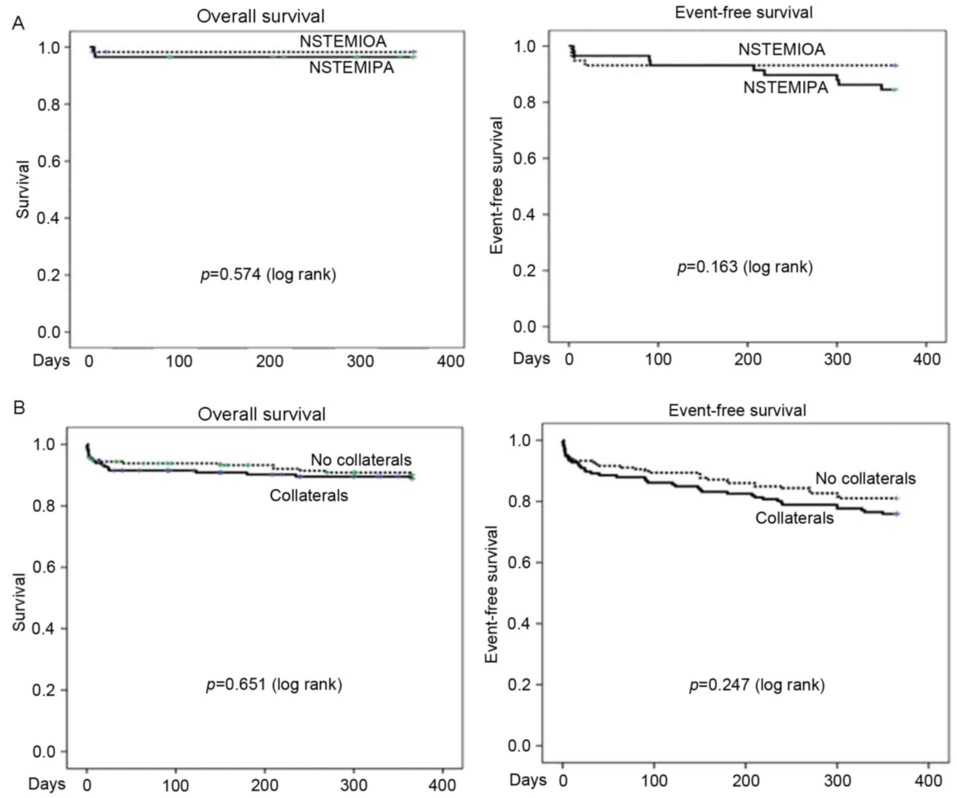
the groups at 30 days and 1 year of follow-up (Table III). Kaplan-Meier curves indicated
that overall survival rates and event-free survival did not differ
significantly between patients with NSTEMIPA and those with
NSTEMIOA (Fig. 1).
 | Table I.Baseline characteristics of patients
with or without an occluded artery. |
Table I.
Baseline characteristics of patients
with or without an occluded artery.
|
| Overall
population | Propensity-score
matched population |
|---|
|
|
|
|
|---|
| Characteristics | NSTEMI with occluded
artery (n=78) | NSTEMI with patent
artery (n=267) | P-values | NSTEMI with occluded
artery (n=58) | NSTEMI with patent
artery (n=58) | P-values |
|---|
| Age, years |
62.1±13.2 |
65.2±12.6 | 0.067 |
60.0±12.7 |
62.2±12.5 | 0.331 |
| Males, (%) | 51 (65.3) | 185 (69.2) | 0.514 | 41(70.6) | 39 (67.2) | 0.688 |
| Height, cm | 162±9 | 163±9 | 0.392 | 162±10 | 164±9 | 0.320 |
| Weight, kg |
64.5±13.4 |
64.8±11.9 | 0.849 |
66.1±14.1 |
65.1±12.0 | 0.696 |
| BMI |
24.3±3.4 |
24.1±3.8 | 0.793 | 24.8±3.4 |
24.0±3.3 | 0.192 |
| Hypertension,
(%) | 41 (52.5) | 168 (62.9) | 0.100 | 28 (48.2) | 35 (60.3) | 0.192 |
| Diabetes (%) | 22 (28.2) | 95
(35.5) | 0.226 | 15 (25.8) | 14 (24.1) | 0.830 |
| Smoking (%) | 44 (56.4) | 159 (59.5) | 0.704 | 36 (62.0) | 33 (56.8) | 0.570 |
| Dyslipidemia (%) | 14 (17.9) | 43
(16.1) | 0.700 | 9 (15.5) | 12 (20.6) | 0.469 |
| Previous PCI (%) | 4 (5.1) | 21 (7.8) | 0.613 | 0 | 4 (6.8) | 0.119 |
| Previous CABG
(%) | 0 (0) | 1
(0.3) | 0.588 | 0 | 1 (1.7) | 0.315 |
| Peripheral artery
disease (%) | 1 (1.2) | 2
(0.7) | 0.538 | 1 (1.7) | 0 | 0.315 |
| Chronic kidney
disease (%) | 6 (7.6) | 31
(11.6) | 0.428 | 4 (6.8) | 5 (8.6) | 0.729 |
| (Creatinine >1.4
mg/dl) |
| Congestive heart
failure (%) | 2 (2.5) | 9
(3.3) | 0.721 | 1 (1.7) | 1 (1.7) | 0.752 |
| Cerebrovacular
disease, (%) | 6 (7.6) | 39
(14.6) | 0.111 | 4 (6.8) | 8
(13.7) | 0.361 |
| ECG finding, (%) |
|
| 0.885 |
|
| 0.784 |
| ST
depression | 27 (11.5) | 87
(13.1) | 0.737 | 18 (31.3) | 18 (31.3) | 0.579 |
| T-wave
inversion | 21 (25.6) | 69
(25.0) | 0.848 | 16 (27.5) | 13 (22.4) | 0.520 |
| No
change | 30 (38.4) | 111 (41.5) | 0.623 | 24 (41.3) | 27 (46.5) | 0.575 |
| Killip class
(%) |
|
| 0.371 |
|
| 0.606 |
| I | 62 (79.8) | 214 (80.1) | 0.898 | 48 (82.7) | 45 (77.5) | 0.485 |
| II | 3 (3.8) | 2
(0.7) | 0.078 | 3 (5.1) | 1 (1.7) | 0.309 |
|
III | 8
(10.2) | 30
(11.2) | 0.808 | 5 (8.6) | 7
(12.0) | 0.542 |
| IV |
5(6.4) | 21(7.8) | 0.668 | 2 (3.4) | 5 (8.6) | 0.242 |
| LVEF, % |
55.8±11.1 |
56.7±11.9 | 0.547 |
56.7±11.0 |
55.0±12.5 | 0.678 |
| LVEF <40% at
index echocardiogram | 8
(10.2) | 23 (8.6) | 0.655 | 5 (8.6) | 7
(12.0) | 0.762 |
| RWMSI |
1.42±0.33 |
1.32±0.36 | 0.038a |
1.39±0.30 |
1.39±0.36 | 0.993 |
| Heart rate at
admission, bpm |
78±19 |
80±21.4 | 0.510 |
75±17 |
80±20 | 0.147 |
| Systolic blood
pressure at admission, mmHg |
131±25 |
138±29 | 0.068 | 131±24 |
133±29 | 0.736 |
| Diastolic blood
pressure at admission, mmHg |
80±14 | 79±15 | 0.599 | 80±14 | 78±16 | 0.443 |
| CKMB, at admission,
mg/dl | 31.6±43.6 | 19.4±42.7 | 0.029a | 35.1±14.3 | 26.5±42.8 | 0.308 |
| CKMB, at peak,
mg/dl | 89.0±85.7 | 52.0±78.1 | <0.001a | 96.5±90.4 | 85.6±99.4 | 0.541 |
| Troponin-I, at
admission, mg/dl | 6.6±9.7 | 3.5±7.2 | 0.002a |
7.5±10.9 | 5.5±9.0 | 0.282 |
| Time from admission
to angiography within 24 h (%) | 60 (76.9) | 214 (80.1) | 0.681 | 45 (77.5) | 46 (80.3) | 0.821 |
| Medication (%) |
|
Aspirin | 73 (93.5) | 249 (93.2) | 0.872 | 57 (98.2) | 56 (96.5) | 0.559 |
|
Thienopyridine | 74 (94.8) | 252 (94.3) | 0.940 | 57 (98.2) | 55 (94.8) | 0.309 |
|
Statins | 73 (93.5) | 246 (92.1) | 0.668 | 54 (94.8) | 53 (91.3) | 0.729 |
|
Beta-blocker | 25 (32.0) | 52
(19.4) | 0.833 | 40 (68.9) | 34 (58/6) | 0.246 |
| ACEi or
ARB | 53 (67.9) | 199 (74.5) | 0.250 | 40 (68.9) | 43 (74.1) | 0.537 |
| Table II.Angiographic characteristics with or
without an occluded artery. |
Table II.
Angiographic characteristics with or
without an occluded artery.
|
| Overall
population | Propensity-score
matched population |
|---|
|
Characteristics | NSTEMI with
occluded artery (n=78) | NSTEMI with patent
artery (n=267) | P-values | NSTEMI with
occluded artery (n=58) | NSTEMI with patent
artery (n=58) | P-values |
|---|
| Lesion
characteristics |
| Culprit coronary
vessel (%) |
| Left
anterior descending | 20 (25.6) | 132 (49.4) |
<0.001a | 14 (24.1) | 17 (29.3) | 0.529 |
| Left
circumflex | 24 (30.7) | 60
(22.4) | 0.133 | 22 (37.9) | 18 (31.3) | 0.435 |
| Right
coronary | 33 (42.3) | 67
(25.0) | 0.003a | 21 (36.2) | 22 (37.9) | 0.848 |
| Left
main | 1 (1.2) | 8
(3.3) | 0.690 |
1(1.7) |
1(1.7) | 0.752 |
| Culprit location
(%) |
|
|
|
|
|
|
|
Proximal | 31 (39.7) | 116 (43.4) | 0.561 | 25 (43.1) | 25 (43.1) | 0.887 |
|
Middle | 17 (21.7) | 91
(34.0) | 0.040a | 12 (20.6) | 22 (37.9) | 0.041 |
|
Distal | 30 (38.4) | 60
(22.4) | 0.005a | 21 (36.2) | 11 (18.9) | 0.038 |
| Number of diseased
vessels (%) | | |
|
|
|
|
|
Single | 33 (42.3) | 88
(32.9) |
0.128 | 25 (43.1) | 27 (46.5) | 0.709 |
|
Double | 21 (31.3) | 67
(25.0) |
0.744 | 17 (29.3) | 19 (32.7) | 0.688 |
|
Triple | 24 (30.7) | 112 (41.9) |
0.076 | 16 (27.5) | 12 (20.6) | 0.385 |
| Collaterals | 54 (69.2) | 78
(29.2) |
<0.001a | 40 (68.9) | 40 (68.9) | 0.579 |
|
Revascularization | 76 (97.4) | 260 (97.3) | 0.978 | 58 (100) | 58 (100) | 0.996 |
| Successful PCI
(%) | 69 (88.4) | 249 (93.2) | 0.305 | 58 (100) | 58 (100) | 0.996 |
| Post-PCI TIMI 3
(%) | 71 (91.8) | 257 (96.2) | 0.221 | 54 (93.1) | 55 (94.8) | 0.697 |
| Quantitative
coronary angiography |
| Reference vessel
diameter, mm |
2.8±0.5 |
2.9±1.2 | 0.339 |
2.8±0.4 |
3.1±0.4 | 0.448 |
| Stent length,
mm | 21.8±7.9 | 22.0±7.7 | 0.800 | 21.6±8.0 | 23.9±8.3 | 0.128 |
| Stent diameter,
mm |
2.7±0.5 |
2.8±0.3 | 0.067 |
2.7±0.5 |
2.7±0.3 | 0.836 |
| Number of
stents |
1.2±0.7 |
1.3±0.6 | 0.068 |
1.3±0.6 |
1.4±0.6 | 0.231 |
| Table III.Clinical outcomes of an occluded
artery in patients over 12 months. |
Table III.
Clinical outcomes of an occluded
artery in patients over 12 months.
|
| Overall
population | Propensity-score
matched population |
|---|
|
|
|
|
|---|
| Clinical
characteristics | NSTEMI with
occluded artery (n=78) | NSTEMI with patent
artery (n=267) | P-values | NSTEMI with
occluded artery (n=58) | NSTEMI with patent
artery (n=58) | P-values |
|---|
| 30-day outcomes
(%) | 4
(3.0) | 22
(8.1) | 0.415 | 1 (1.7) | 2 (3.4) | 0.559 |
| Mortality, 30
days | 4
(3.0) | 21
(7.8) | 0.619 | 1 (1.7) | 2 (3.4) | 0.559 |
| Stroke, 30
days | 0 | 1
(0.3) | 0.588 | 0 | 0 |
|
| 12-month outcomes
(%) | 11
(14.1) | 67
(25.0) | 0.072 | 6
(10.3) | 12 (20.6) | 0.124 |
| Mortality, 12
months | 5
(6.4) | 30
(11.2) | 0.287 | 1 (1.7) | 2 (3.4) | 0.559 |
| MI, 12 months | 2
(2.5) | 11
(3.7) | 0.526 | 1 (1.7) | 2 (3.4) | 0.559 |
| Stroke, 12
months | 3
(3.8) | 5
(1.8) | 0.308 | 2 (3.4) | 0 | 0.496 |
| Readmission for
cardiac cause, 12 months | 5
(6.4) | 39
(14.6) | 0.056 | 2 (3.4) | 8
(13.7) | 0.094 |
Collaterals were present in 166 patients (48.1%).
Baseline characteristics of these patients are present in Table IV. The presence of coronary
collaterals was associated with significantly lower mean left
ventricular ejection fraction (LVEF), mean LVEF <40% and higher
RWMSIs at index admission (P=0.004; P=0.005 and P<0.001,
respectively). There were no other significant differences
regarding clinical variables between the two groups. Angiographic
and procedural characteristics are presented in Table V. The presence of collaterals was
significantly higher in the right coronary artery (RCA; P=0.003)
and in patients with triple vessel artery disease (P=0.001). By
contrast, there was a higher incidence of single vessel disease in
patients with NSTEMI patients that did not have collaterals
(P=0.001). Pre-PCI TIMI 0 was more common in patients with
collaterals (P<0.001). In quantitative coronary analysis,
patients with collaterals exhibited a smaller minimal luminal
diameter (P=0.009), severe diameter stenosis (P=0.002), a longer
stent length (P=0.013) and a smaller stent diameter (P=0.009),
compared with patients with NSTEMI but did not exhibit
collaterals.
| Table IV.Baseline characteristics according to
presence of collaterals. |
Table IV.
Baseline characteristics according to
presence of collaterals.
|
| Overall
population | Propensity-score
matched population |
|---|
|
|
|
|
|---|
|
Characteristics | Collaterals
(n=166) | No collaterals
(n=179) | P-values | Collaterals
(n=120) | No collaterals
(n=120) | P-values |
|---|
| Age, years | 65.2±12.8 | 63.8±12.7 | 0.326 | 60.0±13.0 | 65.4±12.1 | 0.005a |
| Males (%) | 110 (60.2) | 126 (70.3) | 0.492 | 83 (69.1) | 84 (70.0) | 0.888 |
| Height, cm | 162±9 | 163±8 | 0.465 | 162±10 | 163±8 | 0.501 |
| Weight, kg | 64.5±12.9 | 64.9±11.6 | 0.798 | 66.5±14.4 | 64.7±11.7 | 0.360 |
| BMI | 24.4±4.1 | 24.2±3.4 | 0.801 | 24.9±3.5 | 24.0±4.0 | 0.890 |
| Hypertension
(%) | 65 (39.1) | 71 (39.6) | 0.923 | 73 (60.8) | 73 (60.8) | 0.553 |
| Diabetes (%) | 108 (65.0) | 120 (67.0) | 0.698 | 39 (32.5) | 42 (35.0) | 0.682 |
| Smoking (%) | 84 (50.6) | 119 (66.4) | 0.003 | 56 (46.6) | 64 (53.3) | 0.302 |
| Dyslipidemia
(%) | 32 (19.2) | 25 (13.9) | 0.826 | 97 (80.8) | 102 (85.0) | 0.391 |
| Previous PCI
(%) | 17 (10.2) | 9 (5.0) | 0.145 | 7 (5.8) | 8 (6.6) | 0.587 |
| Previous CABG
(%) | 1 (0.6) | 0 | 0.481 | 1 (0.8) | 0 | 0.316 |
| Peripheral artery
disease (%) | 0 | 3 (1.6) | 0.249 | 0 | 2 (1.6) | 0.498 |
| Chronic kidney
disease (%) |
| (Creatinine >1.4
mg/dl) | 19 (11.4) | 18 (10.0) | 0.677 | 14(11.6) | 11 (9.1) | 0.526 |
| Congestive heart
failure (%) | 5 (3.0) | 6 (3.3) | 0.858 | 4 (3.3) | 5 (4.1) | −0.734 |
| Cerebrovacular
disease (%) | 20 (12.0) | 25 (13.9) | 0.597 | 16 (13.3) | 15 (12.5) | 0.847 |
| Killip class
(%) |
| I | 126(75.9) | 150 (83.7) | 0.067 | 93
(77.5) | 104 (86.6) | 0.064 |
| II | 3 (1.8) | 2 (1.1) | 0.592 | 3
(2.5) | 1 (0.8) | 0.313 |
|
III | 22 (13.2) | 16 (8.9) | 0.201 | 15
(12.5) | 11 (9.1) | 0.406 |
| IV | 15 (9.0) | 11 (6.1) | 0.309 | 9
(7.5) | 4 (3.3) | 0.154 |
|
Electrocardiogram |
|
ST-depression | 63 (37.9) | 51 (28.4) | 0.062 | 45 (37.5) | 33 (27.5) | 0.098 |
| T-wave
inversion | 43 (25.9) | 47 (26.2) | 0.940 | 46 (38.3) | 56 (46.6) | 0.192 |
| No
change | 60 (36.1) | 81(45.2) | 0.086 | 29 (24.1) | 31 (25.8) | 0.766 |
| LVEF (%) | 57.5±10.8 | 62.6±8.5 | 0.004a | 55.2±12.4 | 57.4±11.0 | 0.143 |
| LVEF <40% at
index echocardiogram | 22 (13.2) | 9 (5.0) | 0.005a | 17 (14.1) | 9 (7.5) | 0.097 |
| RWMSI | 1.43±0.38 | 1.27±0.32 |
<0.001a | 1.38±0.28 | 1.34±0.38 | 0.394 |
| Heart rate at
admission, bpm 0 | 81±21 | 79±21 | 0.340 | 75±17 | 79±19 | 0.124 |
| Systolic blood
pressure at admission, mmHg | 136±28 | 137±29 | 0.704 | 131±24 | 139±30 | 0.067 |
| Diastolic blood
pressure at admission, mmHg |
79±15 | 79±15 | 0.996 | 80±14 | 79±16 | 0.595 |
| CKMB, at admission,
mg/dl | 17.9±26.7 | 26.0±53.9 |
0.085 | 35.7±48.0 | 14.0±26.1 |
<0.001a |
| CKMB, at peak,
mg/dl | 52.2±70.0 | 67.7±90.0 |
0.080 | 99.5±91.5 | 46.1±69.7 |
<0.001a |
| Troponin-I, at
admission, mg/dl | 4.4±7.7 | 4.0±8.1 |
0.600 | 7.3±10.3 | 2.8±6.0 |
<0.001a |
| Time from admission
to angiography within 24 h, (%) | 135 (81.3) | 141 (78.7) | 0.553 | 101 (84.1) | 97 (80.8) | 0.497 |
| Medications |
|
|
|
|
|
|
|
Aspirin | 156 (93.9) | 167 (93.2) | 0.460 | 98 (81.6) | 93 (77.5) | 0.130 |
|
Thienopyridines | 156 (93.9) | 170 (94.9) | 0.652 | 109 (90.8) | 107 (89.1) | 0.335 |
|
Statins | 154 (92.7) | 165 (92.1) | 0.835 | 112 (93.3) | 113 (94.1) | 0.790 |
| Beta
blocker | 103 (62.0) | 118 (75.9) | 0.550 | 108 (90.0) | 102 (85.0) | 0.242 |
| ACEi or
ARBs | 121 (71.5) | 131 (73.1) | 0.752 | 94 (78.3) | 97 (80.8) | 0.722 |
| Table V.Angiographic characteristics
according to the presence of collaterals. |
Table V.
Angiographic characteristics
according to the presence of collaterals.
|
| Overall
population | Propensity-score
matched population |
|---|
|
|
|
|
|---|
|
Characteristics | Collaterals
(n=166) | No collaterals
(n=179) | P-values | Collaterals
(n=120) | No collaterals
(n=120) | P-values |
|---|
| Lesion
characteristics |
|
|
|
|
|
|
| Culprit
coronary vessel (%) |
|
|
|
|
|
|
|
Left anterior
descending | 65 (39.1) | 87 (48.6) | 0.083 | 51 (42.5) | 62 (51.6) | 0.196 |
|
Left
circumflex | 34 (20.4) | 50 (27.9) | 0.132 | 43 (35.8) | 21 (17.5) | 0.001 |
|
Right
coronary | 61 (36.7) | 39 (21.7) | 0.003a | 25 (20.8) | 36 (30.0) | 0.103 |
|
Left main | 6 (3.6) | 3 (1.6) | 0.259 | 1 (0.8) | 1 (0.8) | 0.751 |
| Culprit
location (%) |
|
|
|
|
|
|
|
Proximal | 67 (40.3) | 80 (44.6) |
0.416 | 44 (36.6) | 55 (45.8) | 0.149 |
|
Middle | 52 (31.3) | 56 (31.2) |
0.994 | 44 (36.6) | 38 (31.6) | 0.414 |
|
Distal | 47 (28.3) | 43 (24.0) |
0.364 | 32 (26.6) | 27 (22.5) | 0.454 |
| Number of diseased
vessel (%) |
|
|
|
|
|
|
|
Single | 43 (25.9) | 78 (43.5) | 0.001a | 101 (84.1) | 97 (80/8) | 0.497 |
|
Double | 42 (25.3) | 46 (25.6) | 0.933 | 15 (12.5) | 19 (15.8) | 0.459 |
|
Triple | 81 (48.7) | 55 (30.7) | 0.001a | 4 (3.3) | 4 (3.3) | 0.701 |
|
Revascularization | 164 (98.7) | 172(96.0) | 0.177 | 120 (100) | 120 (100) | 0.951 |
| Successful PCI
(%) | 153 (92.1) | 165 (92.1) | 0.295 | 120 (100) | 120 (100) | 0.951 |
| Pre-PCI TIMI 0
(%) | 55 (33.1) | 23 (12.8) |
<0.001a | 38 (31.6) | 16 (13.3) | 0.001a |
| Post-PCI TIMI 3
(%) | 77 (91.6) | 214 (99.0) | 0.221 | 112 (93.3) | 112 (93.3) | 0.608 |
| Quantitative
coronary angiography |
|
|
|
|
|
|
|
Reference vessel diameter,
mm | 2.9±1.5 | 2.8±0.4 | 0.669 | 2.8±0.4 | 2.8±0.3 | 0.951 |
| Minimal
luminal diameter, mm | 0.37±0.52 | 0.53±0.55 | 0.009a | 0.49±0.47 | 0.53±0.49 | 0.103 |
|
Diameter stenosis (%) | 90.2±12.2 | 85.9±11.4 | 0.002a | 91.5±11.3 | 88.2±10.4 | 0.157 |
| Stent
length, mm | 23.1±7.8 | 20.9±7.5 |
0.013 | 20.8±7.7 | 22.3±7.6 | 0.277 |
| Stent
diameter, mm | 2.7±0.3 | 2.8±0.4 |
0.009a | 2.7±0.5 | 2.8±0.3 | 0.227 |
| Number
of stents | 1.3±0.7 | 1.3±0.6 |
0.222 | 1.3±0.6 | 1.4±0.6 | 0.208 |
Following propensity score matching for the entire
cohort, 120 matched pairs of patients were identified. There were
no significant differences regarding clinically relevant variables
between patients in the two groups, apart from the culprit coronary
artery (P=0.014), pre-PCI TIMI 0 (P=0.001) and CKMB levels
(Tables V and VI).
| Table VI.Clinical outcomes of collaterals over
12 months. |
Table VI.
Clinical outcomes of collaterals over
12 months.
|
| Overall
population | Propensity-score
matched population |
|---|
|
|
|
|
|---|
| Clinical
characteristics | Collaterals
(n=166) | No collaterals
(n=179) | P-values | Collaterals
(n=120) | No collaterals
(n=120) | P-values |
|---|
| 30-day
outcomes | 14
(8.4) | 12 (6.6) | 0.543 | 3 (2.5) | 3 (2.5) | 0.658 |
| Mortality, 30
days | 14
(8.4) | 11 (6.1) | 0.691 | 3 (2.5) | 2 (1.6) | 0.651 |
| Stroke, 30
days | 0 | 1 (0.5) | 0.335 | 0 | 1 (0.8) | 0.316 |
| 12-month
outcomes | 40
(24.0) | 34 (18.9) | 0.249 | 29 (24.1) | 30 (25.0) | 0.881 |
| Mortality, 12
months | 18
(10.0) | 17 (9.4) | 0.723 | 5 (4.1) | 6 (5.0) | 0.758 |
| MI, 12 months | 6
(3.6) | 7 (3.9) | 0.885 | 3 (2.5) | 6 (5.0) | 0.499 |
| Stroke, 12
months | 5
(3.0) | 3 (1.6) | 0.410 | 4 (3.3) | 2 (1.6) | 0.684 |
| Readmission, 12
months | 23
(13.8) | 22 (12.2) | 0.666 | 17 (14.1) | 16 (13.3) | 0.851 |
There were no significant differences in clinical
outcomes between patients with collaterals and those without
collaterals at 30 days and 1 year of clinical follow-up (Table VI). Kaplan-Meier curves indicated
that survival rates and event-free survival between the two groups
did not differ significantly within 1 year (Fig. 1B).
Univariate analysis indicated that patient age, type
of ECG at admission, weight, hypertension, Killip class, LVEF at
index admission, RWMSI at admission, LAD as the culprit location
and triple vessel disease were associated with mortality at 12
months. Multivariate analysis indicated that age [odds ratio
(OR)=1.165, 95% confidence interval (CI)=1.053–1.290, P=0.003], LAD
as the culprit location (OR=20.359, 95% CI=1.528–271.190, P=0.023)
and hypertension (O=10.407, 95% CI=1.072–101.063, P=0.043) were
independent predictors for patient mortality at 12 months (data not
shown).
Discussion
In the present study, 25% of patients with NSTEMI
exhibited occluded arteries, whereas 50% of patients with NSTEMI
exhibited coronary collaterals. Patients with NSTEMI with occluded
arteries exhibited higher peak biochemical markers and culprit
lesions were located in non-LAD or distal coronary arteries. The
results of the current study demonstrated that NSTEMI with
collaterals have a lower mean LVEF, higher RWMSI and more severe
and extensive coronary artery disease on angiography, including
multivessel disease, pre-PCI TIMI 0, longer stent length and a
smaller diameter, compared with patients that have NSTEMI without
collaterals. However, the clinical outcomes of patients were
similar irrespective of the presence of an occluded culprit artery
or coronary collaterals.
The presence of an occluded culprit artery may be
associated with an increase in mortality and morbidity, as total
artery occlusion is associated with the risk of extensive
myocardial injury and may lead to poor clinical outcomes. Previous
studies have demonstrated that patients with NSTEMIOA not only have
a higher unadjusted rate of mortality at 6–24 months follow-up
(7,17,18), but
also exhibit a higher incidence of heart failure (4) and poor LVEF (5) than those with NSTEMIPA. However, the
results of the current study did not identify any differences
between these two groups of patients regarding clinical outcomes.
Although patients with occluded culprit arteries have significant
disadvantages (19) compared with
those that have non-occluded arteries, the mixed results regarding
clinical outcomes in the current study may be explained by several
factors. Firstly, the treatment of NSTEMI has markedly improved due
to the use of drug-eluting stents and development of novel
medicines, including glycoprotein IIb/IIIa inhibitors. An older
study identified positive clinical outcomes (6); however, more recent studies have
identified mixed results (4,17,20).
Secondly, the culprit artery in patients with NSTEMIOA is more
frequently located in a non-LAD or a distal area in the present
study, which may reduce its adverse effect on patients.
Furthermore, it has been demonstrated that
collaterals in STEMI exert a protective, beneficial effect
(7–10). However, few published studies have
examined the clinical impacts of collaterals in NSTEMI and the
results of previous studies have been controversial (21–24).
Kloepfer et al (25)
prospectively compared the clinical characteristics and outcomes in
patients with NSTEMI with and without collaterals. The results
indicated that those with collaterals had more severe and extensive
coronary artery disease and poorer clinical outcomes. In another
study, patients with NSTEMIOA that had collaterals exhibited better
clinical outcomes compared with NSTEMIOA patients that did not have
collaterals (26). However, this
study was limited; for example, patients with NSTEMIPA that also
had collaterals were excluded. In the present study, the presence
of collaterals was associated with more extensive coronary artery
disease and a lower LVEF; however, the clinical outcomes of
patients with NSTEMI with and without collaterals were similar. It
remains unknown why the presence of coronary collaterals in
patients with NSTEMI is not as protective as in STEMI. It was
hypothesized that in cases of STEMI, which progresses rapidly
within minutes or hours due to abrupt plaque rupture, collateral
circulation may protect the myocardium distal to the total
occlusion site from fatal ischemia. However, in terms of NSTEMI,
the presence of collateral vessels only explains long-standing
severe extensive disease rather than the protection they confer
against abrupt myocardial ischemia. However, further studies are
required to test this hypothesis.
The current study was limited in several ways.
Firstly, the current study was a retrospective, single-center
study. Secondly, locations of the culprit artery lesions were
independently determined by two physicians without central
adjudication and therefore may be subject to bias, particularly
regarding patients with multivessel disease. Thirdly, ~15% of
patients in the current study did not undergo coronary angiography
due to patients' refusal or poor clinical conditions. Despite these
limitations, the results of the current study suggest that the
presence of occluded culprit arteries and coronary collaterals in
patients with NSTEMI are clinically insignificant. A large,
multicenter study is required to evaluate the prognostic impact of
occluded culprit arteries.
In conclusion, the current study demonstrated that
NSTEMIOA is associated with the involvement of non-LAD and distal
arteries. NSTEMI with collaterals is associated with a lower mean
LVEF, as well as more severe and extensive coronary artery disease.
However, the presence of an occluded culprit artery or coronary
collaterals does not significantly affect the clinical outcomes of
patients NSTEMI over a 12-month period. Further studies are
required to evaluate the long-term prognostic impact of an occluded
artery and collaterals.
Acknowledgements
Not applicable.
Funding
No funding was received.
Availability of data and materials
The dataset used and/or analyzed during the current
study are available from the corresponding author on reasonable
request.
Author contribution
PCB designed the study and approved the final
version to be published. KDH and HHJ were involved drafting of the
manuscript. JES, CJM, SSI, KCJ, KDH and HHJ collected and analyzed
the data. All authors reviewed the initial manuscript closely and
revised it critically for important intellectual content.
Ethics approval and consent to
participate
Not applicable.
Patient consent for publication
Not applicable.
Competing interests
The authors declare that they have no competing
interests.
References
|
1
|
Alpert JS, Thygesen K, Antman E and
Bassand JP: Myocardial infarction redefined-a consensus document of
the joint European society of cardiology/American college of
cardiology committee for the redefinition of myocardial infarction.
J AM Coll Cardiol. 36:959–969. 2000. View Article : Google Scholar : PubMed/NCBI
|
|
2
|
Sami S and Willerson JT: Contemporary
treatment of unstable angina and non-ST-segment-elevation
myocardial infarction. Tex Heart Inst J. 37:262–275.
2010.PubMed/NCBI
|
|
3
|
Chan MY, Sun JL, Newby LK, Shaw LK, Lin M,
Peterson DE, Califf RM, Kong DF and Roe MT: Long-term mortality of
patients undergoing cardiac catheterization for ST-elevation and
non-ST-elevation myocardial infarction. Circulaton. 119:3110–3117.
2009. View Article : Google Scholar
|
|
4
|
Soon K, Du HN, Klim S, Zakariyya A and
Kelly AM: Non-ST elevation myocardial infarction with occluded
artery and its clinical implications. Heart Lung Circ.
23:1132–1140. 2014. View Article : Google Scholar : PubMed/NCBI
|
|
5
|
Koyama Y, Hansen PS, Hanratty CG, Nelson
GI and Rasmussen HH: Prevalence of coronary occlusion and outcome
of an immediate invasive strategy is suspected acute myocardial
infarction with and without ST-segment elevation. Am J Cardiol.
90:579–584. 2002. View Article : Google Scholar : PubMed/NCBI
|
|
6
|
Grenne B, Eek C, Sjoli B, Dahlslett T,
Uchito M, Hol PK, Skulstad H, Smiseth OA, Edvardsen T and Brunvand
H: Acute coronary occlusion in non-ST-elevation acute coronary
syndrome: Outcome and early identification by strain
echocardiography. Heart. 96:1550–1556. 2010. View Article : Google Scholar : PubMed/NCBI
|
|
7
|
Wang TY, Zhang M, Fu Y, Amstrong PW, Newby
LK, Gibson CM, Moliterno DJ, Van de Werf F, White HD, Harrington RA
and Roe MT: Incidence, distribution, and prognostic impact of
occluded culprit arteries among patients with non-ST-elevation
acute coronary syndromes undergoing diagnostic angiography. Am
Heart J. 157:716–723. 2009. View Article : Google Scholar : PubMed/NCBI
|
|
8
|
Charney R and Cohen M: The role of the
coronary collateral circulation in limiting myocardial ischemia and
infarct size. Am Heart J. 126:937–945. 1993. View Article : Google Scholar : PubMed/NCBI
|
|
9
|
Habib GB, Heibig J, Forman SA, Brown BG,
Roberts R, Terrin ML and Bolli R: Influence of coronary collateral
vessels on myocardial infarct size in humans. Result of phase I
thrombolysis in myocardial infarction (TIMI) trial. The TIMI
Investigators. Circulation. 83:739–746. 1991. View Article : Google Scholar : PubMed/NCBI
|
|
10
|
Rogers WJ, Hood WP Jr..Mantle JA, Baxely
WA, Kirklin JK, Zorn GL and Nath HP: Return of left ventricular
function after reperfusion in patients with myocardial infarction:
Importance of subtotal stenoses or intact collaterals. Circulation.
69:338–349. 1984. View Article : Google Scholar : PubMed/NCBI
|
|
11
|
Clements IP, Christian TF, Higano ST,
Gibbons RJ and Gersh BJ: Residual flow to the infarct zone as a
determinant of infarct size after direct angioplasty. Circulation.
88:1527–1533. 1993. View Article : Google Scholar : PubMed/NCBI
|
|
12
|
TIMI Study Group, . Comparison of invasive
and conservative strategies after treatment with intravenous tissue
plasminogen activator in acute myocardial infarction. Results of
the thrombolysis in myocardial infarction (TIMI) phase II trial. N
Eng J Med. 320:618–627. 1989. View Article : Google Scholar
|
|
13
|
Folland ED, Parisi AF, Moynihan PF, Jones
DR, Feldman CL and Tow DE: Assessment of left ventricular ejection
fraction and volumes by real-time, two-dimensional
echocardiography. A comparison of cineangiographic and radioneclide
techiniques. Circulation. 60:760–766. 1979. View Article : Google Scholar : PubMed/NCBI
|
|
14
|
Schiller NR, Shah PM, Crawford M, DeMaria
A, Devereux R, Feigenbaum H, Gutqesell H, Reichek N, Sahn D,
Schnittger I, et al: Recommendations for quantitation of the left
ventricle by two-dimentional echocardiography. American society of
echocardiography committee on standards, subcommittee on
quantitation of two-dimensional echocardiograms. J Am Soc
Echocardiogr. 2:358–367. 1989. View Article : Google Scholar : PubMed/NCBI
|
|
15
|
Lang RM, Bierig M, Devereux RB,
Flachskampf FA, Foster E, Pellikka PA, Picard MH, Roman MJ, Seward
J, Shanewise JS, et al: Recommendations for chamber quantification:
A report from the American society of echocardiography's guidelines
and standards committee and the chamber quantification writing
group, developed in conjunction with the European association of
echocardiography, a branch of the European Society of Cardiology. J
Am Soc Echocardiogr. 18:1440–1463. 2005. View Article : Google Scholar : PubMed/NCBI
|
|
16
|
Seung KB, Park DW, Kim YH, Lee SH, Lee CH,
Hong MK, Park SW, Yun SC, Gwon HC, Jeong MH, et al: Stent versus
coronary-artery bypass grafting for left main coronary artery
disease. N Engl J Med. 358:1781–1792. 2008. View Article : Google Scholar : PubMed/NCBI
|
|
17
|
Shin DI, Chang K, Ahn TK, Hwang BH, Park
HJ, Seo SM, Kon YS, Kim PJ, Seung KB and Jeong MH: Impact of
occluded culprit arteries on long-term clinical outcome in patients
with non-ST-elevation myocardial infarction: 48-month follow-up
results in the COREA-AMI registry. J Interv Cardiol. 27:12–20.
2014. View Article : Google Scholar : PubMed/NCBI
|
|
18
|
Pride YB, Tung P, Mohanavelu S, Zorkun C,
Wiviott SD, Antman EM, Giugliano R, Braunwald E and Gibson CM; TIMI
Study Group, : Angiographic and clinical outcomes among patients
with acute coronary syndromes presenting with isolated anterior
ST-segment depression: A TRITON-TIMI 38 (Trial to assess
improvement in therapeutic outcomes by optimizing platelet
inhibition with prasugrel-thrombolysis in myocardial infarction 38)
substudy. JACC Cardiovasc Interv. 3:806–811. 2010. View Article : Google Scholar : PubMed/NCBI
|
|
19
|
Stone GW, Cox D, Garcia E, Brodie BR,
Morice MC, Griffin J, Mattos L, Lansky AJ, O'Neill WW and Grines
CL: Normal flow (TIMI-3) before mechanical reperfusion therapy is
an independent determinant of survival in acute myocardial
infarction: Analysis from the primary angioplasty in myocardial
infarction trials. Circulation. 104:636–641. 2001. View Article : Google Scholar : PubMed/NCBI
|
|
20
|
Warren JW, Mehran R, Yu J, Xu K, Bertrand
ME, Cox DA, Lincoff AM, Manoukian SV, Ohman EM, Pocock SJ, et al:
Incidence and impact of totally occluded culprit coronary arteries
in patients presenting with non-ST-segment elevation myocardial
infarction. Am J Cardiol. 115:428–433. 2015. View Article : Google Scholar : PubMed/NCBI
|
|
21
|
Antoniucci D, Valenti R, Moschi G,
Migliorini A, Trapani M, Santoro GM, Bolognese L, Cerisano G,
Bounamici P and Dovellini EV: Relation between preintervention
angiographic evidence of coronary collateral circulation and
clinical and angiographic outcomes after primary angioplasty or
stenting for acute myocardial infarction. Am J Cardiol. 89:121–125.
2002. View Article : Google Scholar : PubMed/NCBI
|
|
22
|
Sorajja P, Gersh BJ, Mehran R, Lansky AJ,
Krucoff MW, Webb J, Cox DA, Brodie BR and Stone GW: Impact of
collateral flow on myocardial infarction and infarct size in
patients undergoing primary angioplasty for acute myocardial
infarction. Am Heart J. 154:379–384. 2007. View Article : Google Scholar : PubMed/NCBI
|
|
23
|
Gohlke H, Heim E and Roskamm H: Prognostic
importance of collateral flow and residual coronary stenosis of the
myocardial infarct artery after anterior wall Q-wave acute
myocardial infarction. Am J Cardiol. 67:1165–1169. 1991. View Article : Google Scholar : PubMed/NCBI
|
|
24
|
Nicolau JC, Nogueira PR, Pinto MA, Serrano
CV Jr and Garzon SA: Early infarct artery collateral flow does not
improve long-term survival following thrombolytic therapy for acute
myocardial infarction. Am J Cardiol. 83:21–26. 1999. View Article : Google Scholar : PubMed/NCBI
|
|
25
|
Kloepfer AM, Lipson LC and Keeley EC: The
presence of angiographic collaterals in Non-ST elevation myocardial
infarction is a predictor of long-term clinical outcomes. Catheter
Cardiovasc Interv. 83:1–8. 2014. View Article : Google Scholar : PubMed/NCBI
|
|
26
|
Bahrmann P, Rach J, Desch S, Schuler GC
and Thiele H: Incidence and distribution of occluded culprit
arteries and impact of coronary collaterals on outcomes in patients
with non-ST-segment elevation myocardial infarction and early
invasive treatment strategy. Clin Res Cardiol. 100:457–467. 2011.
View Article : Google Scholar : PubMed/NCBI
|