Introduction
Type 2 diabetes is primarily characterized by
insulin resistance, and one of the important causes of insulin
resistance is insulin signal transduction disorder (1,2). Insulin
is the only hormone in the body that is able to lower blood glucose
level. It first binds to the insulin receptor (IR) on the cell
membrane and then activates the phosphoinositide 3-kinase
(PI3K)/protein kinase B (AKT) or Ras/Raf/mitogen-activated protein
kinase signaling pathway (3). The
PI3K/AKT signaling pathway is the primary pathway of insulin
signaling transduction, through which insulin regulates glucose
uptake, glycogen synthesis and degradation (4).
In the liver, insulin binds to the α subunit of IR
on liver cells, and then activates IR substrate (IRS). IRS then
binds to p85, the regulatory subunit of PI3K, and activates p110,
the catalytic subunit of PI3K. The activated PI3K produces the
second messengers Phosphatidylinositol (3,4)-trisphosphate [PtdIns(3,4)P2] and
PtdIns(3,4,5)P3, which promote the activation of AKT (5). Activated AKT regulates the process of
carbohydrate metabolism in hepatocytes via the following pathways:
First, activated AKT promotes the transfer of glucose transporter 4
to cell membranes, which facilitates the transportation of
extracellular glucose into the cells (6). Second, activated AKT mediates glycogen
synthesis through glycogen synthase kinase 3 (GSK3) (7). Third, activated AKT inhibits hepatocyte
gluconeogenesis through peroxisome proliferator-activated
receptor-γ coactivator-α (8).
Therefore, the insulin-PI3K/AKT signaling pathway serves an
important function in the regulation of carbohydrate metabolism in
liver.
At present, chemical drugs, including metformin, are
primarily used for the clinical treatment of diabetes (9). Blood glucose level is significantly
reduced by these drugs, but insulin resistance is not effectively
improved. In addition, long-term use of these drugs can cause a
variety of serious side effects, leading to liver and kidney damage
(10). Therefore, it is necessary to
develop natural drugs with reliable hypoglycemic effects and less
toxic side effects.
Sericin is a water-soluble protein of the silkworm
cocoon, which is composed of 18 different amino acids (11). The content of serine, aspartic acid,
threonine and other polar amino acids (which are rich in hydroxyl,
amino and carboxyl groups) in sericin is >70%, therefore sericin
possesses good water solubility (12). Sericin has been used in wound
healing, tissue regeneration, drug delivery and biomedicine
(11,13). Studies have indicated that the
addition of sericin to the daily diet may reduce the intestinal
intake of cholesterol, triglycerides and other lipids, as well as
improve the body's tolerance to glucose (14,15).
Previous studies by our group indicated that sericin could
effectively reduce blood glucose level in a streptozotocin-induced
type 2 diabetic rat model (16,17).
However, the hypoglycemic mechanism of sericin remains unclear.
In the current study, a type 2 diabetes rat model
was established by administration with high-fat and high-glucose
diet combined with streptozotocin intraperitoneal injection. The
expression levels of IR, IRS-1, PI3K and AKT in the rat liver were
analyzed to investigate whether sericin exerts a hypoglycemic
effect via the insulin-PI3K/AKT signaling pathway. The current
study may provide an experimental basis for the use of sericin in
the treatment of diabetes in the clinic.
Materials and methods
Animals, grouping and sampling
A total of 48 specific-pathogen-free 6-week-old male
Sprague-Dawley rats (purchased from Beijing Vital River Laboratory
Animal Technology Co., Ltd., Beijing, China) were used in the
current study. The rats were housed with a natural light cycle at
18–26°C and relative humidity of 40–70%. They were randomly divided
into four groups: Control group, diabetic model group, high-dose
sericin group and low-dose sericin group, with 12 rats in each
group. The type 2 diabetes rat model was established as follows: by
high-fat and high-glucose diet combined with streptozotocin (35
mg/kg, twice) intraperitoneal injection. The rats were fed with
high-fat and high-glucose diets as previously described (18) for 4 weeks and streptozotocin (35
mg/kg) was administered twice in succession by intraperitoneal
injection. After 72 h, blood glucose was measured and rats with a
fasting blood glucose of 11.1 mmol/l were deemed to have diabetes
(17,19). Model rats were fed with high-fat and
high-glucose diet for another 3 weeks and the diet was changed to
normal diet when they were fed with sericin. The control group was
fed with normal diet throughout the experiment. For the high- and
low-dose sericin groups, the rats were continuously administered
with 2.4 and 1.8 g/kg/day sericin for 35 days, respectively, while
the control rats were administered with the same volume of saline
for 35 days. All animal experiments were approved by the Ethics
Committee of Chengde Medical University (Chengde, China) and
conducted according to the ethical guidelines of Chengde Medical
University.
The rats in each group were fasted for 12 h
following treatment and anesthetized by intraperitoneal injection
of 10% chloral hydrate (300 mg/kg body weight) (20–22).
Then, the thoracic cavity was opened and venous blood samples were
collected from the right atrium of the rats. During blood sample
collection, rats were in deep anesthesia and no signs of
peritonitis were observed. Then, the rats were sacrificed by
decapitation and the left hepatic lobe was collected following
opening of the abdominal cavity. The blood was centrifuged at 800 ×
g for 20 min at 4°C, and the serum was isolated. The liver tissues
were kept in liquid nitrogen until further analysis.
Determination of glucose level
The blood glucose level of rats in each group was
measured using a glucose oxidase method (23) on a Beckman Coulter AU5800 Clinical
Chemistry Analyzer (Beckman Coulter, Inc., Brea, CA, USA).
Hematoxylin and eosin (H&E)
staining
The liver morphology of each group was observed
under an Olympus BH-2 light microscope (Olympus Corporation, Tokyo,
Japan) following H&E staining. Briefly, the liver tissues were
fixed with Bouins fixative [a mixture of picric acid saturated
liquid (1.22%), formaldehyde and glacial acetic acid at a ratio of
15:5:1] for 24 h at room temperature and embedded in paraffin, and
continuously sliced into 5-µm-thick sections. At room temperature,
the sections were stained with hematoxylin for 7 min,
differentiated with 1% HCl in 70% alcohol for 5 sec, stained with
eosin for 1 min and dehydrated in a serial ethanol solution of
increasing concentrations. Then, the sections were deparaffinized
with xylene and sealed.
Periodic acid-Schiff staining
The hepatic glycogen content was determined by
periodic acid-Schiff staining. The liver tissues were fixed as
above described and embedded in paraffin, and continuously sliced
into 5-µm-thick sections. The sections were oxidized with periodic
acid for 10 min and stained with Schiff reagent for 15 min at room
temperature. Cell nuclei were re-stained with hematoxylin for 5 min
at room temperature. Then, sections were deparaffinized with xylene
and sealed. Cells with red or magenta particles in the cytoplasm
were defined as positive for glycogen. Tissue sections digested
with amylase were used as negative controls.
For the quantification of glycogen content, six rat
livers were randomly selected from each group, three sections were
selected from each rat liver, and three views were observed in each
section. The liver lobules with intact tissue structure were
selected for observation using an light microscope (BH-2; Olympus
Corporation, Tokyo, Japan; magnification, ×200). Image-Pro Plus 6.0
image analysis software (Media Cybernetics, Inc., Rockville, MD,
USA) was used to calculate the integral optical density of glycogen
in hepatocyte cytoplasm, and the mean value was determined as the
glycogen content.
Immunohistochemical staining
Protein expression of IR, IRS-1, PI3K and AKT in the
liver was analyzed. Briefly, the sections were dewaxed and
incubated in 3% H2O2-methanol at 37°C for 30
min. Following antigen retrieval by microwaving, the sections were
blocked with 10% goat serum (OriGene Technologies, Inc., Beijing,
China) at 37°C for 30 min. The sections were then incubated with
primary antibodies against IR (1:100; cat. no. ab131238), IRS-1
(1:100; cat. no. ab131487; both Abcam, Cambridge, MA USA), PI3K
(1:100; cat. no. 611398; BD Biosciences, San Jose, CA, USA) and AKT
(1:200; cat. no. ab179463; Abcam) at 4°C overnight. Next, the
sections were incubated with goat anti-rabbit IgG secondary
antibodies, which was provided by the rabbit streptavidin-biotin
assay system (cat. no. SP-9001; Zhongshan Jinqiao Biotechnology
Co., Ltd., Beijing, China) according to the manufacturer's
protocol, and then counterstained with DAB for 5–8 min at room
temperature. Cell nuclei were re-stained with hematoxylin for 10
min at room temperature. PBS was used to substitute the primary
antibody as the negative control. Cells with brownish yellow and/or
brown particles were defined as positive staining.
For quantification, six rat livers were randomly
selected from each group, three sections were selected from each
rat liver, and three views were observed in each section. The liver
lobules with intact tissue structure were selected for observation
by an Olympus BH-2 microscope (magnification, ×200). Image-Pro Plus
6.0 image analysis software was used to calculate the integral
optical density of each protein, and the mean value was determined
as the corresponding protein expression level.
Western blot analysis
Total protein was extracted by RIPA Tissue/Cell
Lysates (Beijing Solarbio & Technology Co., Ltd., Beijing,
China) from 100 mg liver tissues, and the protein concentration was
determined using a BCA protein kit (Kangwei Shiji Biotechnology
Co., Ltd., Beijing, China). Proteins (108 µg/lane) were separated
by 10% SDS-PAGE and transferred on to a PVDF membrane. Following
blocking with 5% skim milk overnight at 4°C, the membrane was
incubated with primary antibodies [IR, IRS-1, AKT (all 1:1,000),
PI3K (1:500) and β-actin (1:1,000; cat no. AF7018; Affinity
Biosciences, Cincinnati, OH, USA)] at room temperature for 2 h.
Then, the membrane was incubated with goat anti-rabbit IgG
(1:5,000; cat. no. 074-1506) or goat anti-mouse IgG (1:5,000; cat.
no. 074-1806; both KPL, Inc., Gaithersberg, MD, USA) for a further
1.5 h at room temperature. The membrane was developed with Super
ECL Plus ultra-sensitive luminescent liquid (Applygen Technologies,
Inc., Beijing, China). The protein bands were analyzed with
Quantity One v4.6.2 software (Bio-Rad Laboratories, Inc., Hercules,
CA, USA). The density of each target band was calculated relative
to the β-actin band to determine relative expression levels.
Reverse transcription-quantitative
polymerase chain reaction (RT-qPCR)
Total RNA was extracted from 100 mg liver tissues
with a TaKaRa MiniBEST Universal RNA Extraction kit (cat. no. 9767)
and reverse transcribed into cDNA using a PrimeScript™ RT Master
mix (cat. no. RR036A; both Takara Biotechnology Co., Ltd., Dalian,
China). The reverse transcription protocol was as follows: 37°C for
15 min and 85°C for 5 sec. The sample was put on ice and the
obtained cDNA was stored at −20°C. Then, the mRNA expression of IR,
IRS-1, PI3K, AKT and GAPDH was determined by qPCR with a SYBR
Premix Ex Taq™ II kit (cat. no. RR820A; Takara Biotechnology Co.,
Ltd.). The primer sequences are listed in Table I. The PCR procedure was as follows:
Pre-denaturation at 98°C for 1 min, followed by 40 cycles of
denaturation at 98°C for 7 sec, annealing and polymerization at
60°C for 30 sec, then final polymerization at 60°C for 5 min. A
relative standard curve method (24)
was used to quantify the mRNA and the relative mRNA expression
level of each target gene was determined relative to the
corresponding GAPDH.
 | Table I.Primer sequences used for quantitative
polymerase chain reaction analysis. |
Table I.
Primer sequences used for quantitative
polymerase chain reaction analysis.
| Gene | Primer sequence
(5′-3′) |
|---|
| IR | F:
TCATGGATGGAGGCTATCTGGA |
|
| R:
TCCTTGAGCAGGTTGACGATTTC |
| IRS-1 | F:
AAGCACCTATGCCAGCATCAAC |
|
| R:
GAGGATTGCTGAGGTCATTTAGGTC |
| PI3K | F:
CCAGAAGAAGGGACAGTGGTATG |
|
| R:
TCGTAGCCAATCAGGGAGGT |
| AKT | F:
ATGGACTTCCGGTCAGGTTCA |
|
| R:
GCCCTTGCCCAGTAGCTTCA |
| GAPDH | F:
GGCACAGTCAAGGCTGAGAATG |
|
| R:
ATGGTGGTGAAGACGCCAGTA |
Statistical analysis
Statistical analysis was performed with the
statistical software SPSS 20.0 (IBM Corp., Armonk, NY, USA). All
data are expressed as mean ± standard deviation. One-way analysis
of variance was used to compare multiple groups, followed by
Tukey's post hoc test. P<0.05 was considered to indicate a
statistically significant difference.
Results
Morphological changes of liver
following sericin treatment
To observe the effect of sericin on the liver
morphology of type 2 diabetic rats, H&E staining was performed.
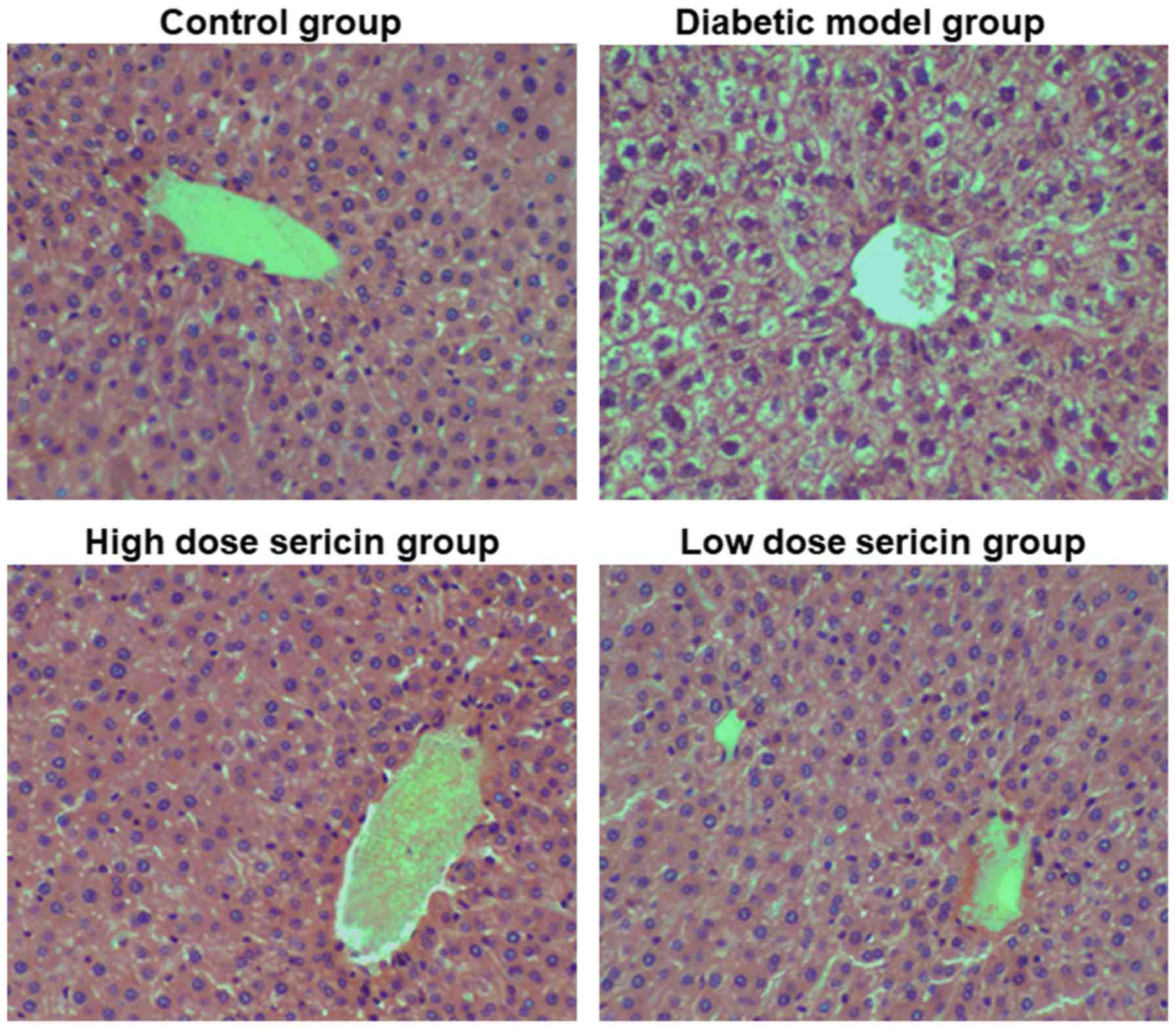
In the control group, the structure of the hepatic lobules was
clear and the hepatocytes were arranged in a cord-like manner
around the central vein (Fig. 1).
The cell nuclei were large and round, located in the center of
cells, and the cytoplasm was stained uniformly. The liver sinus was
clear. In the diabetic model group, the hepatocytes were basically
arranged in a cord-like manner around the central vein, but the
liver cells were swollen, the volume increased, and obvious
vacuolar structure appeared in the cytoplasm. A number of the liver
cells exhibited soluble necrosis, and the hepatic sinus exhibited
stenosis or atresia. Compared with the diabetic model group, the
pathological changes of the rat liver in the high- and low-dose
sericin groups were markedly reduced (Fig. 1). Rat liver lobular structures in
these two sericin treatment groups were clear, and the hepatocytes
were arranged in a cord-like manner around the central vein, with
large round nuclei in the center of the cells. A small number of
hepatocytes exhibited vacuolar structure in the cytoplasm, and the
liver sinus was clear. This indicates that sericin may improve the
liver morphological structure of type 2 diabetic rats.
Glycogen content in liver following
sericin treatment
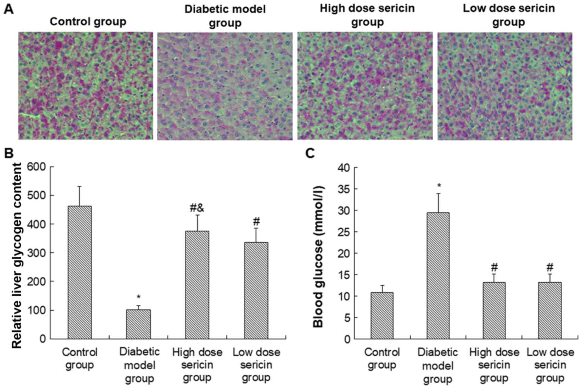
To determine the effect of sericin on the glycogen
content in type 2 diabetic rat livers, periodic acid-Schiff
staining was performed. Hepatic glycogen positive expression was
observed in the liver sections of all groups, indicated by red and
purple particles in the cytoplasm. As indicated in Fig. 2A, the number of positive cells in the
control group was high, and the staining was dark purple. In the
diabetic model group, there were fewer positively stained cells,
and the staining was a lighter reddish color. In the high-dose
sericin group, there was a higher number of positively stained
cells compared with the low-dose sericin group, and the staining
was darker. As indicated in Fig. 2B,
compared with the control group, the glycogen content in the rat
liver of the diabetic model group was significantly decreased
(P<0.05). Compared with the diabetic model group, the glycogen
content in the liver of the high- and low-dose sericin groups was
significantly increased (P<0.01). Furthermore, the glycogen
content in the rat liver of the high-dose sericin group was
significantly higher compared with the low-dose sericin group
(P<0.01). This indicates that sericin may significantly increase
the liver glycogen content of type 2 diabetic rats.
Blood glucose levels following sericin
treatment
To determine the effect of sericin on the blood
glucose level of the type 2 diabetic rats, the rat blood glucose
level was measured. As indicated in Fig.
2C, the blood glucose level in the diabetic model group was
significantly increased compared with the control group
(P<0.05). Compared with the diabetic model group, the blood
glucose levels of the rats in the high- and low-dose sericin groups
were significantly decreased (P<0.01). However, there was no
significant difference in the blood glucose levels between the
high- and how-dose sericin groups. This indicates that sericin may
significantly decrease the blood glucose levels of type 2 diabetic
rats.
Expression of associated factors in
the PI3K/AKT signaling pathway
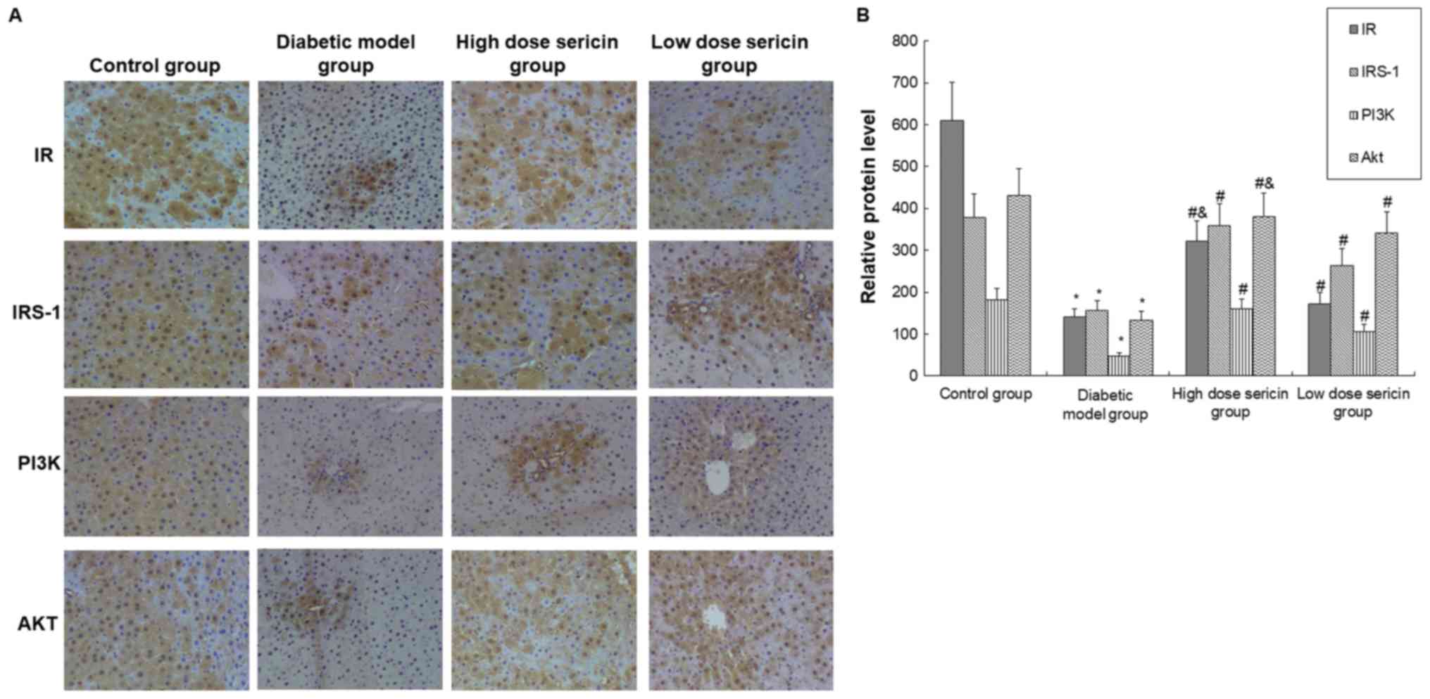
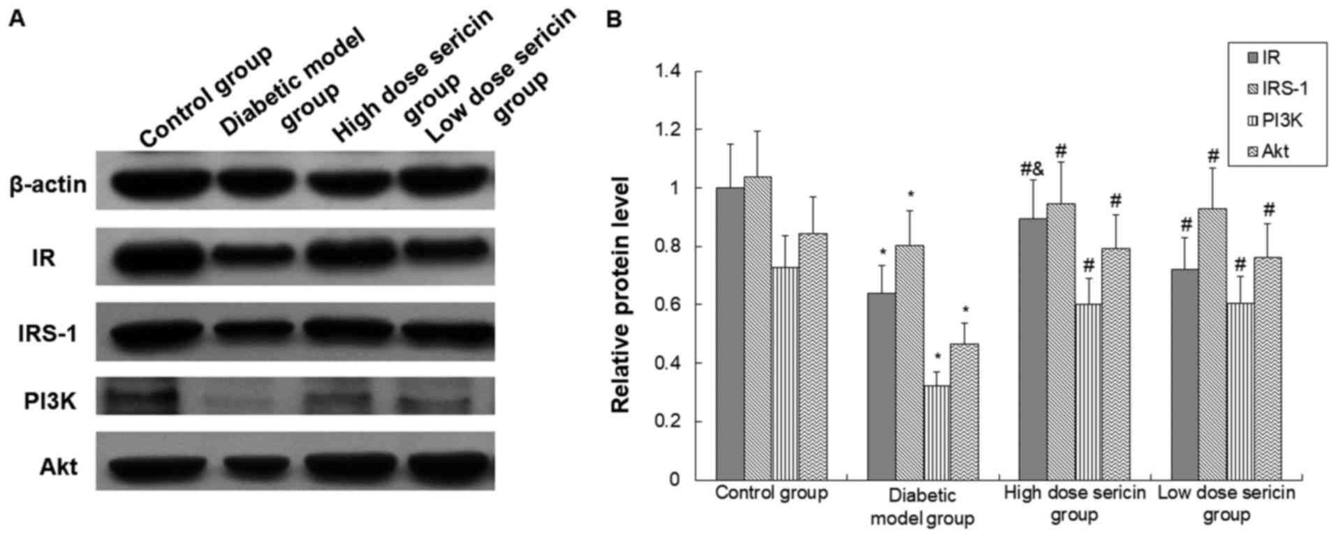
The protein levels of IR, IRS-1, PI3K, and AKT were
detected with immunohistochemical staining and western blot
analysis. Compared with the control group, the protein expression
levels of IR, IRS-1, PI3K and AKT were significantly decreased in
the diabetic model group (P<0.05; Figs. 3 and 4). Compared with the diabetic model group,
the expression levels of these proteins in the liver of the high-
and low-dose sericin groups were significantly increased
(P<0.05; Figs. 3 and 4). Furthermore, the expression levels of
AKT (Fig. 3) and IR (Figs. 3 and 4) in the high-dose sericin group were
significantly higher compared with the low-dose sericin group
(P<0.05).
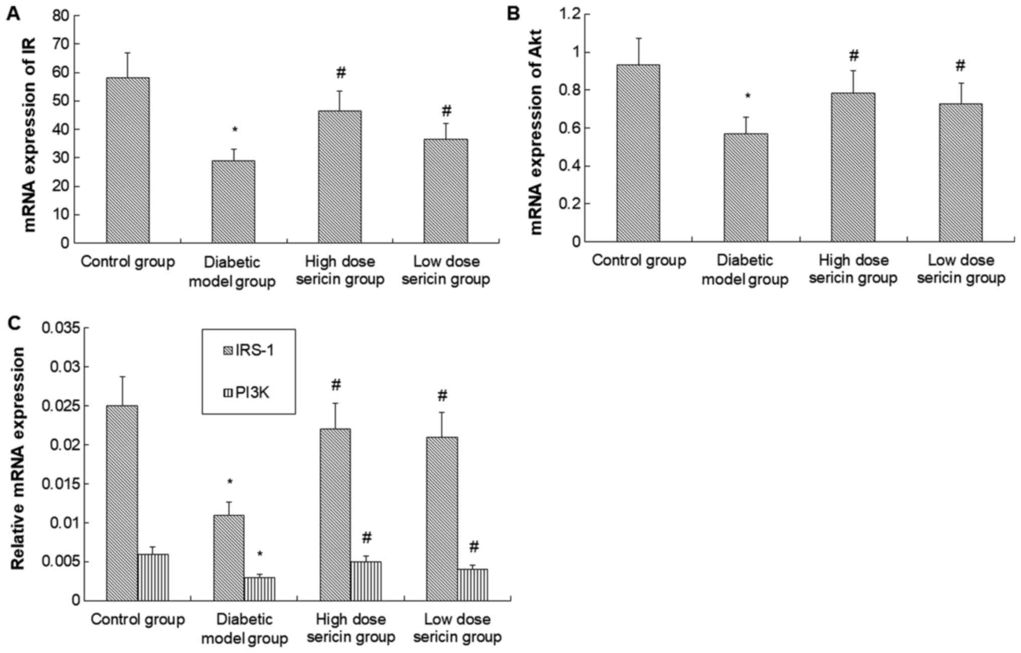
To further verify these results, RT-qPCR was
conducted to detect the mRNA levels of IR, IRS-1, PI3K and AKT. As
indicated in Fig. 5, the mRNA levels
of IR, IRS-1, PI3K and AKT in the diabetic model group were
significantly lower compared with the control group (P<0.05).
However, following sericin treatment in the high- and how-dose
sericin groups, mRNA expression was significantly higher compared
with the diabetic model group (P<0.05). No significant
difference in mRNA levels of these genes was identified between the
high- and low-dose sericin groups.
In summary, these results indicate that sericin may
promote the insulin/PI3K/AKT signaling pathway in type 2 diabetic
rat liver by upregulating IR, IRS-1, PI3K and AKT expression, and
thus may reduce blood glucose levels.
Discussion
Type 2 diabetes is a chronic metabolic disease
characterized by insulin resistance. On a global scale, the
incidence of type 2 diabetes is increasing year by year, with
faster growth rates in developing countries (25,26). The
expected number of patients with type 2 diabetes in 2030 is 552
million (27). At present, diabetes
has become the third most prevalent non-communicable disease that
threatens human life and health after cardiovascular disease and
cancer (28). Therefore, it is of
great importance to explore the pathogenesis of type 2 diabetes and
search for effective and economical treatments.
Insulin resistance is the initiating factor for the
development of type 2 diabetes. Researchers have identified that
the PI3K/AKT signaling pathway is closely associated with insulin
resistance-associated diseases, including diabetes and obesity
(29,30). Furthermore, the PI3K/AKT signaling
pathway is one of the key pathways by which insulin regulates blood
glucose balance. Reduced expression of PI3K regulatory subunit p85,
as well as functional defects of PI3K regulatory subunit p85 and
catalytic subunit p110, may lead to glucose and lipid metabolism
disorder (31,32) which further indicates the important
role of this pathway in regulating glucose and lipid
metabolism.
Hepatic insulin resistance also serves a key
function in the pathophysiology of diabetes. It has been reported
that feeding with high-fat diet and alcohol simultaneously could
induce rat liver insulin resistance through inhibition of mRNA and
protein expression levels of IRS-1 and PI3K (33). Rats with non-alcoholic liver disease
also present typical symptoms of fatty liver, insulin resistance
and glucose metabolism disorder, and silinin, a hepatoprotective
agent, may attenuate hepatic steatosis and insulin resistance
caused by non-alcoholic fatty liver via the IRS-1/PI3K/AKT pathway
(34). The results of the current
study also suggest that there may be associations among the hepatic
insulin PI3K/AKT signaling pathway, hepatic insulin resistance and
diabetes mellitus.
IR is an important mediator of the insulin PI3K/AKT
signaling pathway, and its downregulation or mutation may lead to
reduced insulin sensitivity and insulin resistance (35,36).
Consistent with this, the current study identified that IR protein
and mRNA levels in the type 2 diabetic model rat liver were
significantly reduced. Furthermore, IRS-1, PI3K and AKT exhibited
the same trend as IR. A decrease in AKT level could decrease the
release and transport of glucose transporter in the vesicles
(37). A decrease in AKT could also
interfere with GSK3-mediated hepatic glycogen synthesis, leading to
a significant decrease in hepatic glycogen content in diabetic
model rats. AKT also reduces the inhibition of gluconeogenesis
(7). These three effects of AKT may
finally lead to a significant increase in blood glucose levels and
insulin resistance in the diabetic rats.
Currently, oral hypoglycemic drugs are primarily
used for the treatment of diabetes. Although various types of
hypoglycemic drugs may effectively control blood sugar and delay
the progression of the disease and complications, the majority of
drugs exert side effects to varying degrees, including ineffective
improvement of insulin resistance or islet cell protection, and
drug resistance (38). Therefore, in
recent years, researchers have turned their attention to natural
substances that exert hypoglycemic effects, so as to obtain a drug
that has good absorption and efficacy without obvious side effects.
The ‘Compendium of Materia Medica’ records that cocoons may have
effects on diabetes (39). Sericin
used in the current study is a natural protein in silkworm cocoons,
which is coated on the outside of silk fibroin, accounting for
approximately 25% of the cocoon. Sericin is usually discarded when
reeling. However, cocoons soaked in boiling water have long been
used to control blood glucose in Chinese folk medicine. Modern
research indicates that sericin is a water-soluble protein
consisting of 18 amino acids. It is biocompatible, biodegradable
and non-toxic to humans, and has no obvious side effects (40). Our previous studies also identified
that sericin could effectively reduce blood glucose level (16,17).
However, it is not yet clear whether the PI3K/AKT signaling pathway
is involved in the effect of sericin on blood glucose.
The current study observed the effect of sericin on
associated factors of the PI3K/AKT signaling pathway, including IR,
IRS-1, PI3K and AKT, in the liver of type 2 diabetic rats, and
investigated whether sericin could reduce blood glucose and improve
insulin resistance. Following administration of sericin in diabetic
model rats, the expression levels of IR, IRS-1, PI3K and AKT in the
model rat liver significantly increased, the blood glucose level
significantly reduced and the hepatic glycogen content
significantly increased.
In conclusion, the current findings demonstrate that
sericin is able to increase the expression of IR, IRS-1, PI3K and
AKT in the liver of diabetic rats at the gene and protein level.
Sericin may regulate the PI3K/AKT signaling pathway in diabetes to
enhance the transduction effect of insulin signal and promote the
synthesis of hepatic glycogen. This may be the mechanism by which
sericin is able to lower blood glucose and improve insulin
resistance.
Acknowledgements
Not applicable.
Funding
The current study was supported by the National
Natural Science Foundation of China (grant no. 81441133) and the
Natural Science Foundation of Hebei Province (grant no.
H2013406096).
Availability of data and materials
All data generated or analyzed in the present study
are included in this published article.
Authors' contributions
CS performed all experiments. DL fed the animals,
collected the samples and participated in the blood glucose test.
SY participated in the immunohistochemistry experiments. LC helped
with the hematoxylin and eosin staining and glycogen staining. EX
participated in the polymerase chain reaction analysis. ZC designed
and directed the study.
Ethics approval and consent to
participate
All animal experiments were approved by the Ethics
Committee of Chengde Medical University.
Patient consent for publication
Not applicable.
Competing interests
The authors declare that they have no competing
interests.
References
|
1
|
Chattopadhyay T, Singh RR, Gupta S and
Surolia A: Bone morphogenetic protein-7 (BMP-7) augments insulin
sensitivity in mice with type II diabetes mellitus by potentiating
PI3K/AKT pathway. Biofactors. 43:195–209. 2017. View Article : Google Scholar : PubMed/NCBI
|
|
2
|
Chaudhury A, Duvoor C, Reddy Dendi VS,
Kraleti S, Chada A, Ravilla R, Marco A, Shekhawat NS, Montales MT,
Kuriakose K, et al: Clinical review of antidiabetic drugs:
Implications for type 2 diabetes mellitus management. Front
Endocrinol (Lausanne). 8:62017.PubMed/NCBI
|
|
3
|
Horita S, Nakamura M, Suzuki M, Satoh N,
Suzuki A and Seki G: Selective insulin resistance in the kidney.
Biomed Res Int. 2016:58251702016. View Article : Google Scholar : PubMed/NCBI
|
|
4
|
Gao YF, Zhang MN, Wang TX, Wu TC, Ai RD
and Zhang ZS: Hypoglycemic effect of D-chiro-inositol in type 2
diabetes mellitus rats through the PI3K/Akt signaling pathway. Mol
Cell Endocrinol. 433:26–34. 2016. View Article : Google Scholar : PubMed/NCBI
|
|
5
|
Hou XL, Wang WQ, Shi CY, Tong Q and Fang
JG: Research progress in pharmacological effects of
dihydromyricelin. Chinese Traditional and Herbal Drugs. 46:603–609.
2015.
|
|
6
|
Gandhi GR, Jothi G, Antony PJ, Balakrishna
K, Paulraj MG, Ignacimuthu S, Stalin A and Al-Dhabi NA: Gallic acid
attenuates high-fat diet fed-streptozotocin-induced insulin
resistance via partial agonism of PPARgamma in experimental type 2
diabetic rats and enhances glucose uptake through translocation and
activation of GLUT4 in PI3K/p-Akt signaling pathway. Eur J
Pharmacol. 745:201–216. 2014. View Article : Google Scholar : PubMed/NCBI
|
|
7
|
Liu TY, Shi CX, Gao R, Sun HJ, Xiong XQ,
Ding L, Chen Q, Li YH, Wang JJ, Kang YM and Zhu GQ: Irisin inhibits
hepatic gluconeogenesis and increases glycogen synthesis via the
PI3K/Akt pathway in type 2 diabetic mice and hepatocytes. Clin Sci
(Lond). 129:839–850. 2015. View Article : Google Scholar : PubMed/NCBI
|
|
8
|
Chang S: Progress in studying the
relationship between PDK/Akt signal access and insulin resistance.
Zhong Yi Yao Dao Bao. 14:113–116. 2008.
|
|
9
|
Ahrén B, Masmiquel L, Kumar H, Sargin M,
Karsbøl JD, Jacobsen SH and Chow F: Efficacy and safety of
once-weekly semaglutide versus once-daily sitagliptin as an add-on
to metformin, thiazolidinediones, or both, in patients with type 2
diabetes (SUSTAIN 2): A 56-week, double-blind, phase 3a, randomised
trial. Lancet Diabetes Endocrinol. 5:341–354. 2017. View Article : Google Scholar : PubMed/NCBI
|
|
10
|
Type 2 diabetes and metformin. First
choice for monotherapy: Weak evidence of efficacy but well-known
and acceptable adverse effects. Prescrire Int. 23:269–272.
2014.PubMed/NCBI
|
|
11
|
Chen H, Zhu LJ, Min SJ and Hu GL:
Structure, property and utilization of silk sericin. Journal of
Functional Polymers. 14:344–347. 2001.
|
|
12
|
Takasu Y, Yamada H, Tamura T, Sezutsu H,
Mita K and Tsubouchi K: Identification and characterization of a
novel sericin gene expressed in the anterior middle silk gland of
the silkworm Bombyx mori. Insect Biochem Mol Biol. 37:1234–1240.
2007. View Article : Google Scholar : PubMed/NCBI
|
|
13
|
Aramwit P, Siritientong T and Srichana T:
Potential applications of silk sericin, a natural protein from
textile industry by-products. Waste Manag Res. 30:217–224. 2012.
View Article : Google Scholar : PubMed/NCBI
|
|
14
|
Okazaki Y, Kakehi S, Xu Y, Tsujimoto K,
Sasaki M, Ogawa H and Kato N: Consumption of sericin reduces serum
lipids, ameliorates glucose tolerance and elevates serum
adiponectin in rats fed a high-fat diet. Biosci Biotechnol Biochem.
74:1534–1538. 2010. View Article : Google Scholar : PubMed/NCBI
|
|
15
|
Limpeanchob N, Trisat K, Duangjai A,
Tiyaboonchai W, Pongcharoen S and Sutheerawattananonda M: Sericin
reduces serum cholesterol in rats and cholesterol uptake into
Caco-2 cells. J Agric Food Chem. 58:12519–12522. 2010. View Article : Google Scholar : PubMed/NCBI
|
|
16
|
Chen Z, He Y, Song C, Dong Z, Su Z and Xue
J: Sericin can reduce hippocampal neuronal apoptosis by activating
the Akt signal transduction pathway in a rat model of diabetes
mellitus. Neural Regen Res. 7:197–201. 2012.PubMed/NCBI
|
|
17
|
Song CJ, Yang ZJ, Tang QF and Chen ZH:
Effects of sericin on the testicular growth hormone/insulin-like
growth factor-1 axis in a rat model of type 2 diabetes. Int J Clin
Exp Med. 8:10411–10419. 2015.PubMed/NCBI
|
|
18
|
Hao WJ, Li JY, Jiang H, Li YH, Shen Z, Hou
JJ, Xiong J and Li XR: Advantages of purified high-fat and
high-glucose diet. Wei Sheng Yan Jiu. 46:143–147. 2017.
|
|
19
|
Li N, Liu Q, Li XJ, Bai XH, Liu YY, Jin
ZY, Jing YX, Yan ZY and Chen JX: Establishment and evaluation of a
rat model of type 2 diabetes associated with depression. Zhongguo
Ying Yong Sheng Li Xue Za. 31:23–26. 2015.(In Chinese).
|
|
20
|
Santos EL, Dias BH, Andrade AC, Pascoal
AM, Vasconcelos Filho FE, Medeiros Fd and Guimarães SB: Effects of
acupuncture and electroacupuncture on estradiol-induced
inflammation and oxidative stress in health rodents. Acta Cir Bras.
28:582–588. 2013. View Article : Google Scholar : PubMed/NCBI
|
|
21
|
Silachev DN, Usatikova EA, Pevzner IB,
Zorova LD, Babenko VA, Gulyaev MV, Pirogov YA, Plotnikov EY and
Zorov DB: Effect of anesthetics on efficiency of remote ischemic
preconditioning. Biochemistry. Biokhimiia (Mosc). 82:1006–1016.
2017. View Article : Google Scholar
|
|
22
|
Vollmar B, Janata J, Yamauchi JI and
Menger MD: Attenuation of microvascular reperfusion injury in rat
pancreas transplantation by L-arginine. Transplantation.
67:950–955. 1999. View Article : Google Scholar : PubMed/NCBI
|
|
23
|
Kura RR, Kilari EK and Shaik M: Influence
of aprepitant on the pharmacodynamics and pharmacokinetics of
gliclazide in rats and rabbits. PeerJ. 6:e47982018. View Article : Google Scholar : PubMed/NCBI
|
|
24
|
Wang NN, Liu F, Xu X, Zhang SW, Nie R, Yin
DD, Liu GQ and Wang GJ: Establishment and preliminary application
of fluorescence quantitative RT-PCR detection method for tambus
virus. Zhong Guo Shou Yi Ke Xue. 45:15–19. 2015.
|
|
25
|
Chahardah-Cherik SM, Gheibizadeh MP,
Jahani SP and Cheraghian BP: The relationship between health
literacy and health promoting behaviors in patients with type 2
diabetes. Int J Commun Based Nurs Midwifery. 6:65–75. 2018.
|
|
26
|
Papier K, D'Este C, Bain C, Banwell C,
Seubsman S, Sleigh A and Jordan S: Consumption of sugar-sweetened
beverages and type 2 diabetes incidence in Thai adults: Results
from an 8-year prospective study. Nutr Diabetes. 7:e2832017.
View Article : Google Scholar : PubMed/NCBI
|
|
27
|
Whiting DR, Guariguata L, Weil C and Shaw
J: IDF diabetes atlas: Global estimates of the prevalence of
diabetes for 2011 and 2030. Diabetes Res Clin Pract. 94:311–321.
2011. View Article : Google Scholar : PubMed/NCBI
|
|
28
|
Gautam S and Banerjee M: The macrophage
Ox-LDL receptor, CD36 and its association with type II diabetes
mellitus. Mol Genet Metab. 102:389–398. 2011. View Article : Google Scholar : PubMed/NCBI
|
|
29
|
Hu X, Wang S, Xu J, Wang DB, Chen Y and
Yang GZ: Triterpenoid saponins from Stauntonia chinensis ameliorate
insulin resistance via the AMP-activated protein kinase and
IR/IRS-1/PI3K/Akt pathways in insulin-resistant HepG2 cells. Int J
Mol Sci. 15:10446–10458. 2014. View Article : Google Scholar : PubMed/NCBI
|
|
30
|
Lu J, Chen LN and Ma X: PI3K/Akt signaling
pathway and insulin resistance related diseases. Guo Wai Yi Xue (Yi
Xue Di Li Fen Ce). 33:127–131. 2012.(In Chinese).
|
|
31
|
Winnay JN, Dirice E, Liew CW, Kulkarni RN
and Kahn CR: p85α deficiency protects β-cells from endoplasmic
reticulum stress-induced apoptosis. Proc Natl Acad Sci USA.
111:1192–1197. 2014. View Article : Google Scholar : PubMed/NCBI
|
|
32
|
Nelson VL, Jiang YP, Dickman KG, Ballou LM
and Lin RZ: Adipose tissue insulin resistance due to loss of PI3K
p110alpha leads to decreased energy expenditure and obesity. Am J
Physiol Endocrinol Metab. 306:E1205–E1216. 2014. View Article : Google Scholar : PubMed/NCBI
|
|
33
|
Qu W, Hao L, Chen Y and Zhou S: Effect of
chronic ethanol intake on insulin receptor, insulin receptor
subsrate-1 and phosphoinositide 3-kinase mRNA expression in
skeletal muscle of rats. Wei Sheng Yan Jiu. 36:172–175. 2007.(In
Chinese). PubMed/NCBI
|
|
34
|
Zhang Y, Hai J, Cao M, Zhang Y, Pei S,
Wang J and Zhang Q: Silibinin ameliorates steatosis and insulin
resistance during non-alcoholic fatty liver disease development
partly through targeting IRS-1/PI3K/Akt pathway. Int
Immunopharmacol. 17:714–720. 2013. View Article : Google Scholar : PubMed/NCBI
|
|
35
|
Wu QP, Xiao C, Yang XB and Zhang JM:
Hypoglycemic effects of components extracted from edible and
medicinal fungi and their mechanisms of action. Acta Edulis Fungi.
16:80–86. 2009.
|
|
36
|
Tao MX, Wang F, Liu J, Cheng GY and Jin
BQ: Hypoglycemic Effect of Pleurotus citrinopileats Polysaccharide.
Food Sci. 30:227–230. 2009.
|
|
37
|
Kuai M, Li Y, Sun X, Ma Z, Lin C, Jing Y,
Lu Y, Chen Q, Wu X, Kong X and Bian H: A novel formula
Sang-Tong-Jian improves glycometabolism and ameliorates insulin
resistance by activating PI3K/AKT pathway in type 2 diabetic KKAy
mice. Biomed Pharmacother. 84:1585–1594. 2016. View Article : Google Scholar : PubMed/NCBI
|
|
38
|
Bodmer M, Meier C, Krähenbühl S, Jick SS
and Meier CR: Metformin, sulfonylureas, or other antidiabetes drugs
and the risk of lactic acidosis or hypoglycemia: A nested
case-control analysis. Diabetes Care. 31:2086–2091. 2008.
View Article : Google Scholar : PubMed/NCBI
|
|
39
|
Li SZ: Compendium of Materia Medica.
Beijing: People's Health Publishing House; pp. 1–1052. 1985
|
|
40
|
Zhang HP, Zhu LJ and Hu H: Studies on the
properties of L-asparaginase immobilized on sericin protein powder.
Bulletin of Sericulture. 34:16–19. 2003.
|