Introduction
Reperfusion is believed to be important for the
recovery of ischemic brain injuries and also limits subsequent
infarction development. Cerebral ischemia/reperfusion (I/R) is
characterized by an initial restriction of blood supply to the
brain, followed by restoration of blood flow and re-oxygenation
(1,2). However, IR induced cerebrovascular
dysfunction is also thought to be a significant clinical issue due
to the neurological damage that can occur, such as in patients that
have suffered ischemic strokes (3).
Ischemic stroke are one of the most devastating neurological
diseases world-wide and the third most common cause of death
globally. They can also lead to permanent disability in adults. In
China, approximately 2.5 million people are at risk of stroke and 1
million may die from stroke-related consequences annually (4–6). The
exact pathogenic mechanisms that lead to cerebral IR injury are not
yet completely understood and it is therefore important to find new
effective measures to prevent cerebral I/R injuries and avert
ischemic strokes.
MicroRNAs (miRNAs) are small (18–25 nt) noncoding
single-stranded RNA molecules that act to negatively regulate
transcription and post-transcription by modulating the stability
and/or translational efficiency of target messenger RNAs (mRNAs)
(7). It has previously been
established that most mature miRNAs alter mRNA degradation or
translation through binding the 3′-untranslated region (3′UTR) of
target mRNAs (8,9). Prior research has also revealed that
some miRNAs, including miR-29b, miR-21, miR-200, and miR-497, can
act as potential therapeutic targets. These miRNAs appear to
contribute to cerebral I/R injury by altering key signaling
elements that typically have high expression in the cerebral cortex
after ischemic injury (10). More
recent studies have indicated that miR-29 may also negatively
regulate Bcl-2 family members, including pro-survival proteins such
as BCL-w (BCL2L2) and MCL-1. Despite these proteins being important
regulators of cerebral I/R injury, the mechanisms behind their
effects are unknown (11–13).
Based on prior evidence, we investigated the effects
of miR-29b on cerebral IR injury and aimed to identify any
underlying mechanisms. Our study utilized an OGD/R (Oxygen-glucose
deprivation/reoxygenation) environment as an in vitro model
of induced cerebral IR injury. After administration of miR-29b or
miR-29b inhibitor, we evaluated any differences in neural apoptosis
mediated by MCL-1 and other relevant proteins. Our data contributes
to a better understanding of cerebral IR injury and will aid
efforts to develop new strategies to improve its clinical
treatment.
Materials and methods
Cell culture and treatment
Mouse neuroblastoma (N2a) cells were purchased from
the Chinese Academy of Sciences (Shanghai, China). N2a cells were
cultured in growth media containing high glucose-Dulbecco's
modified Eagle's medium (Hyclone; GE Healthcare Life Sciences,
Logan, UT, USA), supplemented with 10% fetal bovine serum (Hyclone,
GE Healthcare Life Sciences), and 1% penicillin/streptomycin
(Mediatech, Inc., Manassas, VA, USA) in a humidified atmosphere of
5% CO2 at 37°C.
Following dilution into single cell suspensions, N2a
cells were seeded onto 96-well plates (1×10⁴
cells/well). Cells were exposed to an oxygen-glucose deprivation
(OGD) environment for 24 h and the culture medium was then replaced
with deoxygenated glucose-free Hanks' balanced salt solution (HBSS)
for a further 24 h. Cells were then harvested for total RNA
isolation.
MiRNA mimics transfection
miR-29b mimics, miR-29b inhibitor, mimics negative
control (NC), and inhibitor NC were purchased from Guangzhou
RiboBio, Co., Ltd., (Guangzhou, China). The miR-29b mimics sequence
(5′-UAGCACCAUUUGAAAUCAGUGUU-3′), miR-29b inhibitor sequence
(5′-AACACUGAUUUCAAAUGGUGCUA-3′), miR-29b mimics NC sequence
(5′-UUCUCCGAACGUGUCACGUTT-3′); miR-29b inhibitor NC sequence
(5′-CAGUACUUUUGUGUAGUACAA-3′). 2×10⁵ N2a cells were cultured in
6-well plates and then transfected with 200 µl mature miR-29b
mimics, miR-29b inhibitor, mimics NC, and inhibitor NC (GenePharma
Co., Ltd., Shanghai, China) for 72 hrs. All transfections were
completed using Lipofectamine™ 3000 (Invitrogen Life Technologies,
Carlsbad, CA, USA) according to the manufacturer's protocols.
Cell viability detection
N2a R cells were transfected with 1 µg of miR-29b
mimic or an inhibitor using Lipofectamine® 2000
(Invitrogen). Next, 100 µl Cell Counting kit-8 (CCK8) solution
(Dojindo Molecular Technologies, Inc., Kumamoto, Japan) was added
to each well and incubated for 1 h in an incubator. Absorbance at
450 nm was measured using a microplate reader.
Apoptosis assay
Quantification of apoptotic cells was performed
using an Annexin V-propidium iodide (PI) apoptosis kit
(Multiscience Biotech, Ltd., Hangzhou, China). N2a cells were
collected, washed with phosphate buffered saline (PBS) and
re-suspended in 200 µl binding buffer containing 5 µl Annexin V (10
µg/ml). They were then left for 10 min in the dark. Cells were next
incubated with 10 µl PI (20 µg/ml) and analyzed by flow cytometry
(EPICS® XL™; Beckman Coulter, Inc., Brea, CA, USA). Data
acquisition and analysis were performed using CellQuest™ software
(BD Biosciences, Franklin Lakes, NJ, USA).
Protein isolation and western blotting
analysis
In total, 2×10⁶ N2a cells per sample were
lysed for western blotting analysis using conventional procedures.
The primary antibodies used were anti-mouse polyclonal MCL-1
antibody (1:20,000; Sigma-Aldrich, St. Louis, MO, USA), anti-mouse
polyclonal BCL2 antibody (1:1,000, Abcam, Cambridge, UK),
anti-mouse polyclonal caspase-3 (CASP3) antibody (1:10,000, Abcam),
and anti-mouse polyclonal GAPDH antibody (1:1,000, Santa Cruz
Biotechnology, CA, USA). Blots were visualized using
chemiluminescence reagent (Meck Millipore, Billerica, MA, USA) in a
LAS4000 Luminescent Image Analyzer (GE Healthcare, Tokyo, Japan).
TRIzol® reagent (Invitrogen; Thermo Fisher Scientific,
Inc., Waltham, MA, USA) was used to extract total RNA from cells
following the manufacturer's protocol, although ethanol was used
instead of isopropanol for RNA precipitation. RNA quality was
determined using a NanoDrop 1000 spectrophotometer (Thermo Fisher
Scientific, Inc., Wilmington, DE, USA). A total of 1 µg RNA per
sample was reverse-transcribed into cDNA using a DBI
Bestar® qPCR RT kit (DBI Bioscience, Ludwigshafen,
Germany), according to the manufacturer's protocol.
Reverse transcription-quantitative
polymerase chain reaction (RT-qPCR)
RT-qPCR was performed using 96-well optical plates
and a 7500 Fast Real-Time PCR System LightCycler (Applied
Biosystems; Thermo Fisher Scientific, Inc.). Each 20 µl PCR
reaction included 1 µl reverse transcription product (1:5), 0.5 µl
sense primer, 0.5 µl Universal reverse primer, and 10 µl mix buffer
(DBI Bestar® Sybr-Green qPCR master mix, DBI
Bioscience). Reaction conditions were 94°C for 2 min, followed by
40 cycles of 94 and 58°C for 20 sec, and 72°C for 20 sec. All
reactions were completed in triplicate. Primer sequences were
miR-29b forward: 5′UAGCACCAUUUGAAAUCAGUGUU3′ and reverse:
5′CTCAACTGGTGTCGTGGA3′; and U6 forward: 5′CTCGCTTCGGCAGCACA3′; and
reverse: 5′AACGCTTCACGAATTTGCGT3′.
Recombinant plasmid construction
The 3′UTR of mouse MCL-1 mRNA (NM_000286) was
retrieved from the GenBank Database and amplified using synthetic
primers: MCL-1 forward (5′GCTAGCCGCTACTAGGCTCCCC3′) and MCL-1
reverse (5′CGGGTAGTATATACGCGTCGTTAC3′). The product was cloned into
a psiCHECK2 vector (Invitrogen, Carlsbad, CA, USA). Recombinant
vector was then amplified in DH5α Escherichia coli and
purified using an endotoxin-free plasmid purification kit (QIAGEN,
Valencia, CA), according to the manufacturer's instructions. Each
segment was amplified by PCR with Takara LA Taq or Primestar
(Takara Bio, Inc., Otsu, Japan) and cloned into the vector.
Mutation of the miR-29b binding sites in the MCL-1 3′UTR
sequence was performed using a KOD-Plus-Mutagenesis kit (Agilent
Technologies, Santa Clara, CA, USA), according to the
manufacturer's protocol. All constructs were confirmed by
restriction enzyme digest and sequencing (Sangon Biotech, Shanghai,
China).
Dual-luciferase assay
N2a cells were seeded at a density of
1.0×10⁵ cells/ml in 6-well plates to achieve ~50%
confluence the next day. They were first transfected with miR-29b
for 24 h and then psiCHECK2-
MCL-1-3′UTR-WT/psiCHECK2-MCL-1-3′UTR-MUT plasmids using
Lipofectamine 2000 (Invitrogen, Carlsbad, CA) and incubated for an
additional 48 h. Cells were collected and assayed for firefly
luciferase activity, normalized to the activity of Renilla
luciferase, by using a Dual-Luciferase Reporter Assay System
(Promega Corp., Madison, WI, USA) and a Biotek Synergy 4 Microplate
reader (Biotek, Winooski, USA). These results are presented as the
ratio of the luminescence of treated cell samples vs. control
samples and are given as the mean ± SD from three individual
transfections.
Data analysis
For RT-qPCR data analysis, a relative quantification
method was used to determine changes in expression of the target
miRNAs. U6 RNA was used to normalize expression and determine any
changes in amplification. Fold changes in expression were
calculated for each sample using a 2-ΔΔCq method, where ΔΔCq=(Cq
target gene-CqU6) PIH-(Cq target gene-CqU6) control. Values for
2-ΔΔCq >1.5 or <0.67 were considered differentially expressed
miRNAs. Welch t-tests were used to assess the differential
expression of miRNA measured by RT-qPCR.
Other statistical analyses were performed using
SPSS, version 17.0 (SPSS, Inc., Chicago, IL, USA). A one-way
analysis of variance (ANOVA) was used to compare the
log10-transformed relative quantities of target miRNAs
between all groups. A Bartlett's post hoc test was used to assess
the differences in variance between genes. P<0.05 was considered
a statistically significant difference in all experiments.
Results
Interaction between miR-29b and
MCL-1
Based on prior research, overexpression of miR-29b
and its effect on Mcl-1 transcript were already described (14). In our study, we chose to use miR-29b
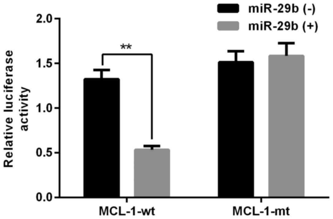
and its target mRNA as candidates for our experiments. To
investigate the effects of miR-29b, two psiCHECK2 luciferase
plasmids containing MCL-1-3′UTR-WT and MCL-1-3′UTR-MUT segments
were co-transfected with miR-29b into N2a cells. Treatment with
both miR-29b and psiCHECK2-MCL-1-3′UTR-WT decreased luciferase
activity in N2a cells compared to cells transfected with
psiCHECK2-MCL-1-3′UTR-MUT (Fig. 1).
As previous studies have shown miR-29b has a complementary site
with the 3′ UTR of Mcl-1, and miR-29b can downregulate the
expression level of Mcl-1 (14).
Therefore, we suggested that miR-29b negatively regulated
Mcl-1.
Effect of exogenous miR-29b mimics and
miR-29b inhibitor in N2a cells
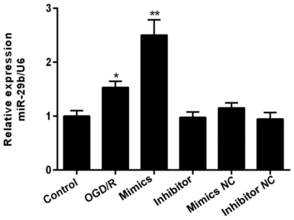
We next explored the effect of miR-29b in
vitro. We found that miR-29b levels were significantly higher
under an OGD/R environment (Fig. 2).
Increased miRNA detection after 48 h confirmed successful
transfection with miR-29b mimics.
Effects of miR-29b mimics and
inhibitor on cell proliferation
The effects of miR-29b mimics and inhibitor on N2a
cell viability were subsequently assessed. In OGD/R pretreatment
N2a cells, cellular viability was significantly reduced by OGD/R,
and then further decreased by miR-29b mimic compared to negative
control (NC) groups. An opposite effect was found for cells treated
with miR-29b inhibitor (Fig. 3).
Effects of miR-29b mimics and
inhibitor on cell apoptosis
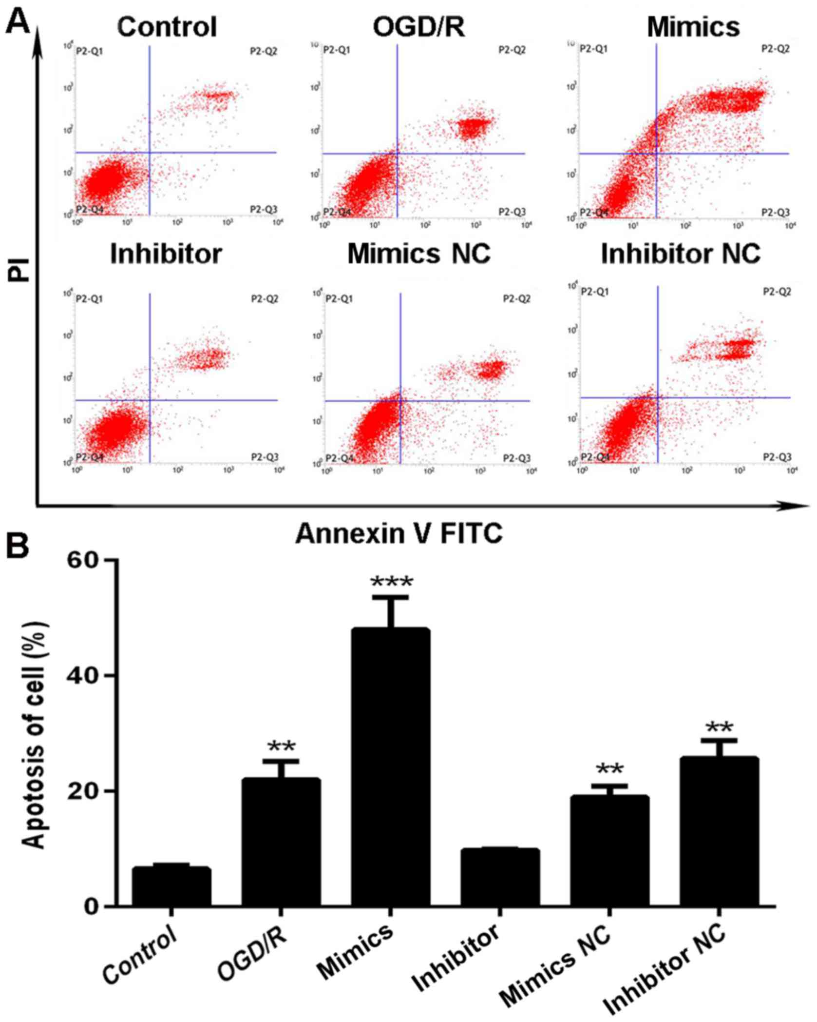
We also assessed rates of apoptosis following
transfection with miR-29b mimics or inhibitor following OGD/R
pre-treatment for 48 h using flow cytometry. Compared to N2a cells
transfected with miR-29b mimics, cells exposed to miR-29b inhibitor
had less cells exhibiting apoptotic morphology, such as nuclear
fragmentation, cell shrinkage, and cellular rupture debris.
Conversely, apoptosis occurred at a significantly higher rate in
cells treated with the miR-29b mimics, compared to the group
treated with OGD/R only (P<0.01; Fig.
4A). The percentage differences in N2a cell apoptosis rates are
shown in Fig. 4B.
Effect of miR-29b mimics and inhibitor
on Wnt-associated proteins in N2a cells
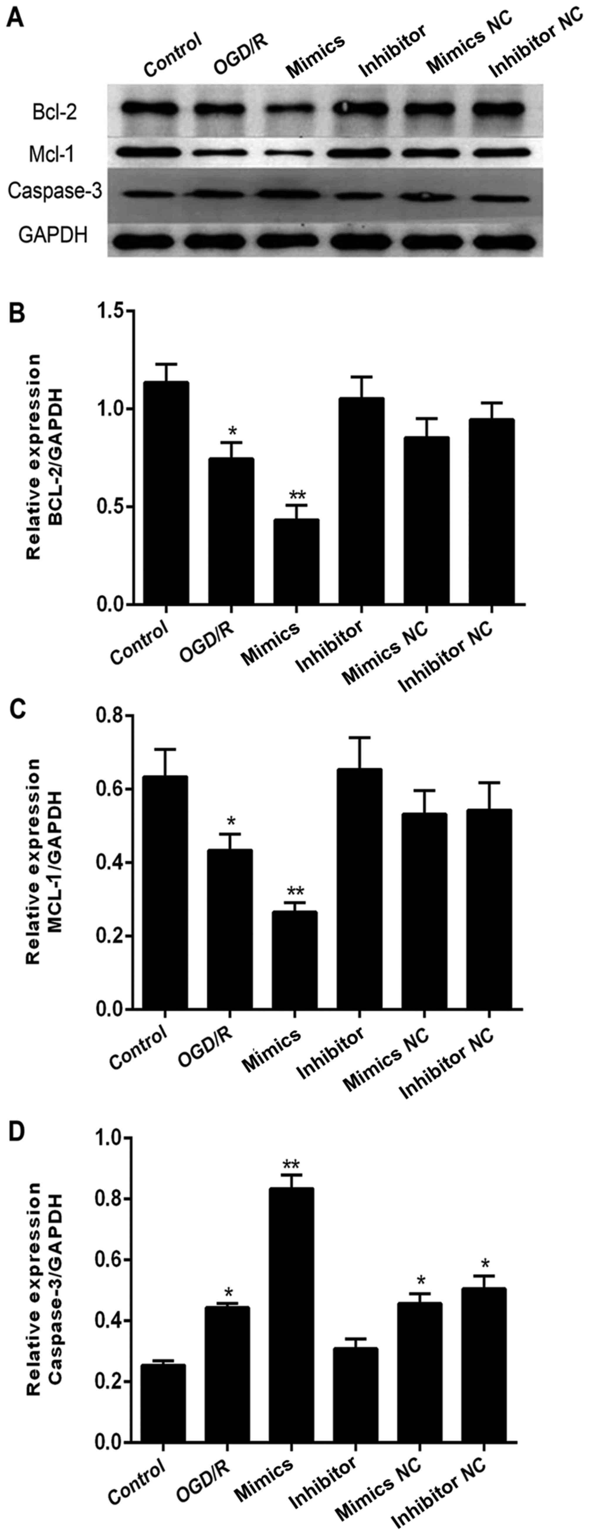
In addition to MCL-1, we also investigated BCL2 and
caspase-3 (CASP3) expression across the same groups. We found that
BCL2 levels (Fig. 5A-B) followed a
similar trend to that of MCL-1 (Fig. 5A
and C) and was downregulated in an OGD/R environment.
Expression was inhibited further in cells transfected with miR-29b
mimics and but higher in cells exposed to miR-29b inhibitor.
Caspase-3 (CASP3) levels showed an opposite effect (Fig. 5A and D).
Discussion
There is an increasing volume of interdisciplinary
research linking cancer and neurodegenerative diseases. In the
present study, we have demonstrated that apoptosis is clearly
increased after exposure to an OGD/R environment. We have also
shown in vitro the neuroprotective effects of miR-29b
inhibitor on I/R injury through promoting cell viability and
suppressing apoptosis. Our data suggests that these effects are
mediated through the targeting of MCL-1. Transfection with miR-29b
mimics led to up-regulated expression of MCL-1 and BCL2 proteins
and a correlating down-regulation of expression for cleaved
caspase-3 (CASP3) proteins in our cerebral I/R injury cell model.
We therefore conclude that the miR-29b inhibitor can act as a
protective regulator against cell apoptosis in cell models that
mimic cerebral I/R injury.
A considerable amount of previous research has
focused on the regulatory role of miR-29 in carcinoma. However,
miR-29b is also significantly up-regulated in neural cells under an
OGD/R environment and in patients with cerebral I/R injury
(10,15). Activated miR-29b has also been
reported to promote neural cell apoptosis targeting BH3 protein
during neuronal maturation (16).
Supporting previous studies, our results indicate that miR-29 binds
the 3′UTR of MCL-1, an important Bcl-2 family protein. Proteins in
the Bcl-2 family are defined by the presence of Bcl-2 homology (BH)
domains but are divided into anti- or pro-apoptotic regulators.
They act through modifying mitochondrial membrane integrity and
function, and also affect apoptotic signaling. The Bcl-2 family
consists of three subgroups, pro-survival proteins (BCL2, BCLxl
[BCL2L1], BCLw [BCL2L2], MCL-1, and A1), multi-domain pro-apoptotic
proteins (BAX and BAK), and BH3 domain-only pro-apoptotic proteins
(BIM, PUMA [BBC3], BID, BAD, BIK, BMF, HRK, and NOXA [PMAIP1])
(17–19). As an important anti-apoptotic protein
in the Bcl-2 family, inhibition of MCL-1 promotes cell death
through the mitochondrial pathway and there is some data suggesting
it is also involved in the apoptosis that occurs during cerebral
ischemic reperfusion (20). Based on
these data and our results, we hypothesize that the interaction
between miR-29b and MCL-1 is a critical factor during neural
cell apoptosis. Finally, recent research has also shown that
several miRNAs directly target the 3′UTR of Bcl-2 family proteins.
For example, miR-15b is highly expressed following permanent middle
cerebral artery occlusion (MCAO) and may directly target
BCL2 (21). Another miRNA,
miR-491-5p, has also been suggested to bind Bcl-xL
(BCL2L1) mRNA, an anti-apoptotic member of the Bcl-2 family
(22). Additionally, up-regulation
of miR-29b promotes neuronal cell death by inhibiting BCL2 after
ischemic brain injury. As many chemotherapeutic drugs induce
apoptosis through down-regulation of MCL-1 expression in tumor
cells, our results suggest that targeting miR-29b may also be a
therapeutic target in cerebral I/R injury by inhibiting MCL-1
expression.
Our experiments have confirmed our initial
hypothesis that miR-29b affects neurocyte apoptosis during cerebral
I/R injury through targeting MCL-1. We have shown this at both the
protein and histological level. However, there were certain
limitations in our experiments. For example, our data was obtained
using miR-29b mimics in an in vitro environment, and
therefore, may not accurately reflect in vivo effects. We
therefore plan to conduct further experiments in MCAO animal models
to explore the relationships between reperfusion and treatment with
miR-29b inhibitor or overexpression of MCL-1. In addition, further
studies are needed to validate the neurodamaging and
neuroprotective effects of miR-29b by targeting MCL-1 during
cerebral ischemia/reperfusion injury. Moreover, it will be
necessary to further explore the functions and mechanisms of
miR-29b in neuronal cells during cerebral ischemia/reperfusion
injury. This will lead to better clinical outcomes for patients
that suffer an ischemic stroke.
Acknowledgements
The present study was supported by funds from the
Natural Science Foundation of China (no. 81460276), the Science and
Technology Development Funds of Guizhou (no. J. [2015] 2090).
Funding
This study was supported by funds from the Natural
Science Foundation of China (no. 81460276), the Science and
Technology Development Funds of Guizhou (no. J. [2015] 2090).
Availability of data and materials
All data generated or analysed during this study are
included in this article.
Authors' contributions
ZH, LL, SAZ and JL designed the study. TPJ, SAZ, YPS
and ZZ performed the experiments. ZH wrote the paper. ASZ, RG and
RL helped perform the analysis with constructive discussions. SZ
revised the manuscript. All authors read and approved the
manuscript.
Consent for publication
Not applicable.
Competing interests
The authors declare that they have no competing
interests.
Ethics approval and consent to
participate
Not applicable.
References
|
1
|
Zhai WW, Sun L, Yu ZQ and Chen G:
Hyperbaric oxygen therapy in experimental and clinical stroke. Med
Gas Res. 6:111–118. 2016. View Article : Google Scholar : PubMed/NCBI
|
|
2
|
Wang W, Zhao L, Bai F, Zhang T, Dong H and
Liu L: The protective effect of dopamine against OGD/R
injury-induced cell death in HT22 mouse hippocampal cells. Environ
Toxicol Pharmacol. 42:176–182. 2016. View Article : Google Scholar : PubMed/NCBI
|
|
3
|
Fluri F, Schuhmann MK and Kleinschnitz C:
Animal models of ischemic stroke and their application in clinical
research. Drug Des Devel Ther. 9:3445–3454. 2015.PubMed/NCBI
|
|
4
|
Ni J, Wang X, Chen S, Liu H, Wang Y, Xu X,
Cheng J, Jia J and Zhen X: MicroRNA let-7c-5p protects against
cerebral ischemia injury via mechanisms involving the inhibition of
microglia activation. Brain Behav Immun. 49:75–85. 2015. View Article : Google Scholar : PubMed/NCBI
|
|
5
|
Zuo XL, Deng HL, Wu P and Xu E: Do
different reperfusion methods affect the outcomes of stroke induced
by MCAO in adult rats? Int J Neurosci. 126:850–855. 2016.
View Article : Google Scholar : PubMed/NCBI
|
|
6
|
Li F, Shi W, Zhao EY, Geng X, Li X, Peng
C, Shen J, Wang S and Ding Y: Enhanced apoptosis from early
physical exercise rehabilitation following ischemic stroke. J
Neurosci Res. 95:1017–1024. 2017. View Article : Google Scholar : PubMed/NCBI
|
|
7
|
De Gasperi R, Graham ZA, Harlow LM, Bauman
WA, Qin W and Cardozo CP: The signature of microRNA dysregulation
in muscle paralyzed by spinal cord injury includes downregulation
of microRNAs that target myostatin signaling. PLoS One.
11:e01661892016. View Article : Google Scholar : PubMed/NCBI
|
|
8
|
Xia HF, Jin XH, Cao ZF, Hu Y and Ma X:
MicroRNA expression and regulation in the uterus during embryo
implantation in rat. FEBS J. 281:1872–1891. 2014. View Article : Google Scholar : PubMed/NCBI
|
|
9
|
Floris I, Kraft JD and Altosaar I: Roles
of MicroRNA across prenatal and postnatal periods. Int J Mol Sci.
17:E19942016. View Article : Google Scholar : PubMed/NCBI
|
|
10
|
Di Y, Lei Y, Yu F, Changfeng F, Song W and
Xuming M: MicroRNAs expression and function in cerebral ischemia
reperfusion injury. J Mol Neurosci. 53:242–250. 2014. View Article : Google Scholar : PubMed/NCBI
|
|
11
|
Jafarinejad-Farsangi S, Farazmand A,
Mahmoudi M, Gharibdoost F, Karimizadeh E, Noorbakhsh F, Faridani H
and Jamshidi AR: MicroRNA-29a induces apoptosis via increasing the
Bax: Bcl-2 ratio in dermal fibroblasts of patients with systemic
sclerosis. Autoimmunity. 48:369–378. 2015. View Article : Google Scholar : PubMed/NCBI
|
|
12
|
Xu L, Xu Y, Jing Z, Wang X, Zha X, Zeng C,
Chen S, Yang L, Luo G, Li B and Li Y: Altered expression pattern of
miR-29a, miR-29b and the target genes in myeloid leukemia. Exp
Hematol Oncol. 3:172014. View Article : Google Scholar : PubMed/NCBI
|
|
13
|
Mott JL, Kobayashi S, Bronk SF and Gores
GJ: mir-29 regulates Mcl-1 protein expression and apoptosis.
Oncogene. 26:6133–6140. 2007. View Article : Google Scholar : PubMed/NCBI
|
|
14
|
Zhang YK, Wang H, Leng Y, Li ZL, Yang YF,
Xiao FJ, Li QF, Chen XQ and Wang LS: Overexpression of microRNA-29b
induces apoptosis of multiple myeloma cells through down regulating
Mcl-1. Biochem Biophys Res Commun. 414:233–239. 2011. View Article : Google Scholar : PubMed/NCBI
|
|
15
|
Altintas O, Ozgen Altintas M, Kumas M and
Asil T: Neuroprotective effect of ischemic preconditioning via
modulating the expression of cerebral miRNAs against transient
cerebral ischemia in diabetic rats. Neurol Res. 38:1003–1011. 2016.
View Article : Google Scholar : PubMed/NCBI
|
|
16
|
Annis RP, Swahari V, Nakamura A, Xie AX,
Hammond SM and Deshmukh M: Mature neurons dynamically restrict
apoptosis via redundant premitochondrial brakes. Febs J.
283:4569–4582. 2016. View Article : Google Scholar : PubMed/NCBI
|
|
17
|
Delgado-Soler L, Del Mar Orzaez M and
Rubio-Martinez J: Structure-based approach to the design of BakBH3
mimetic peptides with increased helical propensity. J Mol Model.
19:4305–4318. 2013. View Article : Google Scholar : PubMed/NCBI
|
|
18
|
Santiveri CM, Sborgi L and de Alba E:
Nuclear magnetic resonance study of protein-protein interactions
involving apoptosis regulator Diva (Boo) and the BH3 domain of
proapoptotic Bcl-2 members. J Mol Recognit. 25:665–673. 2012.
View Article : Google Scholar : PubMed/NCBI
|
|
19
|
Smits C, Czabotar PE, Hinds MG and Day CL:
Structural plasticity underpins promiscuous binding of the
prosurvival protein A1. Structure. 16:818–829. 2008. View Article : Google Scholar : PubMed/NCBI
|
|
20
|
Dewson G: Characterizing Bcl-2 family
protein conformation and oligomerization using cross-linking and
antibody gel-shift in conjunction with native PAGE. Methods Mol
Biol. 1419:185–196. 2016. View Article : Google Scholar : PubMed/NCBI
|
|
21
|
Shi H, Sun BL, Zhang J, Lu S, Zhang P,
Wang H, Yu Q, Stetler RA, Vosler PS, Chen J and Gao Y: miR-15b
suppression of Bcl-2 contributes to cerebral ischemic injury and is
reversed by sevoflurane preconditioning. CNS Neurol Disord Drug
Targets. 12:381–391. 2013. View Article : Google Scholar : PubMed/NCBI
|
|
22
|
Denoyelle C, Lambert B, Meryet-Figuière M,
Vigneron N, Brotin E, Lecerf C, Abeilard E, Giffard F, Louis MH,
Gauduchon P, et al: miR-491-5p-induced apoptosis in ovarian
carcinoma depends on the direct inhibition of both BCL-XL and EGFR
leading to BIM activation. Cell Death Dis. 5:e14452014. View Article : Google Scholar : PubMed/NCBI
|